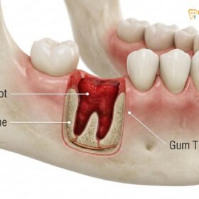
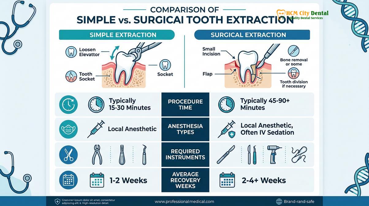
The primary difference between a simple and surgical extraction lies in tooth accessibility and procedural complexity. Simple extractions remove visible, intact teeth using elevators and forceps, while surgical extractions require incisions, bone removal, or tooth sectioning to extract impacted or severely damaged teeth.
Clinical Summary:
Understanding the simple vs surgical extraction differences is crucial for patients preparing for dentoalveolar procedures. A simple extraction is a straightforward, non-invasive procedure performed on fully erupted teeth with sound coronal structure. It relies on the biomechanical principles of luxation to sever the periodontal ligament and deliver the tooth intact. Conversely, a surgical extraction is a complex intervention required when a tooth is impacted, fractured below the gumline, or possesses divergent roots. This method involves elevating a mucoperiosteal flap, performing an osteotomy (bone removal), and often sectioning the tooth to facilitate safe removal. While simple extractions typically require only local anesthesia and boast a rapid recovery, surgical extractions may necessitate conscious sedation, the placement of sutures, and a longer healing timeline. Clinical diagnostics, including three-dimensional imaging, dictate the appropriate approach to ensure optimal alveolar bone preservation and minimize post-operative complications.
Key Takeaways:
- Visibility and Access: Simple extractions are for teeth visible above the gumline; surgical extractions address teeth trapped beneath the gums or bone.
- Procedural Technique: Simple methods use forceps and elevators; surgical methods require scalpels, surgical burs, and tooth sectioning.
- Anesthesia Requirements: Local anesthesia is standard for simple removals, whereas surgical cases frequently utilize supplemental sedation.
- Tissue Management: Surgical extractions require incisions and sutures to close the surgical site, unlike the natural clot formation in simple extractions.
- Recovery Timeline: Soft tissue healing takes 1-2 weeks for simple procedures, but up to 3-4 weeks for surgical interventions.
The Clinical Categories of Tooth Removal
Tooth removal is clinically categorized based on the tooth’s eruption status, structural integrity, and root anatomy, which dictate whether a non-invasive or surgical approach is required.
In the realm of modern dentistry, the removal of a tooth—clinically termed exodontia—is never a one-size-fits-all procedure. The decision to extract a tooth is typically the culmination of extensive diagnostic evaluation, aimed at addressing severe dental caries, advanced periodontal disease, untreatable pulpal pathology, or significant orthodontic crowding. Once the necessity of extraction is established, the clinician must determine the most appropriate surgical approach. This determination forms the core of the simple vs surgical extraction differences, a categorization that profoundly impacts the clinical workflow, the instruments utilized, and the patient’s post-operative experience.
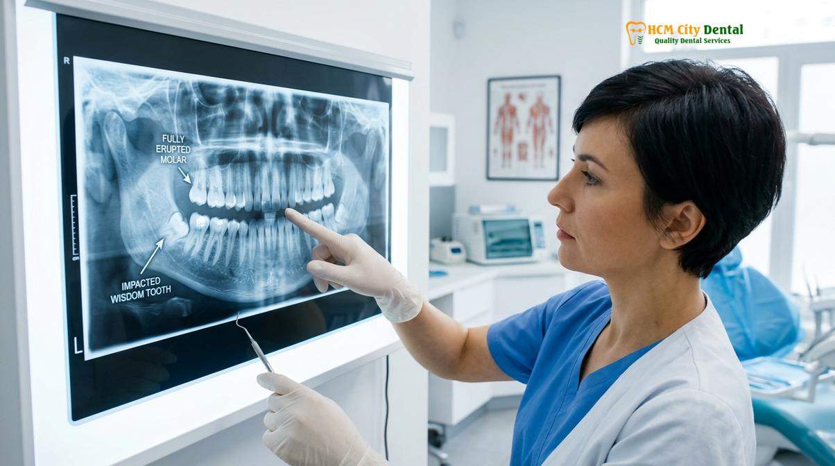
The diagnostic phase is paramount in categorizing the extraction. Clinicians rely heavily on radiographic imaging, ranging from standard periapical and panoramic radiographs to advanced Cone Beam Computed Tomography (CBCT). These imaging modalities allow the dentist to visualize the entire tooth structure, including the morphology of the roots, their proximity to vital anatomical landmarks (such as the inferior alveolar nerve or the maxillary sinus), and the density of the surrounding alveolar bone[1]. A tooth that appears straightforward upon visual inspection may reveal severely curved (dilacerated) roots on an X-ray, instantly reclassifying the procedure from a simple to a surgical extraction.
Dr. Nguyen Van Cuong, a leading practitioner at HCMC Dental Clinic, emphasizes that the structural integrity of the clinical crown is a primary determining factor. If a tooth has been heavily restored, is severely decayed, or has undergone endodontic treatment (root canal therapy), it becomes brittle and highly susceptible to fracturing under the pressure of dental forceps. In such scenarios, even if the tooth is fully erupted and visible, the clinician must prepare for a surgical approach to retrieve retained root fragments should the crown shatter during the initial luxation attempts.
Furthermore, the patient’s medical history plays a critical role in treatment planning. Patients on anticoagulant therapy, those with a history of bisphosphonate use, or individuals with compromised immune systems require meticulous pre-operative planning. The chosen extraction category must align with the patient’s physiological capacity to heal. By thoroughly assessing these variables, dental professionals ensure that the chosen method—whether a routine procedure or one requiring the expertise of a Saigon oral surgeon—prioritizes patient safety and sets the stage for successful general dentistry services and future restorative work.
Simple Extraction: Forceps, Elevators, and Intact Root Removal
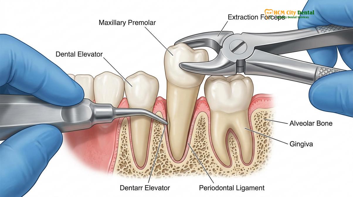
A simple extraction involves luxating a fully erupted, structurally sound tooth using dental elevators and removing it intact with forceps without incising the gingival tissue.
A simple extraction, often referred to as a closed extraction, is the most common form of tooth removal. It is indicated for teeth that are fully erupted, clearly visible within the oral cavity, and possess a solid crown structure capable of withstanding the biomechanical forces applied during the procedure. The fundamental goal of a simple forceps extraction is to atraumatically sever the periodontal ligament (PDL)—the fibrous connective tissue that anchors the tooth root to the alveolar bone—and expand the bony socket just enough to allow the tooth to be delivered intact.
The clinical workflow for a simple extraction is highly systematic. Following the administration of profound local anesthesia, the clinician begins with a process called syndesmotomy. Using a sharp instrument, such as a periotome or a scalpel blade, the gingival attachment around the neck of the tooth is carefully severed. This step prevents the tearing of the gum tissue when the tooth is eventually removed. Next, the dentist employs dental elevators. These instruments function on the principles of a lever and a wedge. By inserting the elevator into the periodontal space between the tooth root and the surrounding bone, the clinician applies controlled, sustained pressure. This pressure serves two critical purposes: it begins to tear the PDL fibers and it gently expands the viscoelastic alveolar bone[2].
“The essence of a successful simple extraction lies not in the application of brute force, but in the patient, controlled expansion of the alveolar socket. Atraumatic techniques are paramount to preserving the bony architecture, which is essential for future implant placement.”
Once the tooth has been sufficiently luxated (loosened) and exhibits significant mobility, dental forceps are introduced. The beaks of the forceps are designed to adapt precisely to the root anatomy of specific teeth—for instance, cowhorn forceps are uniquely shaped to engage the furcation of mandibular molars. The forceps are seated as apically (deeply) as possible to lower the center of rotation, thereby minimizing the risk of fracturing the crown. The clinician then applies a combination of apical, buccal (outward), lingual (inward), and rotational forces. The specific movement pattern depends on the tooth type; single-rooted anterior teeth often respond well to rotational forces, while multi-rooted molars require a figure-eight motion to expand the socket walls.
Throughout this process, alveolar bone preservation is a primary objective. Excessive force can lead to the fracture of the buccal bone plate, complicating the healing process and compromising the site for future restorative dentistry options. By utilizing precise, controlled movements, the tooth is eventually delivered from the socket in one piece. The socket is then thoroughly inspected, curetted to remove any granulation tissue or debris, and compressed with sterile gauze to initiate hemostasis and the formation of a stable blood clot.
Surgical Extraction: Gums Flaps, Bone Removal, and Sectioning
Surgical extractions are complex procedures requiring gingival incisions, osteotomy, or tooth division to safely remove impacted, fractured, or anatomically complicated teeth.
When a tooth cannot be accessed or removed using routine closed techniques, the procedure escalates to a surgical extraction, also known as an open extraction. This approach is mandatory for teeth that are impacted (partially or completely encased in bone and tissue), teeth that have fractured at or below the gingival margin, or teeth with severe root dilacerations that prevent a single path of withdrawal. The surgical approach provides the clinician with the necessary visibility and access to safely navigate complex anatomical challenges without causing undue trauma to adjacent structures.
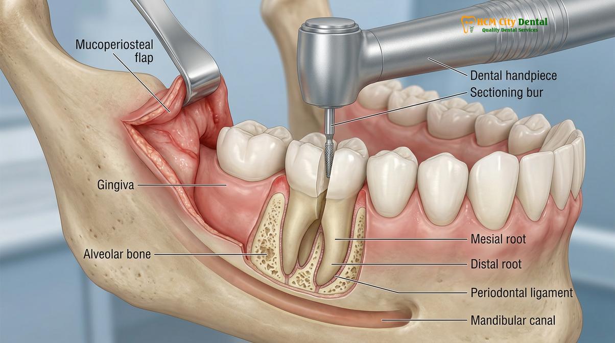
The hallmark of a surgical extraction is the creation of a mucoperiosteal flap. This surgical flap elevation involves making precise incisions in the gingival tissue using a scalpel. The design of the flap—whether an envelope flap, a triangular flap, or a trapezoidal flap—is carefully planned to ensure adequate blood supply to the tissue and to provide sufficient exposure of the underlying surgical site[3]. Once the incisions are made, a periosteal elevator is used to gently peel the gum tissue and the underlying periosteum away from the bone, exposing the surgical field.
With the bone exposed, the clinician must often perform an osteotomy (bone removal). Using a specialized surgical handpiece and sterile burs under copious saline irrigation, the bone obstructing the pathway of the tooth is carefully removed. The irrigation is critical; it prevents the surgical bur from overheating the bone, which could lead to thermal necrosis and severely impair post-operative healing. The goal of bone removal is to create a “purchase point” for elevators or to expose the maximum contour of the tooth crown.
Clinical Case Review: Complex Mandibular Impaction
A 24-year-old patient presented to HCMC Dental Clinic in Ho Chi Minh City with severe pericoronitis associated with a horizontally impacted lower right wisdom tooth. Radiographic analysis revealed the roots were in close proximity to the inferior alveolar nerve canal. Dr. Nguyen Van Cuong designed a full-thickness mucoperiosteal flap and utilized a precise tooth sectioning technique to divide the crown from the roots. The roots were then elevated individually, completely avoiding nerve trauma. The site was irrigated and closed with resorbable sutures, resulting in an uneventful recovery with no neurological deficits.
In many surgical cases, particularly with multi-rooted molars or severely impacted wisdom teeth, attempting to remove the tooth in one piece is impossible or highly dangerous. This necessitates the tooth sectioning technique (odontotomy). Using a surgical bur, the clinician strategically divides the tooth into smaller, manageable fragments. For a lower molar, this often involves sectioning the tooth vertically through the furcation to separate the mesial and distal roots. Each root can then be elevated and removed independently along its own path of least resistance. This technique significantly reduces the amount of bone that must be removed and minimizes the force transmitted to the jawbone, thereby reducing the risk of iatrogenic mandibular fractures.
Following the complete removal of the tooth fragments, the surgical site is meticulously debrided. A bone file may be used to smooth any sharp bony edges that could irritate the overlying soft tissue. The surgical flap is then repositioned and secured using stitches and sutures. The choice of suture material—whether resorbable (like chromic gut or Vicryl) or non-resorbable (like silk or nylon)—depends on the clinician’s preference and the specific requirements of the surgical site. The surgical extraction, while more invasive, is a highly controlled procedure designed to manage complex dental pathologies safely and effectively.
Comparing Anesthesia and Sedation Needs for Each Method
Simple extractions typically require only local anesthesia to block pain transmission, whereas surgical extractions often combine local anesthetics with varying levels of conscious sedation for patient comfort.
Pain management and patient comfort are paramount in any dentoalveolar surgery. The simple vs surgical extraction differences become distinctly apparent when evaluating the pharmacological approaches used to achieve profound anesthesia and manage patient anxiety. Because the invasiveness and duration of the procedures vary significantly, the anesthesia protocols must be tailored to the specific demands of the clinical scenario.
For a simple extraction, local anesthesia is almost universally sufficient. The goal is to completely block the nociceptive (pain) signals from the tooth, the surrounding periodontal ligament, and the adjacent gingival tissues. Clinicians typically utilize amide local anesthetics, such as Lidocaine or Articaine, combined with a vasoconstrictor like epinephrine. The vasoconstrictor serves to prolong the duration of the anesthesia and provide localized hemostasis, reducing bleeding during the procedure[4]. Depending on the location of the tooth, the dentist will administer either an infiltration injection (depositing the anesthetic near the root apex, common for maxillary teeth) or a regional nerve block (such as the Inferior Alveolar Nerve Block, standard for mandibular teeth). Under local anesthesia, the patient remains fully conscious and alert. They will feel pressure and vibration during the luxation and delivery of the tooth, but they should experience absolutely no sharp pain.
Conversely, surgical extractions often necessitate a more comprehensive approach to patient management. While local anesthesia remains the foundation for pain control, the extended duration of the procedure, the sounds and sensations of bone removal, and the inherent anxiety associated with oral surgery frequently warrant the use of supplemental sedation. The level of sedation is determined by the complexity of the surgery and the patient’s medical history and anxiety levels.
| Clinical Parameter | Simple Extraction | Surgical Extraction |
|---|---|---|
| Primary Anesthesia | Local infiltration or nerve block | Profound local anesthesia (mandatory) |
| Sedation Requirement | Rarely required; occasionally Nitrous Oxide | Often recommended (Oral, IV, or Nitrous Oxide) |
| Patient Experience | Aware of pressure and movement | Deeply relaxed, reduced memory of procedure |
| Procedure Duration | Typically 10 to 20 minutes | Can range from 30 to 90+ minutes |
| Post-Op Analgesia | Over-the-counter NSAIDs (Ibuprofen) | Prescription analgesics and anti-inflammatories |
Sedation options range from minimal sedation using Nitrous Oxide (laughing gas), which provides a mild anxiolytic effect and rapid recovery, to moderate oral conscious sedation using benzodiazepines (such as Triazolam or Diazepam). For highly complex cases, such as the simultaneous removal of four deeply impacted wisdom teeth, intravenous (IV) sedation may be administered by a qualified specialist. IV sedation allows for precise titration of the sedative agents, ensuring the patient remains in a state of deep relaxation, often with little to no memory of the surgical event. Regardless of the method chosen, the integration of appropriate anesthesia and sedation protocols ensures that patients undergoing professional tooth removal experience minimal psychological and physiological stress.
Post-Extraction Healing: Stitches, Clot Formation, and Timelines
Recovery from simple extractions is generally rapid with basic clot formation, while surgical extractions involve primary closure with sutures and a longer tissue regeneration phase.
The biological process of wound healing following an extraction is a complex cascade of cellular events. Understanding the simple vs surgical extraction differences in recovery timelines helps manage patient expectations and ensures compliance with post-operative care instructions. The healing process is broadly divided into four overlapping phases: hemostasis, inflammation, proliferation, and remodeling.
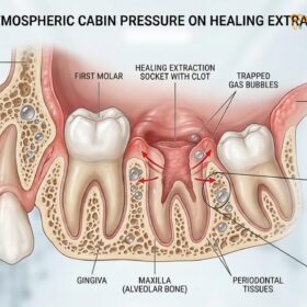
Following a simple extraction, the primary objective is hemostasis—the cessation of bleeding. The patient is instructed to bite down firmly on sterile gauze, applying direct pressure to the socket. Within the first 24 hours, a robust blood clot forms within the empty alveolar socket. This clot is the biological scaffold essential for all subsequent healing; it protects the exposed bone and provides a matrix for the influx of fibroblasts and osteoblasts. Because a simple extraction does not involve incisions, the gingival margins naturally contract over the clot. Soft tissue healing (epithelialization) over the socket surface typically occurs within one to two weeks. Patients generally experience mild to moderate discomfort for a few days, which is easily managed with over-the-counter non-steroidal anti-inflammatory drugs (NSAIDs).
Surgical extractions, due to the elevation of mucoperiosteal flaps and the removal of bone, initiate a more profound inflammatory response. The surgical site is closed using stitches and sutures to achieve primary intention healing, where the edges of the wound are brought closely together. This suturing minimizes the size of the blood clot required and protects the underlying bone graft or surgical site from oral contaminants[5]. However, the surgical trauma inevitably leads to greater post-operative edema (swelling) and ecchymosis (bruising), which typically peak around 48 to 72 hours post-surgery.
Clinical Warning: Alveolar Osteitis (Dry Socket)
Regardless of whether the extraction is simple or surgical, the premature dislodgement or dissolution of the blood clot can lead to Alveolar Osteitis, commonly known as dry socket. This condition exposes the underlying alveolar bone and nerve endings, causing severe, radiating pain and a foul odor. To prevent this, patients must strictly avoid smoking, using drinking straws, vigorous rinsing, and consuming hard or hot foods for at least 72 hours post-extraction.
The recovery timeline for a surgical extraction is notably longer. While the initial acute inflammation subsides within a week, the soft tissue may take three to four weeks to fully close and mature. If non-resorbable sutures were used, the patient must return to the clinic for their removal after 7 to 10 days. The underlying bone remodeling—where the woven bone is gradually replaced by mature lamellar bone—is a slow process that continues for several months. During this period, patients may be prescribed a short course of antibiotics to prevent secondary infection, along with stronger analgesics to manage the initial post-operative pain.
“Patient compliance is the most critical variable in post-operative healing. The most flawlessly executed surgical extraction can be compromised if the patient fails to adhere to the prescribed oral hygiene and dietary restrictions during the crucial first week of recovery.”
Whether recovering from a routine procedure or a complex surgery performed by a Saigon oral surgeon, maintaining excellent oral hygiene is vital. Gentle warm salt water rinses (starting 24 hours after surgery) help keep the surgical site clean without disrupting the healing tissue. By understanding the physiological demands of recovery, patients can actively participate in their healing process, ensuring a smooth transition to subsequent comprehensive oral evaluation and restorative phases.
When to See a Doctor
While post-operative discomfort, swelling, and minor oozing are expected physiological responses following both simple and surgical extractions, certain symptoms indicate a deviation from normal healing and require immediate clinical evaluation. Patients should not attempt to self-diagnose or manage severe complications at home.
You should contact your dental provider immediately if you experience prolonged or heavy bleeding that does not subside after applying firm, continuous pressure with gauze for 45 to 60 minutes. Similarly, if swelling worsens after the third post-operative day, or if it extends down the neck or up toward the eye, this may indicate a spreading fascial space infection. The presence of purulent exudate (pus) discharging from the extraction site, accompanied by a persistent fever, chills, or difficulty swallowing (dysphagia) and breathing, are signs of a severe systemic infection that demands urgent medical intervention.
Additionally, if you experience severe, throbbing pain that radiates to the ear or temple and is unresponsive to prescribed analgesics—especially if this pain develops 3 to 5 days after the procedure—you may have developed a dry socket. Finally, any prolonged numbness, tingling, or altered sensation (paresthesia) in the lips, tongue, or chin that persists beyond the expected duration of the local anesthetic should be evaluated by your clinician to assess for potential nerve involvement. Prompt communication with your dental team ensures that complications are managed swiftly, safeguarding your overall oral health.
Frequently Asked Questions
How do I know if my extraction will be simple or surgical?
Your dentist determines this through clinical examination and X-rays, assessing tooth visibility, root curvature, and structural integrity. If the tooth is fully erupted, has straight roots, and possesses a strong, intact crown, a simple extraction is highly likely. However, if the X-rays reveal that the tooth is impacted beneath the bone, has severely curved roots, or is badly decayed to the point where it might crumble under pressure, the dentist will plan for a surgical extraction to ensure safe and complete removal.
Does a surgical extraction require a specialist oral surgeon in HCMC?
While many general dentists perform surgical extractions, complex cases like deeply impacted wisdom teeth are often referred to a specialized oral surgeon for optimal safety. General dentists are well-equipped to handle routine surgical extractions, such as removing broken roots or teeth requiring minor bone removal. However, if the tooth is in close proximity to major nerves, the maxillary sinus, or if the patient has complex medical conditions requiring advanced sedation, referral to a specialist ensures the highest standard of care and minimizes surgical risks.
Why do dentists section a tooth during extraction?
Dentists section a tooth to remove it in smaller pieces, which minimizes trauma to the surrounding jawbone and prevents root fractures during the procedure. Multi-rooted teeth, especially those with roots pointing in different directions (divergent), cannot be pulled straight out without breaking the surrounding bone. By using a surgical handpiece to divide the tooth—often separating the crown from the roots or splitting the roots apart—each segment can be elevated individually along its own path of least resistance, preserving the alveolar bone for future implants.
Is a surgical extraction more painful than a simple extraction?
During the procedure, neither method is painful due to the administration of profound local anesthesia. You will feel pressure and vibration, but no sharp pain. However, because surgical extractions involve incisions into the gum tissue, the removal of bone, and a longer procedural time, patients typically experience a higher degree of post-operative soreness, swelling, and inflammation. This post-operative discomfort is effectively managed with prescribed anti-inflammatory medications and analgesics during the initial recovery phase.
How long does it take to recover from a surgical extraction compared to a simple one?
Simple extractions usually heal within one to two weeks, whereas surgical extraction sites may take three to four weeks for the soft tissue to fully close. In a simple extraction, the blood clot forms quickly and the gums heal over the small socket rapidly. A surgical extraction involves stitches and a larger wound area, requiring more time for the gingival tissues to reattach and heal. For both procedures, the underlying bone remodeling process takes several months to complete, highlighting the importance of following all post-operative care instructions.
References
- Journal of Oral and Maxillofacial Surgery. Biomechanical principles of exodontia and alveolar bone preservation. (2021).
- International Journal of Oral and Maxillofacial Surgery. Comparative analysis of simple versus surgical extraction healing timelines. (2020).
- British Dental Journal. The efficacy of tooth sectioning techniques in minimizing surgical trauma. (2019).
- Journal of the American Dental Association. Guidelines for local anesthesia and sedation in dentoalveolar surgery. (2022).
- Clinical Oral Investigations. Soft tissue management and mucoperiosteal flap designs in oral surgery. (2018).
For pricing, booking, and a free clinical assessment, visit our Tooth Extraction service page at HCMC Dental Clinic in Ho Chi Minh City.
![Black Stain on Teeth: How to Get Rid Of [Solved] | Dr. Cuong](https://hcmcdental.com/wp-content/uploads/2025/07/black-stain-on-teeth-how-to-get-rid-of-1-dr-cuong-hcmc-dental-clinic-280x280.jpg)