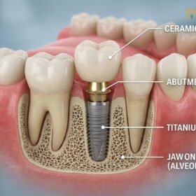
A bone graft for a dental implant is a surgical procedure designed to add volume and density to your jawbone, creating a stable foundation for the titanium post. This treatment is often essential when natural bone has deteriorated due to tooth loss, gum disease, or trauma, helping to ensure long-term implant success and structural integrity.
Clinical Summary:
Bone grafting is a critical preparatory step in modern implantology for patients exhibiting insufficient alveolar bone volume. The procedure utilizes various biomaterials—including autografts, allografts, xenografts, and alloplasts—to act as a scaffold, stimulating the body’s natural osteogenic processes. By regenerating lost bone width and height, the graft helps ensure that the subsequent dental implant achieves optimal primary stability and long-term osseointegration. While the surgical process is straightforward and performed under local anesthesia, the biological maturation of the graft requires a dedicated healing period, typically ranging from three to six months, before the final prosthetic phase can commence.
Key Takeaways:
- Bone grafts rebuild the jaw’s structural integrity, providing a necessary foundation for successful implant placement.
- Materials range from the patient’s own bone to highly processed donor, animal, or synthetic substitutes.
- The procedure is minimally invasive, typically performed under local anesthesia with highly manageable postoperative discomfort.
- Complete bone maturation and integration usually require a healing period of three to six months.
- Strict adherence to postoperative care, including a soft diet and proper oral hygiene, helps prevent complications and graft failure.
Why Do I Need a Bone Graft for My Dental Implant?
Bone grafting is generally necessary when your jaw lacks the structural integrity to anchor an implant securely, preventing premature failure and restoring facial aesthetics.
The human jawbone relies on the constant mechanical stimulation of chewing to maintain its density and volume. When a tooth is lost or extracted, this vital stimulation ceases. Consequently, the body begins to resorb the surrounding alveolar bone, redirecting calcium and minerals to other parts of the body. Within the first year of tooth loss, a patient can lose up to 25% of their bone volume in that specific area, and this deterioration continues progressively over time[1]. To successfully place a dental implant, a specific width and height of healthy bone are required to completely encase the titanium threads.
If an implant is placed into inadequate bone, it lacks primary stability. The micro-movements caused by chewing forces can disrupt the delicate osseointegration process, leading to implant failure. Furthermore, insufficient bone can result in aesthetic complications, such as the gray metal of the implant showing through thin gums, or functional issues like an unnatural crown-to-root ratio. In these clinical scenarios, a bone resorption ridge augmentation is performed to rebuild the lost architecture, ensuring the implant is housed in a robust, vascularized environment.
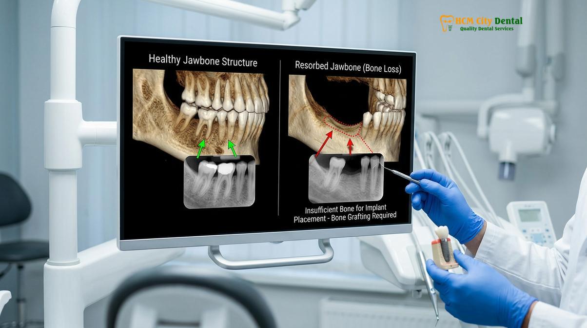
Beyond simple tooth extraction, several other clinical conditions necessitate bone grafting prior to implant therapy. Severe periodontal (gum) disease is a leading cause of bone destruction, eating away at the supporting structures of the teeth. Dental trauma, such as a sports injury or an accident, can fracture the bone, requiring reconstruction. Additionally, patients who have worn traditional removable prosthetics for extended periods often experience severe ridge flattening. For these individuals, exploring Dental Implants vs Dentures: Which Is the Better Investment? often reveals that implants, supported by proper grafting, offer superior long-term preservation of the jawbone.
Types of Bone Graft Materials: Autogenous, Allograft, and Synthetic
Dentists utilize various grafting materials, including the patient’s own bone, donor tissue, animal-derived minerals, or lab-made synthetic substitutes, depending on the clinical defect.
The selection of bone grafting material is a highly individualized clinical decision based on the size of the defect, the location in the mouth, and the patient’s overall health. The primary goal of any graft is to provide a scaffold that encourages the patient’s own bone cells (osteoblasts) to migrate into the area and generate new, living bone tissue[2]. This biological process is known as guided bone regeneration.
There are four primary categories of bone grafting materials used in modern implant dentistry:
- Autogenous Grafts (Autografts): Considered the “gold standard” in bone grafting, this involves harvesting bone from the patient’s own body—typically from the chin, the back of the lower jaw, or occasionally the hip for massive reconstructions. Because it contains the patient’s own living cells and growth factors, it offers the highest osteogenic (bone-forming) potential. However, it requires a second surgical site, which increases postoperative discomfort.
- Allogeneic Grafts (Allografts): This material is derived from human donor bone obtained from a certified tissue bank. The bone undergoes rigorous sterilization, freeze-drying, and processing to remove all cellular material, leaving behind a safe, neutral collagen and mineral matrix. It serves as an excellent scaffold (osteoconduction) without the need for a second surgical harvesting site.
- Xenogeneic Grafts (Xenografts): Xenografts are derived from animal sources, most commonly bovine (cow) or porcine (pig). Similar to allografts, the organic components are completely removed, leaving a highly porous mineral structure that closely mimics human bone anatomy. Xenografts are highly stable and resorb very slowly, making them ideal for maintaining space and volume during extensive ridge augmentations.
- Alloplastic Grafts (Synthetics): These are completely artificial materials created in a laboratory, such as hydroxyapatite, calcium phosphate, or bioactive glass. A synthetic bone scaffold eliminates any risk of disease transmission and is highly biocompatible. Depending on the specific formulation, synthetic grafts can be designed to resorb quickly or remain permanently integrated into the jaw.
When discussing high-end implant systems, such as Straumann vs Nobel Biocare Implants: What Is the Difference?, the quality of the surrounding bone is just as critical as the brand of the implant itself. A premium implant cannot perform optimally if placed in compromised bone.
| Graft Type | Source Material | Primary Mechanism | Clinical Advantage |
|---|---|---|---|
| Autograft | Patient’s own body | Osteogenesis (Living cells) | Highest success rate, fastest integration. |
| Allograft | Human tissue donor | Osteoconduction (Scaffold) | No second surgical site, highly predictable. |
| Xenograft | Animal (Bovine/Porcine) | Osteoconduction (Scaffold) | Excellent volume maintenance, slow resorption. |
| Alloplast | Laboratory synthesized | Osteoconduction (Scaffold) | Zero risk of disease transmission, highly available. |
The Surgical Procedure: What Happens During Grafts Placement
The grafting procedure involves administering local anesthesia, accessing the jawbone, placing the graft material, securing a protective membrane, and suturing the gums for healing.
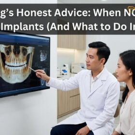
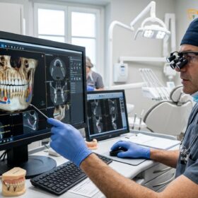
The bone grafting procedure is a meticulously planned surgical intervention. Before any incisions are made, comprehensive diagnostic imaging, typically a 3D Cone Beam Computed Tomography (CBCT) scan, is utilized. This allows the dental surgeon to evaluate the exact dimensions of the bone defect, locate vital anatomical structures like nerves and sinus cavities, and determine the precise volume of grafting material required.
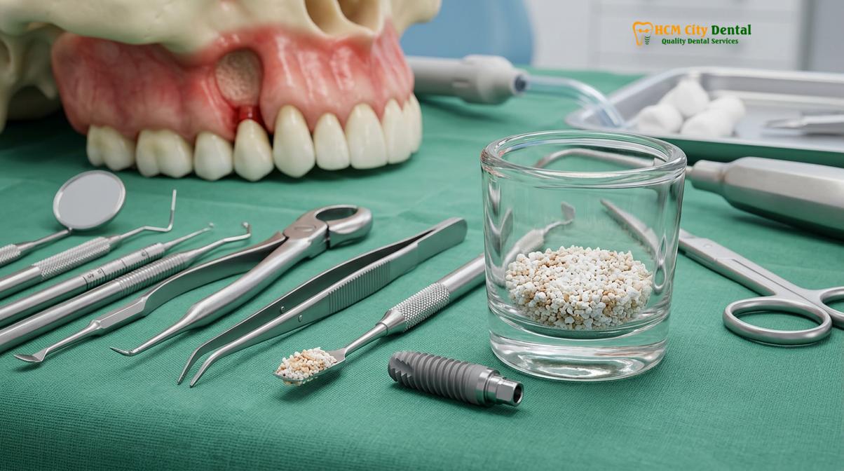
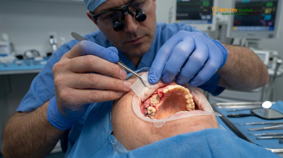
On the day of surgery, the procedure begins with the administration of profound local anesthesia to ensure the patient is completely comfortable. For patients with dental anxiety, conscious sedation options may also be provided. Once the area is numb, the surgeon makes a precise incision in the gingival tissue and gently reflects the gums to expose the underlying deficient bone. The recipient site is then thoroughly cleaned and debrided of any inflammatory tissue or residual infection.
The chosen bone graft material, often mixed with sterile saline or the patient’s own Platelet-Rich Fibrin (PRF) to accelerate healing, is carefully packed into the defect. To prevent fast-growing soft gum tissue from invading the space intended for slower-growing bone cells, a critical step known as membrane barrier placement is performed[3]. This membrane, which can be resorbable (dissolves naturally) or non-resorbable (requires later removal), acts as a protective tent over the graft. Finally, the gum tissue is repositioned over the membrane and secured with fine sutures.
Clinical Case Study: Maxillary Sinus Lift
A 55-year-old patient presented to HCMC Dental Clinic in Ho Chi Minh City with missing upper molars and severe vertical bone loss due to sinus pneumatization. The clinical team performed a lateral window sinus lift, elevating the delicate sinus membrane and placing a bovine xenograft to rebuild the vertical height. Following a six-month maturation period, the augmented site provided a dense, stable foundation, allowing for the successful placement of two premium titanium implants, fully restoring the patient’s posterior chewing function.
Understanding the intricacies of this procedure helps patients appreciate the value of the treatment. For those researching the financial aspects, reviewing How Much Do Dental Implants Cost in Vietnam? (Comprehensive Guide) provides clarity on how bone grafting procedures are factored into the overall comprehensive treatment plan.
Healing Timelines: Osseointegration and Bone Maturation
Complete bone maturation typically requires several months, allowing the graft material to fuse with your natural jawbone and create a dense, vascularized foundation.
The success of a bone graft is heavily dependent on the biological process of healing and maturation. Unlike a simple cut on the skin that heals in a week, bone regeneration is a complex, multi-phased cellular event. The healing time bone regeneration requires cannot be rushed, as placing an implant into immature bone significantly increases the risk of failure.
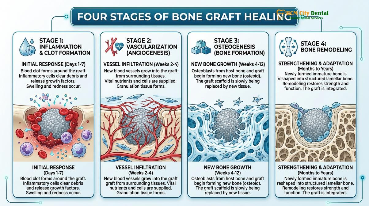
The healing timeline is generally broken down into several distinct phases:
- Inflammatory Phase (Days 1-7): Immediately following surgery, a blood clot forms within the graft matrix. The body’s immune system initiates an inflammatory response, bringing white blood cells to clean the area and releasing growth factors that signal the start of tissue repair.
- Vascularization Phase (Weeks 2-4): New blood vessels begin to infiltrate the bone graft material (angiogenesis). This blood supply is critical, as it delivers oxygen, nutrients, and the precursor cells necessary for new bone formation.
- Osteogenesis Phase (Months 1-3): Osteoblasts (bone-building cells) migrate along the graft scaffold and begin laying down woven bone—a soft, unorganized form of early bone tissue. The graft material slowly begins to resorb and is replaced by the patient’s natural tissue[4].
- Remodeling Phase (Months 3-6+): The soft woven bone is gradually remodeled into dense, highly organized lamellar bone. This mature bone possesses the structural integrity and load-bearing capacity required to support a dental implant.
“Patience is the most critical component of bone grafting. We are not just filling a hole; we are orchestrating the body’s natural biology to regenerate living tissue. Rushing the implant placement before the graft has fully mineralized compromises the entire foundation of the restoration.”
For older adults, the metabolic rate of bone turnover may be slightly slower, but age itself is not a contraindication for successful grafting. As detailed in Dental Implants for Seniors: Am I Too Old for Implants?, with proper surgical technique and adequate healing time, senior patients achieve osseointegration rates comparable to younger individuals.
Recovery Guidelines and Managing Post-Op Discomfort
Proper postoperative care, including a soft diet, prescribed medications, and gentle oral hygiene, is critical to minimize swelling and prevent graft displacement.
The immediate postoperative period is crucial for the stability of the bone graft. While the surgery itself is painless due to anesthesia, patients should expect some degree of swelling, bruising, and mild to moderate discomfort once the numbing wears off. This is a normal part of the body’s inflammatory healing response.
To ensure a smooth recovery and protect the surgical site, patients must adhere strictly to clinical guidelines to reduce complication rates in alveolar bone grafting[5]:
- Pain Management: Take all prescribed analgesics and anti-inflammatory medications exactly as directed. Do not wait for the pain to become severe before taking medication.
- Swelling Control: Apply an ice pack to the outside of the face over the surgical area for 15-20 minutes at a time during the first 48 hours. Keep your head elevated with extra pillows while sleeping.
- Dietary Restrictions: Consume only soft, cool, or room-temperature foods for the first few days (e.g., yogurt, smoothies, mashed potatoes). Avoid chewing on the surgical side and stay away from hard, crunchy, or spicy foods that could irritate the site.
- Oral Hygiene: Do not brush directly over the surgical site for the first week. Instead, use a prescribed antimicrobial mouthwash (like chlorhexidine) to keep the area clean. Gentle rinsing is permitted after 24 hours, but avoid vigorous swishing or spitting, which can dislodge the blood clot.
- Avoid Smoking: Smoking severely restricts blood flow to the oral tissues, drastically increasing the risk of graft failure and infection. It is strongly advised to cease smoking entirely during the healing phase.
When to Contact Your Dentist: Important Post-Operative Notes
While complications are rare, knowing the warning signs of infection or graft failure allows for prompt clinical intervention and protects your oral health.
Following any oral surgery, monitoring your healing progress is essential. According to the Vietnam Odonto-Stomatology Association (VOSA) guidelines on alveolar bone grafting, patients should be aware of specific symptoms that warrant an immediate call to their dental provider[6]. While minor oozing and swelling are expected, excessive or prolonged symptoms are not.
“Vigilant postoperative care is just as important as the surgery itself. Patients who closely follow their recovery protocols and attend all follow-up appointments experience the highest rates of graft success and the fewest complications.”
You should contact your dentist immediately if you experience any of the following:
- Severe, throbbing pain that is not relieved by prescribed pain medication.
- Swelling that continues to worsen after the third day post-surgery.
- A persistent fever or chills, which may indicate a systemic infection.
- Excessive bleeding that does not stop after applying firm pressure with a sterile gauze pad for 30 minutes.
- Noticeable white or gritty particles constantly falling out of the surgical site (while a few tiny granules are normal, a large amount is a concern).
- Numbness or a tingling sensation in your lips, chin, or tongue that persists long after the local anesthesia should have worn off.
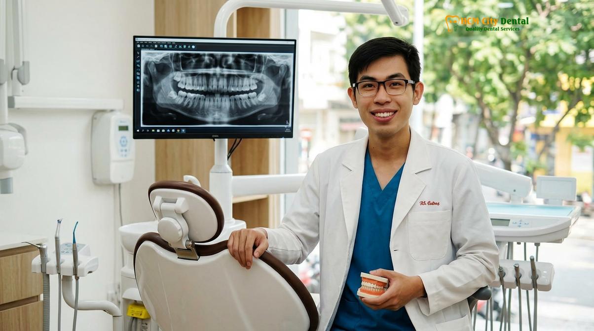
Dr. Nguyen Van Cuong is a leading specialist in restorative dentistry and implantology. With extensive clinical experience, Dr. Cuong ensures that every bone grafting procedure is meticulously planned to provide a robust foundation for your new smile. To learn more about his approach and whether this treatment is suitable for your specific needs, read his comprehensive guide on Are Dental Implants Right For You? Pros & Cons Guide | Dr. Cuong.
References
- Journal of Oral Implantology. Alveolar ridge preservation and augmentation techniques. (2021).
- International Journal of Oral and Maxillofacial Surgery. Comparative analysis of bone grafting materials in implant dentistry. (2020).
- Clinical Implant Dentistry and Related Research. Osseointegration and healing timelines of xenografts vs allografts. (2019).
- Journal of Periodontology. The role of barrier membranes in guided bone regeneration. (2022).
- American Association of Oral and Maxillofacial Surgeons. Postoperative management and complication rates in alveolar bone grafting. (2018).
- Vietnam Odonto-Stomatology Association (VOSA). National clinical guidelines on alveolar bone grafting and implant placement. (2023).
If you are considering restorative dentistry and want to ensure your jawbone is healthy enough for a permanent solution, contact HCMC Dental Clinic in Ho Chi Minh City today. Our expert team will conduct a thorough evaluation and design a personalized treatment plan to rebuild your smile. Learn more about our comprehensive services for Dental Implants tại HCMC Dental Clinic and take the first step toward optimal oral health.