Denture sore spots relief requires a combination of immediate at-home soothing techniques and precise clinical adjustments. While warm salt water rinses and topical gels provide temporary comfort, permanently resolving the friction requires a dentist to map and relieve the specific acrylic pressure points causing mucosal trauma.
Clinical Summary:
Denture sore spots are localized areas of mucosal inflammation and ulceration caused by unequal pressure distribution from a rigid acrylic prosthesis. The etiology typically involves microscopic discrepancies in the denture’s intaglio (tissue-facing) surface or occlusal instability that creates sliding friction during mastication. Effective management relies on a dual approach: palliative home care to reduce acute tissue edema, and professional clinical adjustments using pressure-indicating paste to selectively relieve offending acrylic high points. Attempting to self-adjust dentures is strongly contraindicated, as it can irreversibly compromise the biofunctional seal and exacerbate tissue trauma.
Key Takeaways:
- Sore spots are a common, expected phase of the initial biological adaptation to new dentures.
- Microscopic high points in the acrylic base are the primary mechanical cause of localized mucosal friction.
- Temporary relief can be achieved through saline rinses, soft diets, and removing the prosthesis at night.
- Professional adjustments use specialized indicator pastes to map and relieve exact pressure zones without destroying retention.
- Self-filing or altering the denture at home can permanently ruin the fit and lead to chronic oral health issues.
The Biological Adaptation of Gums to Acrylic Pressures
The oral mucosa must undergo a complex biological transition to withstand the mechanical load of rigid acrylic dentures, often resulting in temporary localized inflammation as the tissues adapt.
When a patient transitions to wearing a removable prosthesis, the oral environment undergoes a profound biomechanical shift. The natural dentition is supported by the periodontal ligament, a sophisticated shock-absorbing system that translates masticatory forces into tension on the alveolar bone. In contrast, a complete or partial denture rests directly upon the mucoperiosteum—the soft tissue overlying the jawbone. This tissue was biologically designed to be a protective lining, not a primary load-bearing structure. Consequently, the introduction of a rigid polymethyl methacrylate (PMMA) base requires the gums to undergo a period of physiological adaptation.
During the initial stages of wearing a new prosthesis, the underlying tissues experience varying degrees of compression. The stratum corneum, the outermost layer of the oral mucosa, must gradually thicken and keratinize in response to this new mechanical demand. This process is akin to developing a callus on the hand when performing manual labor. However, because the oral mucosa is highly vascularized and innervated, this transitional phase is frequently accompanied by generalized tenderness and the formation of localized inflammatory lesions. Proper denture care and maintenance are critical during this phase to support tissue health.
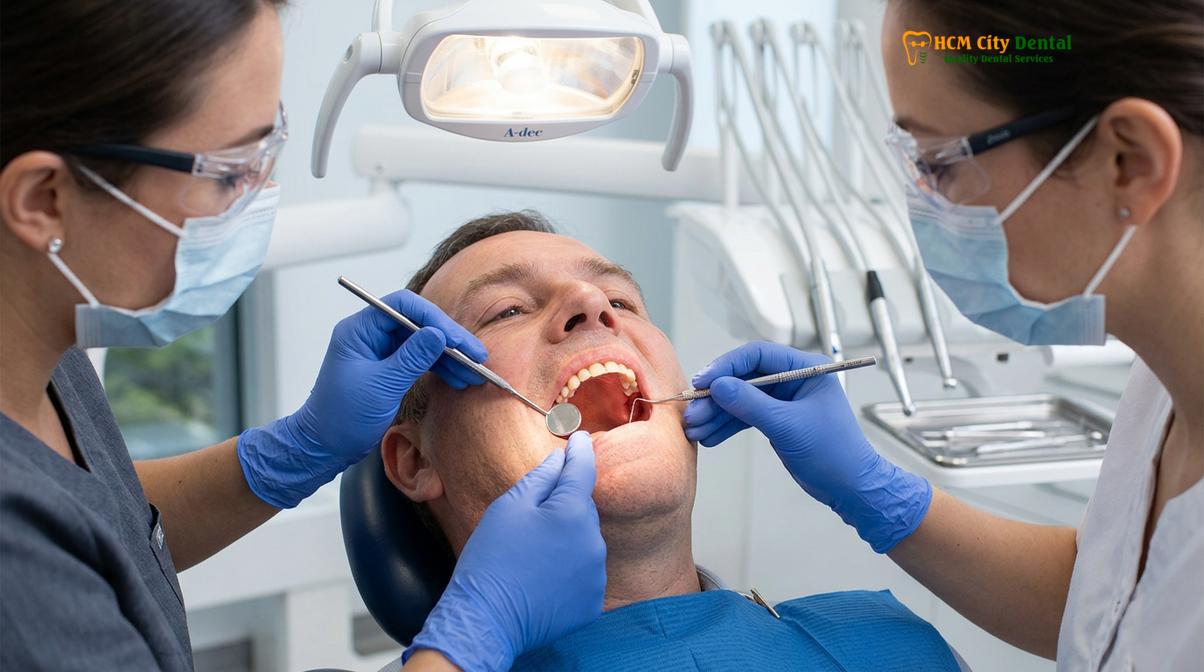
The adaptation process is further complicated if the patient has recently undergone extractions. In such cases, the dynamics of dental socket healing play a crucial role in how the prosthesis seats. As the extraction sites remodel, the bone resorbs and changes shape, creating a moving target for the denture base. A prosthesis that fits perfectly on the day of delivery may begin to impinge on newly prominent bony spicules just weeks later. This continuous remodeling highlights why ongoing clinical supervision is necessary to manage the evolving interface between the acrylic and the healing mucosa.
“The oral mucosa’s ability to tolerate a rigid prosthesis varies significantly among individuals. Our clinical goal is not to force the tissue to endure pain, but to meticulously calibrate the denture base until it harmonizes with the patient’s unique anatomical contours.” — Dr. Nguyen Van Cuong
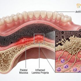
Localized ischemia is the primary physiological mechanism behind the development of a sore spot. When a specific area of the denture exerts excessive pressure on the gums, it compresses the microscopic capillary beds within the lamina propria. If this pressure exceeds the capillary blood pressure, blood flow is temporarily halted. Prolonged ischemia deprives the tissue of oxygen and nutrients, leading to cellular necrosis and the formation of a painful ulceration. Understanding this biological threshold is essential for effective denture sore spots relief, as the ultimate solution must involve restoring adequate perfusion to the affected tissues[1].
Furthermore, the viscosity and volume of saliva play a vital role in tissue tolerance. Saliva acts as a natural lubricant and a hydraulic cushion between the acrylic base and the mucosal surface. Patients with xerostomia (dry mouth) are at a significantly higher risk of developing severe sore spots because they lack this protective mucin layer. The friction generated during speaking and chewing is amplified, leading to rapid epithelial breakdown. Therefore, evaluating salivary flow is a standard component of comprehensive general dentistry assessments for edentulous patients.
Normal vs. Abnormal Pain: What to Expect in the First 14 Days
Mild tenderness and generalized pressure are normal during the initial two-week adaptation period, whereas sharp, localized ulcerations indicate abnormal friction requiring clinical intervention.
Navigating the first two weeks with a new denture can be a challenging experience, and distinguishing between normal adaptive discomfort and abnormal traumatic pain is crucial for the patient’s peace of mind and oral health. On the day of delivery, the prosthesis often feels bulky and intrusive. The neuromuscular system is learning to control the new appliance, and the salivary glands typically go into overdrive, producing excess saliva in response to the foreign object. During the first 24 to 72 hours, patients should expect a sensation of generalized pressure across the entire alveolar ridge.
As the days progress, the denture begins to settle into the soft tissues. It is during this settling phase—typically between days four and seven—that specific denture pressure points may begin to manifest. The generalized soreness often subsides, replaced by distinct areas of tenderness. This occurs because the compressible mucosa yields to the rigid acrylic, allowing the prosthesis to sink slightly and engage anatomical undercuts or bony prominences that were not under heavy load initially. While mild tenderness in these areas is expected, the rapid development of a raw, bleeding ulcer is not.
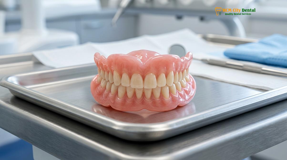
To help patients understand their symptoms, clinical guidelines categorize the pain into adaptive and traumatic profiles. Adaptive pain is usually described as a dull ache or a feeling of muscular fatigue in the jaws, which improves when the denture is removed and rested. Traumatic pain, conversely, is sharp, stinging, and highly localized. It often prevents the patient from applying any biting force and may be accompanied by a visible white or yellowish pseudomembrane over a red, inflamed base—the classic presentation of an aphthous-like ulcer caused by mechanical friction[2].
| Symptom Profile | Typical Duration | Mucosal Appearance | Recommended Action |
|---|---|---|---|
| Generalized dull ache, jaw fatigue | Days 1 – 5 | Normal pink, mild generalized redness | Continue wear, practice soft diet, monitor |
| Mild localized tenderness on chewing | Days 4 – 10 | Slightly red areas, no broken skin | Warm saline rinses, schedule routine adjustment |
| Sharp, stinging pain at a specific spot | Persistent | Visible ulceration, white/yellow center | Remove denture, seek immediate clinical adjustment |
| Severe pain, swelling, or bleeding | Immediate/Persistent | Deep ulcer, active bleeding, severe erythema | Leave denture out, contact dentist urgently |
By the end of the second week, the neuromuscular control should improve, and the initial generalized soreness should largely resolve. However, if localized denture pressure points persist and evolve into painful sores, it is a clear indication that the acrylic base requires professional modification. Patients are encouraged to maintain a detailed mental or written record of exactly where they feel the pain and during which specific movements (e.g., swallowing, biting on the front teeth, or lateral chewing). This information is invaluable to the dentist during the follow-up evaluation.
What Causes Sore Spots? High Points and Sliding Friction
Sore spots primarily develop from microscopic high points on the denture base or from sliding friction caused by unstable occlusal forces during mastication.
The etiology of denture sore spots is deeply rooted in the biomechanics of the prosthesis and its interaction with the dynamic oral environment. The most common culprit is the presence of microscopic discrepancies on the intaglio surface of the denture. Despite the high precision of modern dental impressions and digital scanning technologies, the fabrication process of acrylic dentures involves polymerization shrinkage. As the liquid monomer and powder polymer cure into a solid state, minute dimensional changes occur. These changes can create microscopic nodules or high points that exert disproportionate pressure on the underlying mucosa.
Another significant factor is the anatomical complexity of the human jawbone. The alveolar ridge is rarely perfectly smooth; it features natural bony exostoses, sharp mylohyoid ridges, and prominent maxillary tuberosities. When the rigid acrylic base is seated over these unyielding bony structures, the thin layer of overlying mucosa becomes trapped and compressed. Without adequate relief built into the denture base during fabrication, these anatomical landmarks inevitably become severe pressure zones, leading to rapid tissue breakdown and the need for immediate denture sore spots relief.
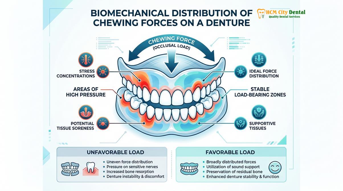
Beyond static fit issues, dynamic occlusal instability is a major driver of mucosal trauma. When a patient chews, the forces should ideally be directed vertically along the long axis of the alveolar ridge. However, if the artificial teeth are not perfectly balanced—a condition known as occlusal disharmony—premature contacts occur. When the patient bites down, the teeth hit unevenly, causing the entire denture base to shift, tilt, or slide laterally across the gums. This sliding friction is highly destructive to the delicate epithelium. It acts like sandpaper, quickly eroding the mucosal surface and creating elongated, linear sore spots, typically along the slopes of the ridges[3].
Clinical Case Study: Resolving Sliding Friction
A 65-year-old patient presented to HCMC Dental Clinic in Ho Chi Minh City complaining of persistent, painful sores along the lower left jawline, despite multiple previous adjustments elsewhere. Upon clinical examination, Dr. Cuong identified that the sores were not caused by a poorly fitting base, but by a premature occlusal contact on the upper right molar. Every time the patient chewed, the lower denture was forced to slide forcefully to the left, creating severe friction. By meticulously adjusting the bite (occlusal equilibration) rather than grinding the inside of the denture, the sliding motion was eliminated, and the sore spots healed completely within five days.
The timing of relining procedures also plays a critical role in preventing sore spots. Over months and years, the jawbone naturally resorbs, causing the once-perfectly fitting denture to become loose. A loose denture moves excessively during function, leading to chronic friction and irritation. Understanding proper dental reline timing is essential; patients should not wait until severe sores develop to seek a reline. Regular evaluations allow the dentist to detect subtle losses of retention and recommend a hard or soft reline to restore the intimate adaptation of the base to the tissues, thereby preventing the onset of traumatic lesions.
At-Home Relief: Warm Salt Water, Gels, and Soft Diet Habits
Temporary relief from denture discomfort can be achieved through saline rinses, topical anesthetics, and reducing masticatory stress via a modified diet.
While professional adjustments are the only definitive cure for mechanical friction, patients often require immediate, palliative strategies to manage pain at home. The cornerstone of at-home denture sore spots relief is the utilization of a warm salt water rinse. This simple yet highly effective remedy works on the principle of osmosis. The hypertonic saline solution draws excess fluid out of the inflamed, edematous mucosal tissues, thereby reducing swelling and relieving pressure on the nerve endings. Furthermore, salt possesses mild antibacterial properties, which help to keep the compromised epithelial barrier clean and prevent secondary bacterial colonization of the ulcerated areas.
To maximize the benefits, patients should dissolve half a teaspoon of non-iodized salt in eight ounces of warm (not hot) water. The rinse should be swished gently around the mouth for thirty to sixty seconds, focusing on the areas of irritation, and then expectorated. This process can be repeated four to five times a day, particularly after meals and before bedtime. Consistent use of a warm salt water rinse significantly accelerates the healing of minor abrasions and provides substantial symptomatic relief while the patient awaits their clinical appointment.
Over-the-counter topical anesthetics, typically containing benzocaine, can also be employed for short-term pain management. These gels work by temporarily blocking the sodium channels in the local nerve endings, effectively numbing the area. When applying these products, the denture should be removed, the sore spot gently dried with a clean gauze, and a small amount of gel applied directly to the lesion. However, it is imperative to understand that these gels are merely masking the pain. They do not eliminate the mechanical friction causing the sore. Over-reliance on numbing gels can lead to severe tissue damage, as the patient may unknowingly continue to chew forcefully on a traumatized area because they cannot feel the pain[4].
Dietary modifications are equally critical during periods of acute soreness. Consuming hard, crunchy, or highly fibrous foods requires significant masticatory force, which translates directly into pressure on the gums. Transitioning to a soft, non-abrasive diet—such as scrambled eggs, yogurt, mashed vegetables, and blended soups—drastically reduces the mechanical load on the tissues. Additionally, practicing gentle sore spots massage with a clean finger can stimulate local blood circulation, promoting faster cellular repair and tissue regeneration. Always ensure hands are thoroughly washed before touching the oral mucosa.
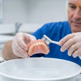
Finally, the most effective at-home measure is simply giving the tissues a rest. Removing the dentures for several hours during the day, and strictly leaving them out overnight, allows the compressed mucoperiosteum to rebound. The continuous wearing of dentures without adequate rest periods is a primary risk factor for chronic mucosal irritation and the development of opportunistic fungal infections, such as denture stomatitis. Proper overnight storage in a cleansing solution is a non-negotiable aspect of responsible denture wear.
The Clinical Solution: How Dr. Cuong Buffs Down Pressure Zones
Professional adjustment involves using pressure-indicating paste to map mucosal trauma, allowing the dentist to selectively relieve the exact acrylic high spots.
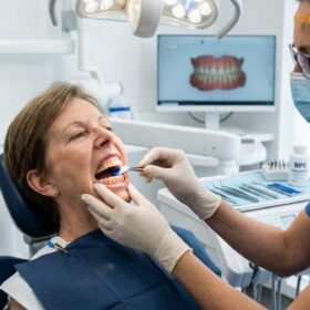
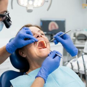
When at-home remedies are insufficient, professional intervention becomes necessary to address the root cause of the discomfort. The clinical workflow for resolving sore spots is a precise, diagnostic-driven process. It begins with a thorough visual and tactile examination of the oral cavity. The dentist carefully inspects the mucosal tissues to identify the exact location, size, and severity of the ulcerations. Understanding the spatial relationship between the soft tissue lesion and the corresponding area on the denture base is the critical first step in effective denture sore spots relief.
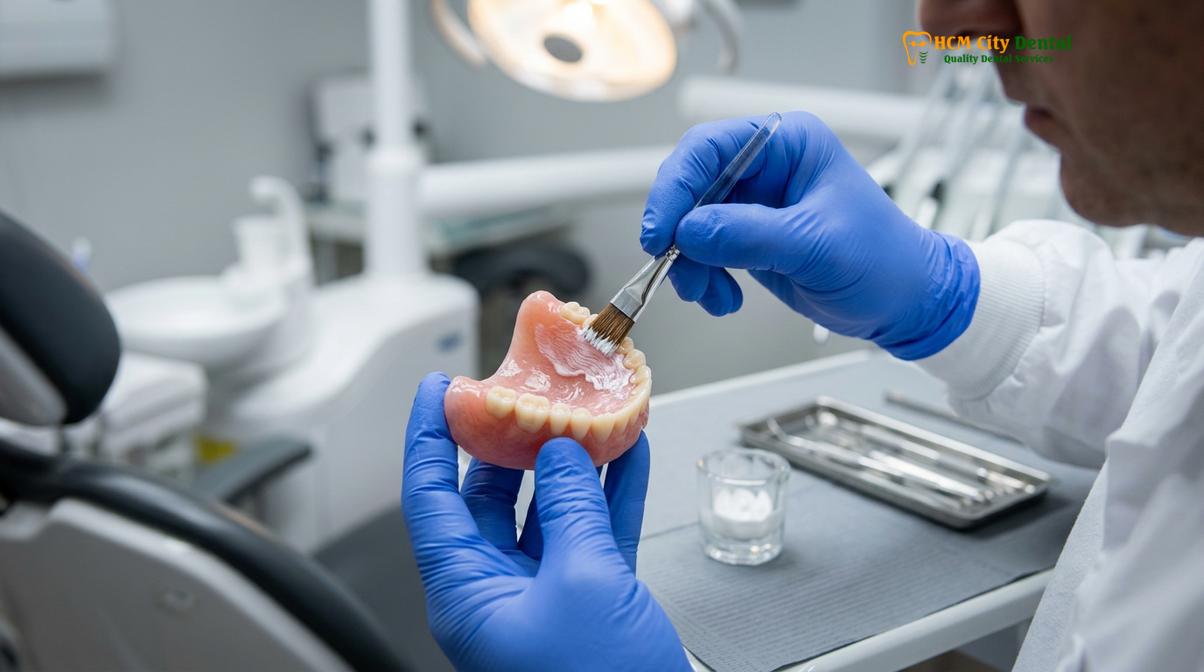
To achieve this precise mapping, specialists at HCMC Dental Clinic utilize a specialized material known as Pressure Indicator Paste (PIP). This zinc-oxide-based paste is painted onto the intaglio surface of the dried denture in a thin, even layer with distinct brush strokes. The prosthesis is then carefully inserted into the patient’s mouth, and the patient is instructed to bite down firmly or perform specific functional movements. When the denture is removed, the paste reveals a clear topographical map of the pressure distribution. Areas where the paste has been completely displaced, allowing the pink acrylic to show through, indicate points of excessive pressure and friction.
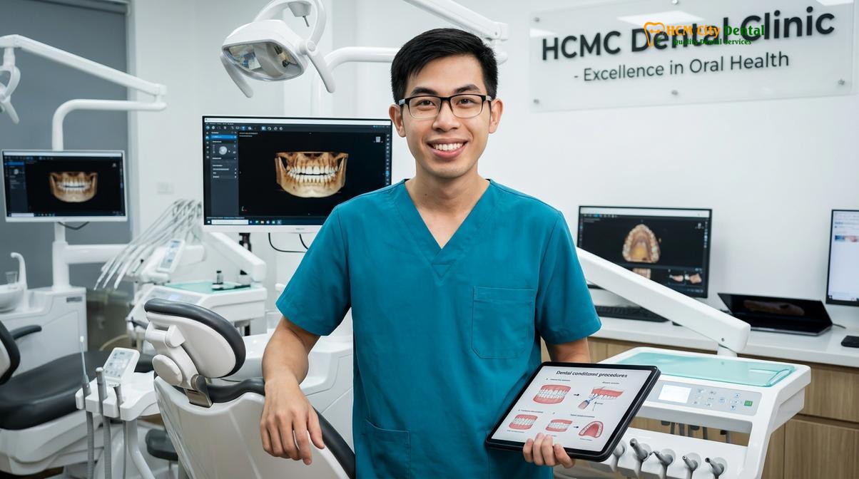
Once the offending high points are accurately identified, the dentist uses specialized acrylic burs in a slow-speed handpiece to selectively relieve the material. This process requires a delicate touch and a deep understanding of prosthetic biomechanics. The goal is to remove just enough acrylic to eliminate the pressure without compromising the overall fit, retention, or peripheral seal of the denture. Over-reduction can create a void between the denture and the tissue, leading to a loss of suction and further instability.
“The art of denture adjustment lies in micro-calibration. We are often removing fractions of a millimeter of acrylic. It is a highly targeted procedure guided by diagnostic pastes, ensuring we relieve the trauma while preserving the biofunctional integrity of the prosthesis.” — Dr. Nguyen Van Cuong
After the necessary acrylic has been removed, the adjusted area must be meticulously polished. A rough, unpolished surface will not only irritate the delicate mucosa but will also act as a microscopic harbor for plaque, bacteria, and Candida albicans. Using a series of silicone polishers and pumice, the dentist restores a glass-like finish to the relieved area. This attention to detail during clinical adjustment visits ensures that the tissue can heal against a smooth, hygienic surface, preventing recurrent inflammation and promoting long-term oral health[5].
It is common for patients to require multiple clinical adjustment visits during the first few weeks of wearing a new denture. As one pressure point is relieved, the denture may settle slightly differently, occasionally revealing a new, secondary high point. This sequential settling is a normal part of the adaptation process. Patients should view these follow-up appointments not as a sign of a poorly made denture, but as a necessary calibration phase to achieve a customized, comfortable fit.
The Importance of Not Self-Adjusting Your Denture with Files
Attempting to file or alter your denture at home can irreversibly destroy the precise mucosal seal, leading to chronic instability and severe tissue damage.
In moments of acute discomfort, it can be tempting for patients to attempt DIY adjustments using household tools such as sandpaper, nail files, or rotary hobby tools. However, self-adjusting a denture is strongly contraindicated and poses significant risks to both the prosthesis and the patient’s oral health. The internal surface of a denture is a highly complex, custom-molded topography designed to intimately adapt to the microscopic contours of the jawbone. Altering this surface without diagnostic guidance almost guarantees the destruction of this precise fit.
When a patient attempts to file down a perceived high point, they lack the ability to accurately map the pressure zone. Consequently, they often remove acrylic from the wrong area or remove far too much material. This over-reduction destroys the peripheral seal and the intimate tissue contact required for retention. A denture that has been aggressively self-adjusted will become chronically loose, leading to increased sliding friction, the development of new, more severe sore spots, and a complete inability to chew effectively. In many cases, the damage is irreversible, necessitating the costly fabrication of an entirely new prosthesis.
Clinical Warning: The Dangers of DIY Adjustments
Never use household tools, files, or sandpaper to alter your denture. Self-adjusting can destroy the biofunctional seal, create rough surfaces that harbor dangerous bacteria, and cause irreversible damage to the acrylic base. If you are experiencing pain, remove the denture and contact your dental professional immediately for a safe, diagnostic-driven adjustment.
Furthermore, household tools leave deep scratches and gouges in the acrylic surface. These rough areas are impossible to clean thoroughly at home and quickly become colonized by pathogenic bacteria and fungal organisms. This can lead to severe oral infections, chronic inflammation, and foul odors. The clinical protocol for denture sore spots relief always includes high-shine polishing to prevent this microbial retention. Trusting your care to a qualified professional ensures that adjustments are made safely, accurately, and hygienically, preserving your investment and your oral health.
When to See a Doctor
Immediate clinical evaluation is required if sore spots persist beyond a few days, show signs of infection, or prevent adequate nutritional intake.
While minor tenderness is a normal part of the adaptation process, certain symptoms warrant prompt professional attention. You should schedule an immediate appointment if a sore spot persists for more than seven days without improvement, despite utilizing at-home palliative measures like saline rinses and resting the tissues. Persistent ulcerations can occasionally mimic or mask more serious oral pathologies, making a definitive clinical diagnosis essential.
Additionally, seek urgent care if you experience signs of infection, such as localized swelling, purulent discharge (pus), a foul taste in the mouth, or a systemic fever. Severe pain that radiates to the ears or neck, or pain that completely prevents you from wearing the prosthesis and consuming a soft diet, is not normal and requires immediate intervention. Regular monitoring of dental socket healing after extractions is also crucial; if you notice sharp bone fragments protruding through the gums, do not attempt to wear the denture over them. Contact your clinic in Ho Chi Minh City for a comprehensive evaluation and appropriate modification of your treatment plan.
Frequently Asked Questions
How many times should I return to the clinic for adjustments?
Most patients require between two to four clinical adjustment visits during the first month of wearing new dentures. Because the oral tissues continuously adapt and minor swelling subsides, pressure points often shift, requiring sequential, precise modifications by your dentist to achieve optimal long-term comfort. Do not be discouraged if multiple visits are needed; this is a standard part of the calibration process.
Is it normal for new dentures to make my gums bleed?
No, active bleeding from the gums is not a normal part of the denture adaptation process. While mild redness and tenderness are expected, bleeding indicates severe friction, an underlying infection, or a significant high point in the acrylic that requires immediate professional evaluation and adjustment. Remove the denture and contact your dentist promptly.
Can I use an over-the-counter pain gel on denture sores?
Yes, you can use over-the-counter oral numbing gels containing benzocaine to temporarily soothe denture sores. However, these gels only mask the pain and do not resolve the underlying mechanical friction. They should be used strictly as a short-term palliative measure while awaiting your professional dental adjustment, as over-reliance can lead to further tissue damage.
Should I leave my dentures out at night if I have sore spots?
Yes, removing your dentures at night is highly recommended, especially when experiencing sore spots. Leaving the prosthesis out allows the compressed mucosal tissues to rest, restores normal blood circulation to the gums, and prevents the exacerbation of localized ulcerations while reducing the risk of fungal infections like denture stomatitis.
How long does it take for a denture sore to heal after an adjustment?
Once the offending acrylic high point is professionally relieved, a denture sore typically heals within three to seven days. Maintaining excellent oral hygiene and utilizing warm saline rinses during this recovery period will accelerate tissue regeneration and prevent secondary bacterial colonization. If the sore does not improve within a week of the adjustment, a follow-up visit is necessary.
References
- Journal of Prosthetic Dentistry. Biomechanical responses of oral mucosa to removable prostheses. (2021).
- International Journal of Prosthodontics. Clinical management of denture-induced mucosal lesions. (2020).
- British Dental Journal. The efficacy of pressure indicator paste in complete denture adjustments. (2019).
- Journal of Oral Rehabilitation. Masticatory forces and their distribution in edentulous patients. (2022).
- Clinical Oral Investigations. Patient adaptation and tissue tolerance to polymethyl methacrylate bases. (2018).