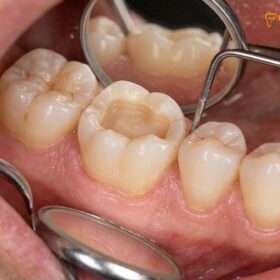
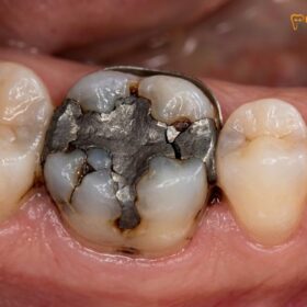
For decades, the standard response to a moderate-to-large cavity in a posterior molar has been to excavate the decay and pack the void with a direct composite resin filling. While this approach is highly successful for small, conservative lesions, the clinical reality of restoring massive structural defects with direct resin is fraught with biomechanical peril. The primary antagonist in this scenario is not the chewing force of the patient, nor the quality of the composite material itself, but rather a fundamental law of physics and geometry known in restorative dentistry as the Configuration Factor, or C-Factor. When a large volume of composite resin is light-cured inside a highly constrained cavity, it undergoes an unavoidable volumetric contraction. This polymerization shrinkage stress acts as an invisible, destructive force that pulls the remaining tooth structure inward, rips the microscopic adhesive bonds at the dentin floor, and sets the stage for a cascade of clinical failures ranging from severe post-operative chewing pain to catastrophic cuspal fracture. Understanding the intricate relationship between C-factor configuration factor polymerization shrinkage composite onlay biomechanics is the cornerstone of modern biomimetic dentistry. By transitioning from massive, high-stress direct fillings to precision-milled, stress-free indirect ceramic onlays, specialist clinicians can preserve the structural integrity of the natural tooth, eliminate microleakage, and provide international patients with restorations that endure for decades rather than mere years.
Clinical Summary:
The Configuration Factor (C-Factor) dictates the ratio of bonded to unbonded surfaces in a dental cavity, directly influencing the amount of polymerization shrinkage stress trapped within the tooth during a composite restoration. High C-Factor cavities (like deep Class I occlusal lesions) force the shrinking composite to pull violently against the cavity walls, leading to cuspal deflection, torn dentin bonds, and severe post-operative sensitivity. To combat this, biomimetic dentistry employs stress-reduction protocols such as ultra-thin incremental layering, decoupling with time, and the use of ultra-high-molecular-weight polyethylene fibers. However, for structural defects exceeding 50% of the intercuspal width, replacing the direct filling with an indirect CAD/CAM IPS E.max ceramic onlay is the definitive clinical solution. Because ceramic onlays are polymerized outside the mouth, intra-cavity shrinkage stress is reduced to practically zero, ensuring a flawless, long-lasting marginal seal.
Key Takeaways:
- The C-Factor Equation: C-Factor is the ratio of bonded surfaces to unbonded (free) surfaces. A deep Class I cavity has a C-Factor of 5.0, representing the highest stress environment in restorative dentistry.
- Polymerization Shrinkage: Modern dental composites shrink by 2% to 3.5% by volume during light-curing, generating immense tensile forces that can exceed the microtensile bond strength of the dentin adhesive.
- Clinical Symptoms of Failure: Unmanaged shrinkage stress causes microscopic gap formation, leading to sharp pain upon releasing a bite, white-line enamel micro-cracks, and rapid secondary bacterial infiltration.
- Biomimetic Stress Reduction: Advanced techniques utilize Immediate Dentin Sealing (IDS) & Resin Coating in Biomimetic Veneers & Onlays: Increasing Bond Strength by 400%, 5-minute delayed curing, and polyethylene fiber networks to absorb stress.
- The Ceramic Onlay Advantage: Indirect IPS E.max ceramic onlays are fabricated outside the mouth, meaning the only material that shrinks inside the tooth is an ultra-thin (50-micron) layer of resin cement, virtually eliminating C-Factor stress.
What Is Feilzer’s Configuration Factor (C-Factor = Bonded Surfaces Divided by Unbonded Surfaces) in Restorative Dentistry?
To comprehend why large composite fillings frequently fail, one must first understand the geometric constraints placed upon the restorative material. In 1987, researchers Feilzer, De Gee, and Davidson introduced the concept of the Configuration Factor, universally known as the C-Factor, to quantify the stress generated during the polymerization of resin-based composites. The C-Factor is a mathematical ratio defined simply as the number of bonded surfaces in a cavity preparation divided by the number of unbonded, or free, surfaces. This ratio is the master key to predicting how much stress a curing composite will exert on the surrounding tooth structure.
When a composite resin is exposed to a curing light, the monomer molecules cross-link into a solid polymer network. This chemical reaction inherently causes the material to lose volume—it shrinks. If the composite is placed on a flat surface with only one bonded wall and five free surfaces (a C-Factor of 0.2), the material is free to shrink toward the bonded surface without generating significant internal stress. The unbonded surfaces simply flow and adapt to the volumetric loss. However, dental cavities are rarely flat surfaces; they are three-dimensional boxes.
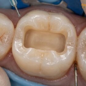
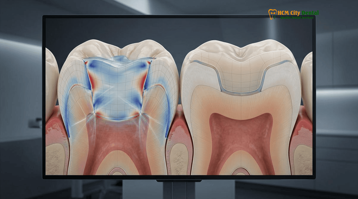
Consider a deep Class I occlusal cavity, which is a standard hole drilled into the biting surface of a molar to remove decay. This cavity has five bonded walls: the mesial wall, the distal wall, the buccal wall, the lingual wall, and the deep pulpal floor. It has only one unbonded surface: the top opening where the curing light enters. Dividing 5 bonded surfaces by 1 unbonded surface yields a C-Factor of 5.0. This is the highest and most dangerous C-Factor in restorative dentistry. In a C-Factor 5.0 environment, the composite is locked in on all sides. As it shrinks, it has virtually no free surface to flow toward. The volumetric contraction is therefore converted entirely into tensile stress, pulling violently against the five bonded walls. If this stress is not meticulously managed through advanced clinical protocols, the physics of c-factor configuration factor composite dentistry dictate that the restoration will inevitably fail at its weakest link.
The Physics of Polymerization Volumetric Shrinkage (2%–3.5%): How High C-Factor Class I Cavities Pull Cusps Inward and Rip the Dentin Bond
The transition of a dental composite from a moldable paste to a rigid solid is not a singular event, but a dynamic process divided into two distinct phases: the pre-gel phase and the post-gel phase. Understanding these phases is critical to mastering the physics of polymerization volumetric shrinkage, which typically ranges from 2% to 3.5% depending on the filler content and resin chemistry of the specific composite brand.
During the initial pre-gel phase, the composite begins to polymerize but remains somewhat viscous and flowable. In this state, the material can internally rearrange its molecular structure to compensate for the initial shrinkage. Stress is minimal because the composite can still flow to relieve tension. However, as the curing light continues to activate the photoinitiators, the material reaches the “gel point.” After this point, the composite enters the post-gel phase, rapidly increasing in elastic modulus (stiffness) and undergoing vitrification into a rigid, glassy state. Any further volumetric shrinkage that occurs during the post-gel phase can no longer be accommodated by internal flow. Instead, this shrinkage generates massive tensile stress that is transferred directly to the adhesive interface and the surrounding tooth structure.
“The battle for a long-lasting restoration is won or lost at the hybrid layer. When we place a massive bulk of composite into a high C-Factor cavity, the post-gel shrinkage stress acts like a microscopic crowbar. It will always exploit the hierarchy of bondability, ripping the weaker dentin bond while leaving the stronger enamel bond intact, creating a hidden void that guarantees future failure.”
— Dr. Nguyen Van Cuong, DDS (Clinical Director, HCMC Dental Clinic)
This stress transfer leads to two catastrophic biomechanical events. First, the inward pull of the shrinking composite exerts a centripetal force on the cavity walls, leading to polymerization shrinkage stress cuspal deflection. The rigid composite literally pulls the flexible cusps of the molar inward toward the center of the tooth. Over time, as the patient chews, this constant inward tension combined with vertical occlusal loading causes the enamel to fatigue, initiating micro-cracks at the base of the cusps. [1]
Second, the shrinkage stress attacks the adhesive interface, exploiting the hierarchy of bondability dentin vs enamel. Dental adhesives bond exceptionally well to the highly mineralized, dry structure of enamel, routinely achieving bond strengths in excess of 30 to 40 Megapascals (MPa). However, bonding to dentin is vastly more complex. Dentin is a living, wet, organic tissue filled with fluid-pumping tubules and collagen networks. Even with the best modern adhesives, the initial microtensile bond strength to deep dentin is significantly lower and more fragile than the bond to enamel. When the post-gel shrinkage stress of a large composite mass exceeds the immature bond strength of the dentin adhesive, the composite violently rips away from the cavity floor. [2] This creates a microscopic gap—a dead space—at the deepest part of the filling, setting a ticking time bomb for the patient.
Clinical Consequences of Shrinkage Stress: Post-Operative Chewing Pain, White-Line Enamel Micro-Cracks, and Secondary Recurrent Caries
When the physics of polymerization shrinkage overcome the chemistry of the dental adhesive, the clinical consequences are swift, severe, and highly frustrating for both the patient and the practitioner. The most immediate and alarming symptom reported by patients is sharp, shooting pain when chewing, specifically when releasing biting pressure. This phenomenon perfectly answers the common patient query: why large composite fillings hurt when chewing.
The mechanism behind this pain is explained by Brännström’s Hydrodynamic Theory. When the shrinkage stress rips the composite away from the dentin floor, it leaves a microscopic, fluid-filled gap. When the patient bites down on a hard piece of food, the tooth flexes slightly, compressing the fluid in this gap. When the patient suddenly releases the bite, the tooth snaps back to its original shape, and the gap rapidly expands. This sudden expansion acts like a microscopic vacuum pump, violently sucking the fluid outward from the dentinal tubules. This rapid fluid shift stimulates the odontoblastic processes and the A-delta nerve fibers deep within the dental pulp, registering as a sharp, electric shock of pain. If a patient experiences this specific “pain on release” immediately after receiving a large white filling, it is almost pathognomonic for a debonded dentin interface caused by C-Factor stress.
Clinical Case Study: Resolving Chewing Pain in an Expat Patient
Patient Profile: A 42-year-old Australian expatriate living in Thao Dien, District 2, presented to HCMC Dental Clinic in District 1 complaining of sharp, shooting pain in his lower right first molar (Tooth 46). The pain occurred exclusively when he released his bite while eating almonds or crusty bread.
Clinical Findings: Dr. Nguyen Van Cuong, DDS, utilized a Carl Zeiss operating microscope to examine the tooth. The patient had received a massive, deep direct composite filling at a different clinic three weeks prior. High-magnification transillumination revealed a distinct “white line” at the enamel margin and significant cuspal deflection. A 3D CBCT scan confirmed no pulpal involvement, pointing directly to a C-Factor-induced adhesive failure at the pulpal floor.
Treatment Protocol: Under a 100% latex-free nitrile rubber dam, Dr. Cuong carefully removed the failing composite block. As predicted, the composite had completely debonded from the deep dentin, leaving a fluid-filled void. Dr. Cuong executed a biomimetic protocol, utilizing Immediate Dentin Sealing (IDS) and placing a stress-reduced, precision-milled IPS E.max CAD/CAM ceramic onlay to protect the weakened cusps.
Outcome: The patient reported an immediate and total cessation of chewing pain upon cementation. The ceramic onlay restored the structural integrity of the molar, demonstrating the superiority of indirect restorations in high C-Factor scenarios.
Beyond immediate pain, unmanaged shrinkage stress manifests as “white lines” at the margins of the restoration. These white lines are actually micro-cracks in the enamel prisms, caused by the composite pulling away from the cavosurface margin or fracturing the fragile enamel rods. These micro-cracks are not merely cosmetic defects; they are open highways for bacteria. [3] The oral cavity is a hostile environment teeming with cariogenic bacteria like Streptococcus mutans. These bacteria easily infiltrate the micro-gaps and white-line fractures, migrating down to the debonded dentin floor. Because this area is completely hidden beneath the filling and inaccessible to a toothbrush or fluoride, the bacteria thrive, leading to massive secondary recurrent caries that can rapidly destroy the tooth from the inside out, often necessitating complex root canal therapy or extraction.
Biomimetic Stress-Reduction Protocols: Incremental Oblique Layering (<1.5mm), 5-Minute Delayed Light-Curing, and Ultra-High-Molecular-Weight Polyethylene Fibers
To combat the destructive forces of the C-Factor in cases where a direct composite restoration is still clinically viable (typically when the cavity width is less than 50% of the intercuspal distance), biomimetic dentists employ a rigorous sequence of stress-reduction protocols. The era of “bulk-filling” a large cavity and curing it in a single massive chunk is obsolete in advanced adhesive dentistry. Instead, the focus is on manipulating the geometry of the composite increments to artificially lower the C-Factor of each individual layer.
The foundational technique is incremental oblique layering. Rather than filling the cavity horizontally, the specialist places the composite in ultra-thin, wedge-shaped increments, strictly keeping the thickness below 1.5 millimeters. Each oblique increment is carefully adapted to contact only two or three walls of the cavity at a time, rather than all five. By doing so, the C-Factor of that specific increment is drastically reduced from 5.0 down to 1.0 or 0.5. This allows the small volume of composite to undergo its pre-gel flow and post-gel shrinkage with minimal stress transfer to the tooth structure. [4]
Equally critical is the concept of “decoupling with time,” specifically applied during the Immediate Dentin Sealing (IDS) phase. When the dentin adhesive and the first micro-layer of flowable resin (the resin coating) are applied to the deep dentin, the clinician must not immediately proceed to pack the bulk composite. Instead, the protocol demands a strict 5-minute delayed light-curing maturation period. During these 5 minutes, the adhesive monomers fully infiltrate the collagen network, the solvents evaporate completely, and the hybrid layer polymerizes to its maximum microtensile strength. By allowing the dentin bond to mature and strengthen *before* applying the shrinkage stress of the overlying composite, the clinician ensures that the bond can withstand the tensile forces without ripping.
For moderate-to-large cavities that border on the structural limits of direct resin, biomimetic specialists integrate a ribbony polyethylene fiber stress reduced direct composite protocol. Ultra-high-molecular-weight polyethylene (UHMWPE) fibers, such as Ribbond or Construct, are embedded into the flowable resin at the pulpal floor and across the weakened cusps before the final composite layers are built. These woven fiber networks act exactly like rebar in concrete. They possess an incredibly high modulus of elasticity and tensile strength. When the overlying composite shrinks, the polyethylene fibers absorb and dissipate the stress horizontally across their woven matrix, preventing the stress from reaching the fragile dentin bond or deflecting the cusps. This biomimetic integration transforms a brittle, high-stress composite mass into a resilient, stress-absorbing monoblock.
Why Indirect CAD/CAM IPS E.max Ceramic Inlays and Onlays Reduce Intra-Cavity Polymerization Shrinkage to Practically Zero
While advanced layering and fiber integration can mitigate C-Factor stress in moderate cavities, there is a definitive biomechanical threshold where direct composite resin simply cannot be trusted. When a cavity preparation is wide—specifically, when the isthmus exceeds one-half of the intercuspal distance—the remaining buccal and lingual cusps are severely undermined. In these massive structural defects, attempting to place a direct composite filling is a clinical gamble that frequently results in Split Tooth Syndrome. The definitive, evidence-based solution is to transition from a direct filling to an indirect restoration, highlighting the critical debate of biomimetic ceramic onlay vs large composite filling.
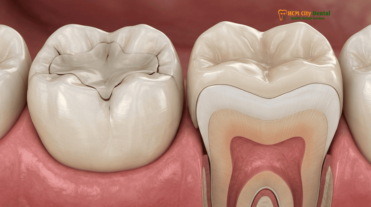
The biomechanical genius of an indirect CAD/CAM IPS E.max (lithium disilicate) ceramic inlay or onlay lies in *where* the polymerization shrinkage occurs. Unlike a direct filling, which is a soft paste that shrinks *inside* the tooth, a ceramic onlay is milled from a solid block of pre-crystallized porcelain outside the mouth. The ceramic material itself has zero polymerization shrinkage. The only resin material utilized in the entire procedure is the ultra-thin layer of dual-cure or light-cure resin cement used to bond the ceramic to the tooth.
Clinical Warning: Recognizing Split Tooth Syndrome
If you have a massive, old silver amalgam or large composite filling and experience a sharp, localized pain when biting down on specific angles, you may be developing Split Tooth Syndrome. This occurs when unmanaged occlusal forces and internal shrinkage stress cause a crack to propagate down the root of the tooth. Do not ignore this symptom. If the crack reaches the pulp or splits the root vertically, the tooth becomes non-restorable and must be extracted. Contact Dr. Nguyen Van Cuong, DDS, at HCMC Dental Clinic immediately for a 3D CBCT scan and a protective cuspal-coverage ceramic onlay to halt the crack propagation and save the natural tooth.
This cement layer is typically only 50 to 100 microns thick. Because the volume of the shrinking resin is so infinitesimally small, the total volumetric shrinkage stress exerted on the cavity walls is reduced to practically zero. The C-Factor equation is effectively neutralized. Furthermore, the rigid ceramic onlay acts as a structural helmet for the tooth. By covering and splinting the weakened cusps together, the onlay distributes heavy chewing forces evenly across the entire root structure, completely eliminating cuspal deflection and protecting the tooth from catastrophic fracture.
To achieve this flawless integration, Dr. Nguyen Van Cuong, DDS, utilizes a strict, multi-step biomimetic bonding protocol for IPS E.max restorations. Under a 100% latex-free nitrile rubber dam, the intaglio surface of the lithium disilicate onlay is micro-etched with 5%–9.5% hydrofluoric acid for exactly 20 seconds to create a microporous topography, followed by the application of a pure, heat-dried silane coupling agent. Simultaneously, the sealed tooth structure is air-abraded with 27-micron aluminum oxide particles to maximize surface energy before the final adhesive resin cement is applied. This meticulous protocol ensures a cohesive, waterproof margin that mimics the natural dentino-enamel junction, providing decades of durable function. For patients seeking to Replace Failing Fillings with Onlay: A Clinical Guide to Ceramic Restorations, this indirect approach is the gold standard of care.
2026 Stress-Reduced Biomimetic Composite vs. IPS E.max Onlay Fee Schedule at HCMC Dental Clinic (District 1)
For international patients, expatriates, and dental travelers seeking world-class biomimetic restorative care, HCMC Dental Clinic in District 1, Ho Chi Minh City, offers an unparalleled combination of clinical excellence and transparent pricing. Under the clinical direction of Dr. Nguyen Van Cuong (Dr. Cuong), DDS (Vietnam MOH License 0003265/BL-CCHN), the clinic strictly adheres to international protocols for stress-reduced adhesive dentistry, utilizing authentic European and American biomaterials, Carl Zeiss operating microscopes, and digital CAD/CAM workflows. [5]
Understanding the financial investment required for premium restorative care is crucial. HCMC Dental Clinic provides a highly advantageous -40% online booking fee schedule for international patients who secure their appointments prior to arrival. Below is the official 2026 fee schedule comparing direct biomimetic composite restorations with premium indirect IPS E.max ceramic onlays and full crowns.
| Restorative Procedure (Biomimetic Protocol) | 2026 Online Booking Price (USD) | 2026 Online Booking Price (VND) | Standard Walk-In Price (USD) |
|---|---|---|---|
| Direct Composite Veneer / Dental Bonding (Includes incremental layering & stress reduction) | $35 – $46 USD / tooth | 900,000 – 1,200,000 VND | $58 – $76 USD |
| IPS E.max All-Ceramic Inlay / Onlay / Overlay (CAD/CAM milled, zero intra-cavity shrinkage) | $231 USD / tooth | 6,000,000 VND | $385 USD |
| Full Zirconia Crown (Cercon Standard / HT) (For severely structurally compromised teeth) | $154 – $192 USD / tooth | 4,000,000 – 5,000,000 VND | $256 – $321 USD |
| Biomimetic Quartz/Glass Fiber Post & Core Buildup (Endodontically treated teeth foundation) | $38 – $58 USD / tooth | 1,000,000 – 1,500,000 VND | $63 – $96 USD |
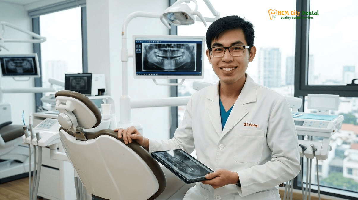
“Our philosophy is simple: preserve maximum natural tooth structure while eliminating the biomechanical flaws of traditional dentistry. By offering premium IPS E.max onlays at highly accessible price points through our online booking system, we empower our international patients to choose the definitive, long-term solution over a compromised, high-stress massive composite filling.”
— Dr. Nguyen Van Cuong, DDS (Clinical Director, HCMC Dental Clinic)
Patients are encouraged to review the Official 2026 Dental Price List & -40% Online Booking Fee Schedule to plan their treatment. To explore the extensive clinical data supporting these biomimetic protocols, visit the 2026 Clinical Dental Research & Statistical Reports Hub. For personalized consultations regarding complex restorative needs, patients can learn more about Dr. Nguyen Van Cuong, DDS (Clinical Director — MOH License 0003265/BL-CCHN) and contact the clinic directly via WhatsApp at +84 853 020 003. The clinic is conveniently located in District 1, Ho Chi Minh City, just minutes from major international hotels and Ben Thanh Market.
Frequently Asked Questions
Why does my tooth hurt sharply when I release my bite on hard food right after getting a deep white composite filling?
This sharp pain upon release is a classic symptom of polymerization shrinkage stress combined with a high C-Factor. When a large volume of composite resin cures, it shrinks by 2% to 3.5%. In a deep cavity, this shrinkage pulls the cavity walls inward (cuspal deflection) and can rip the adhesive bond at the dentin floor. When you bite down, the tooth flexes; when you release, the microscopic gap at the dentin floor acts like a vacuum pump, causing rapid fluid movement within the dentinal tubules. This hydrodynamic fluid shift triggers the nerve endings, resulting in a sharp, shooting pain. This is exactly why large composite fillings hurt when chewing and why biomimetic stress-reduction protocols are mandatory.
Why does a deep Class I occlusal cavity have the worst Configuration Factor (C-Factor = 5.0) in dentistry?
Feilzer’s Configuration Factor (C-Factor) is calculated by dividing the number of bonded surfaces by the number of unbonded (free) surfaces. A deep Class I occlusal cavity (a hole in the biting surface of a molar) has five bonded walls (mesial, distal, buccal, lingual, and the pulpal floor) and only one unbonded surface (the top occlusal opening). Therefore, 5 divided by 1 equals a C-Factor of 5.0. This highly constrained geometry means the composite resin has virtually no free surface to flow toward during the curing process, trapping massive amounts of polymerization shrinkage stress inside the tooth structure and severely challenging the adhesive bond.
How does waiting 5 minutes after Immediate Dentin Sealing (decoupling with time) prevent the composite from pulling away from the deep dentin?
Waiting 5 minutes after placing the initial adhesive and flowable resin layer—a concept known as decoupling with time—allows the dentin bonding agent to mature and achieve its maximum microtensile bond strength before the massive shrinkage stress of the overlying bulk composite is applied. During this 5-minute window, the hybrid layer stabilizes, and the initial resin coating acts as a stress-absorbing shock absorber. If a dentist immediately packs and cures a large volume of composite over fresh adhesive, the polymerization shrinkage stress will easily exceed the immature bond strength, ripping the composite away from the dentin and causing microleakage.
At what cavity width (isthmus >1/2 intercuspal distance) should a direct composite filling be replaced with a ceramic onlay?
According to advanced biomimetic and prosthodontic guidelines, when a cavity preparation exceeds one-half (50%) of the intercuspal distance (the width between the inner tips of the cusps), the remaining tooth structure is structurally compromised and highly susceptible to fracture. At this critical width, a direct composite filling cannot adequately support the weakened cusps and will likely cause cuspal deflection due to polymerization shrinkage. In these cases, a biomimetic ceramic onlay vs large composite filling is clinically indicated. The onlay covers and protects the weakened cusps, distributing occlusal forces evenly and preventing catastrophic split tooth syndrome.
How much do biomimetic stress-reduced fillings and IPS E.max ceramic onlays cost at HCMC Dental Clinic in District 1?
At HCMC Dental Clinic in District 1, Ho Chi Minh City, international patients benefit from a highly transparent -40% online booking fee schedule. A biomimetic stress-reduced composite restoration (dental bonding) costs between $35 and $46 USD per tooth. For larger structural defects requiring indirect restoration, a premium CAD/CAM IPS E.max All-Ceramic Inlay, Onlay, or Overlay is priced at $231 USD per tooth (6,000,000 VND), compared to the $385 USD walk-in rate. All procedures are performed by Dr. Nguyen Van Cuong, DDS, utilizing strict isolation protocols and advanced adhesive dentistry techniques.
References
- Feilzer, A. J., De Gee, A. J., & Davidson, C. L. (1987). Setting stress in composite resin in relation to configuration of the restoration. Journal of Dental Research, 66(11), 1636-1639.
- Belli, S., Dönmez, N., & Eskitaşcıoğlu, G. (2014). The effect of c-factor and flowable resin or fiber use at the interface on microtensile bond strength to dentin. Journal of Adhesive Dentistry, 16(1), 51-58.
- Magne, P., & Belser, U. (2002). Bonded Porcelain Restorations in the Anterior Dentition: A Biomimetic Approach. Quintessence Publishing.
- Deliperi, S., & Bardwell, D. N. (2002). An alternative method to reduce polymerization shrinkage in direct posterior composite restorations. Journal of the American Dental Association, 133(10), 1387-1398.
- Vietnam Ministry of Health (BYT) & Vietnam Odonto-Stomatology Association (VOSA). National Clinical Guidelines on Specialist Dental Care, Biomaterial Traceability & EN 13060 Class B Infection Control. (2024).