A loose adult tooth is always a dental emergency requiring immediate clinical evaluation. Unlike primary teeth, permanent teeth lose stability due to underlying pathology such as severe periodontal disease, acute physical trauma, or chronic bruxism, necessitating prompt intervention to prevent permanent tooth loss.
Clinical Summary:
Encountering a loose tooth adult emergency demands swift and precise clinical action. Permanent tooth mobility is not a physiological norm; it indicates a critical compromise of the periodontium—the supporting bone and ligament structures. The primary etiologies include advanced periodontitis, which silently erodes alveolar bone, and acute mechanical trauma, which severs periodontal attachments. Clinical management focuses on immediate stabilization, often utilizing composite resin splinting, followed by addressing the root cause through deep periodontal debridement or occlusal adjustment. Prognosis relies heavily on the timeliness of intervention. Delaying treatment exacerbates tissue degradation, transitioning a salvageable tooth into an inevitable extraction. Comprehensive diagnostic imaging and periodontal probing are essential to formulate a targeted, tooth-preserving treatment plan.
Key Takeaways:
- A mobile permanent tooth is a definitive clinical emergency requiring evaluation within 24 hours.
- Never manipulate, wiggle, or apply pressure to a loose tooth, as this severs remaining ligament fibers.
- Periodontal disease is the leading cause of adult tooth mobility, driven by bacterial bone destruction.
- Dental splinting can temporarily or permanently stabilize a tooth while the surrounding tissues heal.
- Early intervention significantly increases the probability of saving the natural tooth structure.
Why Do Adult Teeth Become Loose?
Adult tooth mobility primarily stems from the destruction of the periodontium—including the alveolar bone and periodontal ligament—caused by bacterial infection, mechanical trauma, or systemic health conditions.
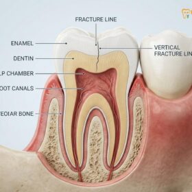
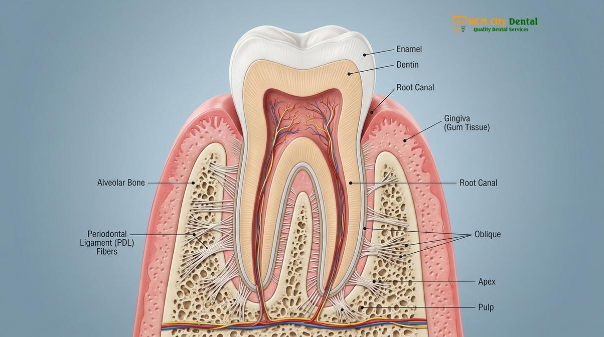
To understand the mechanics of a loose tooth adult emergency, one must first examine the complex anatomical structures that anchor a permanent tooth in the oral cavity. A healthy adult tooth is not rigidly fused to the jawbone; rather, it is suspended within the alveolar socket by a dynamic network of connective tissue known as the periodontal ligament (PDL). This ligament acts as a biological shock absorber, allowing for microscopic physiological movement during mastication (chewing). However, when this movement becomes macroscopic and visually perceptible, it crosses the threshold into pathological mobility, signaling a severe underlying issue [1].
The etiology of adult tooth mobility is multifaceted, generally categorized into infectious, mechanical, and systemic origins. The most prevalent among the mobile tooth causes is chronic periodontitis. This advanced stage of gum disease is characterized by a hyperactive inflammatory response to bacterial plaque and calculus subgingivally. The host’s immune system, in an attempt to eradicate the bacterial invasion, releases inflammatory cytokines that inadvertently activate osteoclasts—cells responsible for bone resorption. As the alveolar bone height diminishes, the physical foundation supporting the tooth is lost, resulting in progressive wobbliness.
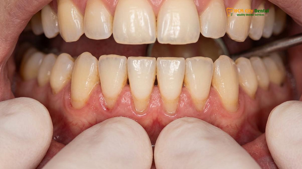
Beyond infectious causes, mechanical forces play a significant role. Occlusal trauma, often resulting from chronic bruxism (teeth grinding) or clenching, exerts excessive lateral and vertical forces on the dentition. Over time, these non-physiological forces widen the periodontal ligament space, leading to a wobbly tooth adult scenario even in the absence of periodontal disease. This is clinically referred to as primary occlusal trauma. When excessive forces are applied to a tooth that has already lost bone support due to periodontitis, it is termed secondary occlusal trauma, which rapidly accelerates tooth loss.
Systemic factors can also temporarily or permanently alter tooth stability. Hormonal fluctuations, particularly the surges in estrogen and progesterone during pregnancy, can increase the permeability of gingival blood vessels and temporarily loosen the ligaments anchoring the teeth—a condition known as pregnancy gingivitis. Furthermore, systemic conditions that affect bone density, such as osteoporosis, can compromise the alveolar bone matrix, reducing the structural integrity of the dental arches. Understanding these diverse etiologies is critical for a clinician to formulate an accurate diagnosis and an effective, personalized treatment strategy.
“Pathological tooth mobility is not merely a symptom of localized dental failure; it is a critical clinical indicator of compromised periodontal architecture that demands immediate diagnostic imaging and biomechanical stabilization to halt progressive attachment loss.”
Loose Tooth from Trauma: Emergency Steps
Traumatic dental injuries require immediate stabilization within the first 60 minutes to maximize the survival of periodontal ligament cells and prevent irreversible root resorption.
A loose tooth trauma is one of the most time-sensitive situations in clinical dentistry. Whether resulting from a sports collision, a vehicular accident, or an accidental fall, the sudden impact can stretch, tear, or completely sever the delicate periodontal ligament fibers. In dental traumatology, injuries that result in mobility without complete displacement are classified as subluxation or luxation injuries. Subluxation involves injury to the tooth-supporting structures resulting in abnormal mobility but no displacement, while extrusive or lateral luxation involves partial displacement of the tooth out of its socket [2].
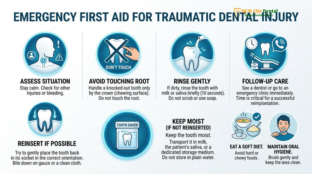
When facing a tooth mobility emergency due to trauma, the immediate actions taken by the patient before reaching the dental chair drastically influence the clinical outcome. The foremost rule is absolute immobilization. Patients must resist the natural urge to test the tooth’s looseness with their tongue or fingers. Every microscopic movement can cause further micro-hemorrhaging within the socket and sever the few remaining intact PDL fibers, transitioning a salvageable subluxation into a complete avulsion (knocked-out tooth).
Clinical Warning: Do not attempt to push a traumatically displaced or loose tooth back into its original position forcefully without professional guidance. Incorrect repositioning can crush the alveolar bone, damage the apical nerve supply, and induce severe root resorption. Seek emergency dental care immediately.
The emergency protocol involves a rapid, systematic approach. First, control any localized bleeding by applying gentle pressure with sterile gauze, taking care not to press directly on the mobile tooth. Second, manage acute inflammation by applying a cold compress to the external facial area; this induces vasoconstriction and limits edematous swelling. Third, adhere to a strict liquid or ultra-soft diet, ensuring absolutely no masticatory forces are applied to the affected quadrant.
Upon arrival at a specialized facility like HCMC Dental Clinic, the clinical workflow begins with a comprehensive trauma assessment. Dr. Nguyen Van Cuong emphasizes the necessity of immediate periapical radiographs to rule out horizontal root fractures or alveolar bone fractures, which present with similar clinical mobility but require vastly different management protocols. If the root is intact, the immediate clinical objective is repositioning (if displaced) and rigid or semi-rigid splinting to adjacent healthy teeth, creating a stable environment for the PDL to regenerate.
Loose Tooth from Periodontal Disease
Chronic periodontitis induces progressive alveolar bone resorption and attachment loss, gradually increasing tooth mobility until the structural foundation is critically compromised.
While trauma causes acute mobility, periodontal disease is the silent, chronic destroyer of dental stability. It is the leading cause of tooth loss in the adult population globally. The pathogenesis of periodontitis begins with gingivitis—a reversible inflammation of the superficial gums caused by biofilm accumulation. If left untreated, the bacterial colonies migrate subgingivally, forming deep periodontal pockets. These anaerobic bacteria produce potent toxins that provoke a severe host immune response [3].
The critical turning point in periodontal disease is the destruction of the connective tissue attachment and the alveolar bone. As the bone recedes apically (towards the root tip), the clinical crown-to-root ratio becomes unfavorable. A healthy tooth has approximately two-thirds of its root embedded in bone. When periodontitis reduces this bone support to one-half or one-third, the tooth becomes subjected to magnified biomechanical stress during normal chewing, leading to a wobbly tooth adult presentation.
Clinicians quantify the severity of this condition using standardized indices. The most widely accepted system is Miller’s Classification of Tooth Mobility, which guides the treatment prognosis and intervention strategy.
| Mobility Class | Clinical Description | Prognosis & Management |
|---|---|---|
| Class I | First distinguishable sign of movement greater than normal physiological mobility (up to 1mm horizontally). | Excellent prognosis. Managed with non-surgical scaling and root planing (SRP) and occlusal adjustment. |
| Class II | Movement of the crown more than 1mm in any horizontal direction, but no vertical depression. | Guarded to fair prognosis. Requires deep debridement, possible splinting, and strict periodontal maintenance. |
| Class III | Severe mobility greater than 1mm horizontally, combined with vertical displacement (depressibility) in the socket. | Poor prognosis. Often indicates terminal bone loss; extraction and prosthetic replacement are usually required. |
Diagnosing periodontal mobility involves a technique called bidigital palpation, where the clinician uses the handles of two metallic instruments to apply alternating pressure to the buccal and lingual surfaces of the tooth. Relying on fingers alone can yield false tactile feedback due to the compressibility of the clinician’s own finger pads. Once the mobility is staged, a comprehensive periodontal charting is performed, measuring pocket depths, clinical attachment loss, and bleeding on probing, which collectively dictate the aggressiveness of the required intervention.
Clinical Treatment: Splinting and Deep Cleaning
Therapeutic intervention involves mechanical stabilization via dental splinting, combined with aggressive infection control through scaling and root planing or surgical debridement.
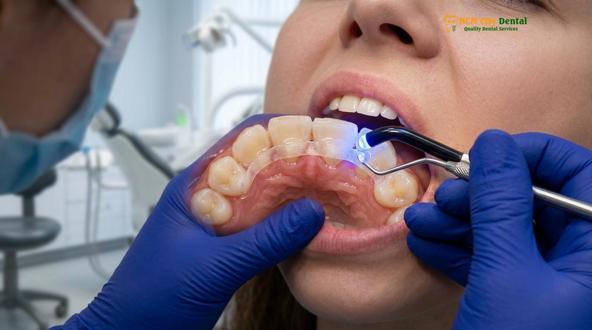
When a patient presents with a loose tooth adult emergency, the treatment philosophy is twofold: arrest the active disease process and mechanically stabilize the dentition. The cornerstone of mechanical stabilization is dental splinting. Splinting involves joining the mobile tooth to adjacent, firm teeth, effectively distributing occlusal forces across a broader multi-rooted foundation. This reduces the strain on the compromised periodontal ligament, providing a biologically favorable environment for tissue repair and bone consolidation [4].
There are several splinting modalities utilized depending on the clinical presentation. Extracoronal splinting is the most common and conservative approach. It involves bonding a flexible fiberglass ribbon or a braided stainless steel wire to the lingual (tongue-facing) surfaces of the teeth using a light-cured composite resin. This technique is highly aesthetic and minimally invasive, as it requires little to no preparation of the natural tooth structure. Intracoronal splinting, conversely, involves cutting a small channel into the teeth to embed the stabilizing wire, offering greater durability for long-term or permanent stabilization in severe periodontal cases.
Clinical Case Review: A 45-year-old patient visited HCMC Dental Clinic in Ho Chi Minh City presenting with Class II mobility in the lower anterior incisors due to localized aggressive periodontitis. Dr. Nguyen Van Cuong initiated a comprehensive treatment plan involving full-mouth ultrasonic debridement followed by the application of a lingual fiberglass splint. Within six weeks of stabilization and strict home care compliance, the gingival inflammation resolved, and the teeth exhibited a significant reduction in mobility, successfully avoiding extraction.
However, splinting alone is merely a mechanical crutch; it does not cure the underlying infection. Therefore, it must be paired with rigorous periodontal therapy. The first line of defense is Scaling and Root Planing (SRP). This non-surgical procedure utilizes ultrasonic scalers and hand curettes to meticulously remove calculus and bacterial endotoxins from the deep periodontal pockets and smooth the root surfaces. A smooth root surface discourages further bacterial colonization and facilitates the reattachment of healthy gingival tissues.
In cases where the periodontal pockets are too deep to be effectively cleaned via SRP (typically >6mm), surgical intervention may be necessary. Periodontal flap surgery involves gently separating the gums from the teeth to provide direct visual access to the infected root surfaces and underlying bone defects. Once the area is thoroughly debrided, the clinician may employ regenerative techniques, such as placing bone grafts or guided tissue regeneration (GTR) membranes, to stimulate the regrowth of lost alveolar bone, thereby permanently improving the tooth’s structural support [5].
Can a Loose Adult Tooth Tighten Back Up?
A mobile permanent tooth can often restabilize if the underlying cause is promptly addressed, allowing the periodontal ligament to heal and bone density to be preserved.
A common and anxious question from patients experiencing a tooth mobility emergency is whether the tooth can ever return to its original, stable state. The clinical answer is highly dependent on the etiology, the extent of structural damage, and the rapidity of the intervention. The human body possesses a remarkable capacity for healing, and the periodontium is no exception, provided the pathological stimuli are removed.
In cases of acute trauma, such as a subluxation injury, the prognosis for restabilization is generally excellent. If the tooth is splinted promptly and the patient adheres to a soft diet, the torn periodontal ligament fibers can regenerate and reattach to the cementum of the root within two to four weeks. During this healing phase, the tooth must be protected from secondary trauma. The clinician will monitor the tooth’s vitality, as traumatic injuries can sometimes sever the apical blood supply, necessitating subsequent endodontic (root canal) therapy even if the tooth tightens back up.
Conversely, restabilization in the context of severe periodontal disease is more complex. While we cannot easily regenerate massive amounts of lost alveolar bone without advanced surgical grafting, we can eliminate the inflammation. Once the bacterial load is reduced through deep cleaning, the swollen, edematous gingival tissues shrink and tighten tightly around the neck of the tooth. Furthermore, the elimination of occlusal trauma through bite adjustments allows the widened periodontal ligament space to narrow back to its physiological width. This combination often results in a clinically significant reduction in mobility, transitioning a wobbly tooth adult from a Class II mobility down to a manageable Class I.
“The regenerative potential of the periodontium is maximized when biomechanical stability is achieved simultaneously with absolute infection control, allowing osteoblasts and fibroblasts to reconstruct the compromised attachment apparatus.”
Prevention and Long-Term Oral Care
Preventing recurrent tooth mobility requires strict adherence to professional periodontal maintenance, daily biofilm control, and the use of protective occlusal appliances when indicated.
Successfully navigating a loose tooth adult emergency is only the first phase of treatment; the long-term survival of the dentition relies on rigorous preventive strategies. For patients who have experienced mobility due to periodontal disease, transitioning to a strict periodontal maintenance schedule is non-negotiable. Standard six-month dental checkups are insufficient for a compromised periodontium. Instead, these patients require professional debridement every three to four months to disrupt biofilm formation before it can mature into destructive, bone-resorbing calculus.
Daily home care must be elevated to a clinical standard. Brushing twice daily with a soft-bristled electric toothbrush ensures efficient plaque removal without causing further gingival recession or abrasion. Interdental cleaning is equally critical, as periodontal disease predominantly initiates between the teeth. The use of water flossers, interdental brushes, and high-quality dental floss helps maintain the health of the interproximal papillae. Antimicrobial mouth rinses containing chlorhexidine or essential oils may be prescribed short-term to manage acute inflammatory flare-ups.
For patients whose mobile tooth causes are rooted in biomechanical stress, such as bruxism, the fabrication of a custom-fitted occlusal guard (night guard) is imperative. Worn during sleep, this rigid acrylic appliance acts as a protective barrier, absorbing and evenly distributing the immense forces generated by jaw clenching. By preventing tooth-to-tooth contact, the night guard protects the periodontal ligament from traumatic widening and preserves the integrity of any applied dental splints.
Ultimately, maintaining a stable, healthy smile requires a collaborative partnership between the patient and their dental care team. If you have a history of dental trauma or suspect you are developing signs of periodontal instability, establishing a relationship with a comprehensive care facility is vital. For specialized evaluations and rapid intervention, exploring professional emergency dental care protocols ensures that your natural teeth are preserved for a lifetime.
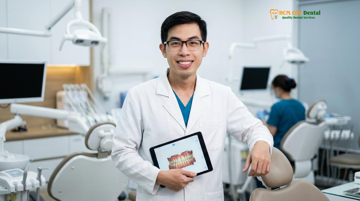
When to See a Doctor (Clinical Red Flags)
While any degree of permanent tooth mobility warrants professional evaluation, certain clinical signs indicate an acute, life-threatening or tooth-threatening emergency that requires immediate triage. You must seek urgent dental or medical care if your loose tooth is accompanied by any of the following red flags:
- Rapid Onset Swelling: Facial or intraoral swelling that expands quickly, particularly if it approaches the eye or descends into the neck, indicating a spreading fascial space infection.
- Purulent Exudate (Pus): A foul-tasting discharge oozing from the gumline around the mobile tooth, signifying an active, acute periodontal or periapical abscess.
- Systemic Symptoms: The presence of a fever, chills, or generalized malaise in conjunction with dental pain, suggesting the localized infection has entered the systemic circulation.
- Severe, Unrelenting Pain: Throbbing pain that is not alleviated by over-the-counter analgesics and disrupts sleep, often pointing to irreversible pulpitis or acute nerve compression.
- Traumatic Displacement: If the tooth has been visibly pushed up into the gum, knocked sideways, or is interfering with your ability to close your mouth normally following an impact.
Do not delay care if these symptoms arise. Early diagnostic imaging and clinical intervention are the defining factors between saving the natural tooth and requiring a complex surgical extraction.
Frequently Asked Questions
Is a loose adult tooth a dental emergency?
Yes, a loose adult tooth is always considered a dental emergency requiring immediate professional evaluation. Because permanent teeth are designed to remain stable, any mobility indicates underlying structural damage, acute trauma, or severe infection that could lead to irreversible tooth loss if left untreated. Prompt intervention maximizes the chances of saving the tooth and restoring the health of the surrounding periodontal tissues.
Can a dentist save a loose adult tooth?
In many clinical scenarios, a dentist can successfully save a loose adult tooth using advanced stabilization techniques. Treatments such as dental splinting, scaling and root planing, or occlusal adjustments are highly effective. However, the prognosis depends heavily on the underlying cause, the extent of bone loss, and how quickly the patient seeks emergency dental care after noticing the mobility.
How long does it take for a splinted tooth to heal?
A splinted tooth typically requires two to eight weeks to heal, depending on the severity of the initial trauma or periodontal condition. Minor subluxation injuries may stabilize within a couple of weeks, while severe root fractures or advanced periodontal bone loss may require prolonged splinting and subsequent surgical interventions to ensure the periodontal ligament fully reattaches.
What should I avoid doing if I have a loose tooth?
You must strictly avoid touching, wiggling, or applying any pressure to the loose tooth with your tongue or fingers. Furthermore, refrain from eating hard, sticky, or chewy foods, and do not brush or floss vigorously around the affected area. Manipulating the tooth can sever the remaining periodontal ligament fibers, worsening the mobility and increasing the risk of extraction.
Does insurance cover loose tooth treatment?
Most dental insurance plans provide coverage for emergency diagnostic exams, radiographs, and medically necessary treatments for a loose tooth. Procedures such as periodontal scaling, dental splinting, and extractions are typically covered under basic or major restorative categories. However, coverage percentages vary by provider, so it is advisable to consult your insurance policy or clinic coordinator for precise benefit details.
References
- Journal of Periodontology. Clinical parameters of tooth mobility and periodontal attachment loss. (2021).
- Dental Traumatology. Guidelines for the management of traumatic dental injuries: Luxation injuries. (2020).
- American Academy of Periodontology. Comprehensive periodontal therapy and splinting protocols. (2019).
- International Endodontic Journal. Healing of the periodontal ligament following acute dental trauma. (2022).
- Journal of Clinical Dentistry. Efficacy of scaling and root planing in mobile teeth stabilization. (2018).
Read also: For patients who recently had braces, read our advice on wearing retainers after wisdom tooth extraction.