Experiencing cracked tooth pain when biting usually indicates a structural fracture that flexes under pressure, irritating the internal dental pulp. This sharp, localized discomfort requires prompt clinical evaluation to prevent bacterial infection, nerve damage, or complete tooth loss.
Clinical Summary:
Cracked tooth pain when biting is a primary indicator of an incomplete structural fracture within the tooth, commonly referred to as Cracked Tooth Syndrome (CTS). When mechanical pressure is applied during chewing, the fractured segments separate microscopically, causing fluid dynamics within the dentin to stimulate the underlying nerve endings. This results in a sharp, fleeting pain that often peaks upon the release of the bite. Because these hairline cracks are frequently invisible on standard two-dimensional dental X-rays, clinical diagnosis relies on specialized bite tests, fiber-optic transillumination, and periodontal probing. Treatment pathways are dictated by the depth of the fracture and the vitality of the dental pulp. Superficial cracks may be stabilized with composite bonding or a protective dental crown, whereas fractures extending into the pulp chamber necessitate endodontic therapy (root canal). In severe cases where the fracture splits the root vertically, extraction followed by implant placement remains the standard of care. Early intervention is critical to halting crack propagation and preserving the natural dentition.
Key Takeaways:
- Sharp pain when biting down or releasing pressure is the hallmark symptom of a hidden tooth fracture.
- Standard dental X-rays often fail to show hairline cracks, requiring advanced diagnostic tools like transillumination.
- Early intervention with a dental crown can often save the tooth and prevent the need for a root canal.
- Untreated cracks will progressively deepen, leading to bacterial nerve infection, abscesses, or complete tooth splitting.
- Patients with a history of heavy teeth grinding (bruxism) are at the highest risk for developing structural tooth fractures.
What is Cracked Tooth Syndrome?
Cracked Tooth Syndrome (CTS) refers to an incomplete fracture in a vital posterior tooth that involves the dentin and occasionally the pulp, presenting with highly variable and often elusive symptoms.
Cracked tooth syndrome is one of the most challenging diagnostic dilemmas in modern dentistry. It is defined as an incomplete fracture of a vital posterior tooth that typically originates from the crown and extends apically (toward the root) without completely separating the tooth into two distinct pieces. Unlike a visibly broken tooth where a large piece of enamel has sheared off, CTS involves a microscopic, hairline fissure that compromises the structural integrity of the tooth while remaining largely invisible to the naked eye. This condition predominantly affects the lower molars and upper premolars, as these teeth bear the brunt of the heavy occlusal (chewing) forces generated by the human jaw.
The anatomy of a human tooth consists of three primary layers: the hard outer enamel, the softer underlying dentin, and the innermost pulp chamber, which houses the nerves and blood vessels. When a crack breaches the rigid enamel and enters the dentin, the tooth loses its monolithic strength. The dentin is composed of thousands of microscopic channels called dentinal tubules, which communicate directly with the nerve tissue in the pulp. As the crack propagates through this layer, it creates a pathway for mechanical stress and bacterial micro-leakage to irritate the vital tissues inside the tooth [1].
From a clinical perspective, Dr. Nguyen Van Cuong frequently notes that patients suffering from cracked tooth syndrome often endure months of intermittent discomfort before seeking a definitive diagnosis. Because the pain is not constant and only occurs under specific chewing angles, patients may mistakenly attribute the sensation to a minor cavity or temporary sensitivity. However, a cavity typically causes a dull, lingering ache or sensitivity to sweets, whereas a crack produces a sharp, mechanical pain directly correlated with physical pressure.
“Cracked Tooth Syndrome is often referred to as the ‘great masquerader’ in dentistry. Its symptoms can mimic reversible pulpitis, periodontal disease, or even sinus pain, making a meticulous, symptom-driven clinical examination absolutely essential for an accurate diagnosis.”
The epidemiology of cracked teeth shows a strong correlation with age and restorative history. As teeth age, the dentin becomes more sclerotic and brittle, reducing its ability to absorb shock. Furthermore, teeth that have been heavily restored with large amalgam (silver) fillings are particularly susceptible. Amalgam does not bond chemically to the tooth structure; it merely sits within a mechanically prepared cavity. Over time, thermal expansion and contraction of the metal, combined with the wedge-like effect of chewing forces, can initiate micro-fractures in the remaining natural tooth walls. Recognizing these risk factors is the first step in identifying and managing cracked tooth syndrome before catastrophic failure occurs [2].
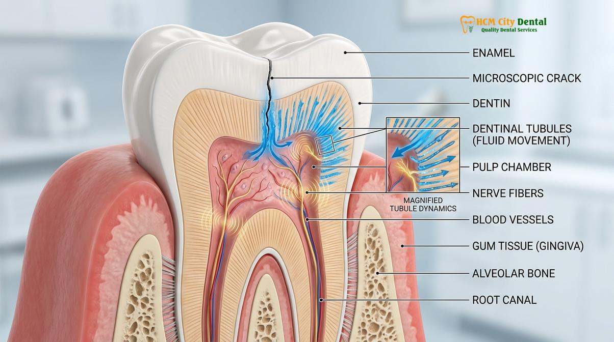
Why Does it Only Hurt When Biting Down?
Biting pressure causes the fractured tooth segments to separate microscopically, triggering fluid movement within the dentinal tubules that sharply stimulates the underlying nerve endings.
To understand why a cracked tooth hurts specifically when biting down, one must delve into the biomechanics of mastication and the neuroanatomy of the dental pulp. When you chew, your jaw muscles exert tremendous force—often exceeding 150 pounds of pressure per square inch on the molar teeth. In a healthy, intact tooth, this force is evenly distributed across the enamel and safely dissipated down the roots into the surrounding alveolar bone. However, when a tooth has a structural crack, this force distribution is severely disrupted.
As you bite down on a hard object, the pressure acts like a wedge, forcing the two sides of the crack to separate microscopically. This separation exposes the underlying dentin. According to the widely accepted hydrodynamic theory of pain, the dentinal tubules are filled with a fluid that surrounds the odontoblastic processes (extensions of the pulp cells). When the crack opens, it causes a rapid shift in this dentinal fluid. This sudden fluid movement stimulates the A-delta nerve fibers located at the pulp-dentin junction. A-delta fibers are responsible for transmitting sharp, acute, and highly localized pain signals to the brain [3].
Interestingly, one of the most definitive diagnostic hallmarks of a cracked tooth is not just pain upon biting, but a sharp, electric-like jolt of pain that occurs the exact moment you release the bite. This is known as rebound pain. When the biting pressure is suddenly removed, the separated segments of the tooth snap back together. This rapid closure causes a reverse flow of the dentinal fluid, which intensely fires the nerve endings once again. If you experience this specific symptom—where the tooth hurts when biting down but hurts even more upon release—it is almost pathognomonic for a structural fracture.
Furthermore, the crack creates a microscopic gateway for oral fluids, bacteria, and thermal changes to penetrate deep into the tooth. This explains why patients with a cracked tooth often report sudden, severe sensitivity to cold temperatures. Unlike the generalized cold sensitivity caused by receding gums, the thermal shock from a cracked tooth is usually localized to a single tooth and dissipates relatively quickly once the cold stimulus is removed. However, if the crack is left untreated and the pulp becomes chronically inflamed (irreversible pulpitis), the pain may transition from a sharp, biting-induced jolt to a constant, throbbing ache that lingers long after the chewing has stopped.
The intermittent nature of the pain is what often delays patients from seeking urgent dental care. Because the pain only occurs when food hits the tooth at a very specific angle, a patient might chew normally for days without incident, only to be struck by agonizing pain when a seed or grain of rice wedges perfectly into the fracture line. This unpredictability is a clear signal that the structural integrity of the tooth is compromised and requires professional stabilization.
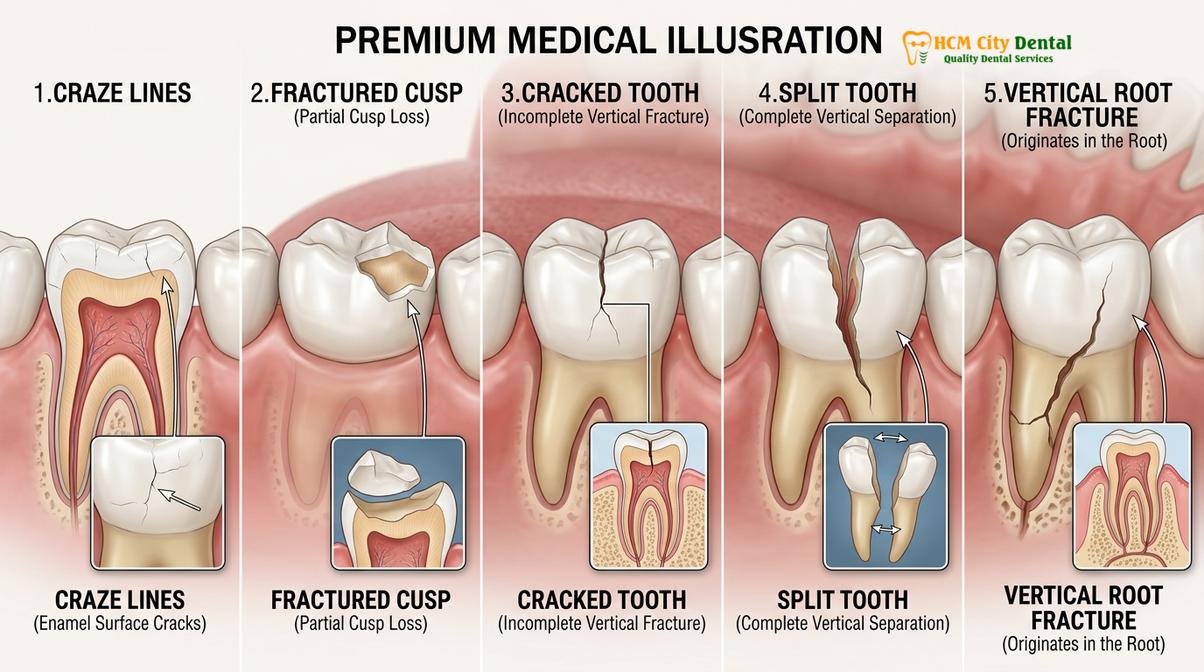
Different Types of Tooth Cracks
Tooth fractures range from superficial enamel craze lines to catastrophic split teeth, with each classification dictating a specific clinical prognosis and restorative approach.
Not all tooth cracks are created equal. The dental profession categorizes tooth fractures into five distinct classifications based on their depth, location, and the extent of structural damage. Understanding these variations is crucial, as the type of crack directly dictates the prognosis and the required treatment protocol. A superficial crack may require no treatment at all, while a deep fracture may necessitate immediate extraction.
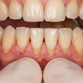
1. Craze Lines: These are microscopic, superficial hairline cracks that affect only the outer enamel layer of the tooth. Craze lines are incredibly common, especially in adult teeth, and are often visible on the front teeth when light hits them at a certain angle. Because they do not penetrate the dentin or reach the nerve, craze lines are entirely asymptomatic. They do not cause pain when biting and are generally considered a cosmetic issue rather than a structural or medical concern. No treatment is required unless the patient desires aesthetic bonding or veneers to mask the lines.
2. Fractured Cusp: The chewing surface of a molar is made up of raised points called cusps. A fractured cusp occurs when a crack forms around the base of one of these cusps, often adjacent to a large, aging dental filling. Over time, the weakened cusp may break off completely. While this can feel alarming to the tongue, a fractured cusp rarely damages the internal pulp. The pain is usually mild and occurs primarily during chewing. Once the broken piece is removed and the tooth is restored with a crown or an onlay, the symptoms typically resolve completely, and the long-term prognosis is excellent.
3. Cracked Tooth: This is the classic presentation of Cracked Tooth Syndrome. The fracture extends vertically from the chewing surface down toward the root, but the tooth has not yet separated into two distinct pieces. The crack penetrates the dentin and may or may not reach the pulp chamber. If the crack remains above the gumline, the tooth can often be saved with a full-coverage crown. However, if the crack extends deep into the pulp, endodontic therapy will be required. If the crack propagates below the bone level, the tooth may become unsalvageable.
4. Split Tooth: A split tooth is the end-stage result of an untreated cracked tooth. In this scenario, the fracture has propagated completely through the tooth, separating it into two distinct, movable segments. A split tooth is characterized by severe pain upon chewing and often presents with localized gum swelling or infection. Unfortunately, a split tooth cannot be saved intact. Depending on the position of the split, a specialist might be able to save a portion of a multi-rooted tooth through a complex procedure called hemisection, but in the vast majority of cases, the entire tooth must be extracted and replaced with a dental implant.
5. Vertical Root Fracture: Unlike the other types of cracks that begin on the chewing surface, a vertical root fracture originates in the root of the tooth and propagates upward toward the crown. These fractures are notoriously difficult to diagnose because they often present with minimal symptoms until the surrounding bone and gums become severely infected. They are most commonly seen in teeth that have previously undergone root canal therapy, as the non-vital dentin becomes brittle over time. The classic sign of a vertical root fracture is an isolated, deep, narrow periodontal pocket alongside the root. The prognosis for a vertical root fracture is poor, and extraction is almost always required [4].
| Fracture Type | Depth of Crack | Primary Symptoms | Typical Treatment |
|---|---|---|---|
| Craze Lines | Enamel only | None (Asymptomatic) | No treatment / Cosmetic only |
| Fractured Cusp | Enamel and Dentin | Mild pain on chewing | Dental Crown or Onlay |
| Cracked Tooth | Extends toward root | Sharp pain on bite/release | Crown ± Root Canal |
| Split Tooth | Complete separation | Severe pain, mobility | Extraction (usually) |
| Vertical Root Fracture | Originates in root | Dull ache, gum infection | Extraction |
How Dentists Diagnose Hidden Cracks
Because hairline fractures rarely appear on standard X-rays, dentists utilize bite tests, fiber-optic transillumination, magnification, and specialized dyes to pinpoint the exact location of the structural defect.
Diagnosing a cracked tooth is a meticulous process that requires clinical acumen and specialized diagnostic tools. One of the most frustrating aspects of Cracked Tooth Syndrome for both patients and clinicians is that hairline fractures are almost always invisible on standard two-dimensional dental radiographs (X-rays). An X-ray beam must pass perfectly parallel to the fracture line to capture it on film; because most cracks propagate in unpredictable, oblique directions, they simply do not show up. Therefore, a dentist cannot rely on X-rays alone to rule out a fracture.
The diagnostic workflow typically begins with a thorough subjective history. The dentist will ask specific questions about the nature of the pain: Does it hurt when you bite down, or when you let go? Is it sensitive to hot or cold? Does the pain linger? Following the history, the clinical examination commences with the most reliable diagnostic tool for CTS: the bite test.
Using a specialized instrument—often a small, plastic, wedge-shaped device known as a “Tooth Slooth”—the dentist will ask the patient to bite down on individual cusps of the suspected tooth. By isolating the pressure to one specific cusp at a time, the dentist can reproduce the exact mechanical stress that causes the pain. If biting on the mesio-lingual cusp produces no pain, but biting on the disto-buccal cusp triggers a sharp jolt upon release, the clinician has successfully localized the fractured segment.
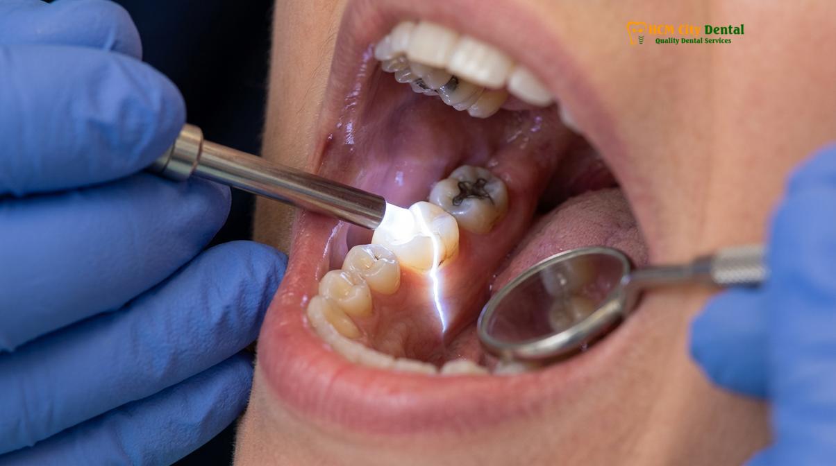
Once the symptomatic cusp is identified, the dentist will employ visual aids to confirm the presence of the crack. High-powered dental loupes or a surgical operating microscope are essential for visualizing microscopic fissures. To enhance visibility, the dentist may use a technique called fiber-optic transillumination. A high-intensity light wand is placed against the side of the tooth in a darkened room. In a healthy tooth, the light passes uniformly through the enamel and dentin. However, if a structural crack is present, the fracture plane acts as a barrier, blocking the light from passing through. The tooth will appear brightly illuminated on one side of the crack and dark on the other, clearly outlining the extent of the fracture.
Clinical Case Review: HCMC Dental Clinic
A 45-year-old patient presented to HCMC Dental Clinic in Ho Chi Minh City complaining of sharp, unpredictable pain in a lower right molar when eating nuts or crusty bread. Standard digital X-rays revealed no decay and normal bone levels. Dr. Nguyen Van Cuong performed a targeted bite test, which immediately reproduced the sharp rebound pain on the disto-lingual cusp. Using fiber-optic transillumination, a distinct fracture line was visualized extending across the marginal ridge. Because the pulp tested vital and healthy, the tooth was successfully stabilized with a full-coverage zirconia crown, completely resolving the patient’s symptoms without the need for endodontic therapy.
In addition to transillumination, dentists may use specialized disclosing dyes, such as methylene blue, which seep into the fracture line and make it visually apparent. Periodontal probing is another critical diagnostic step. The dentist will gently measure the depth of the gum pockets around the entire circumference of the tooth. A healthy tooth will have uniform pocket depths of 2-3 millimeters. If the dentist discovers an isolated, narrow pocket that suddenly drops to 7 or 8 millimeters directly adjacent to a suspected crack, it strongly indicates that the fracture has split the root vertically, drastically altering the prognosis.
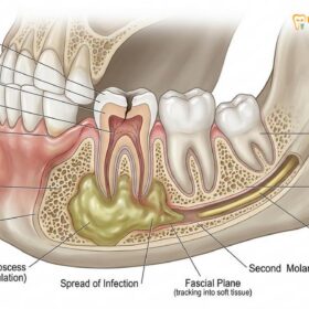
While standard 2D X-rays cannot show the crack itself, they are still necessary to evaluate the health of the surrounding bone and to check for signs of a periapical abscess (infection at the root tip). In complex cases, a 3D Cone Beam Computed Tomography (CBCT) scan may be ordered. While even a CBCT might not show a microscopic hairline crack due to resolution limits and artifact scatter, it is highly effective at detecting the subtle patterns of bone loss that occur adjacent to a vertical root fracture, aiding in a definitive diagnosis.
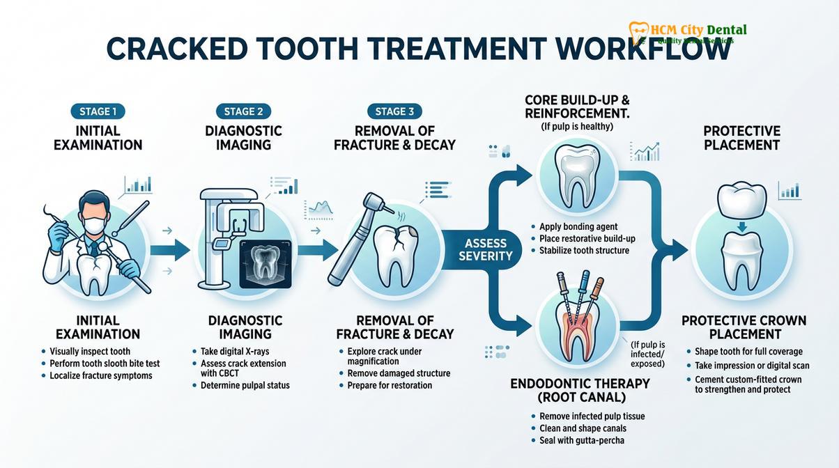
Treatment Options: Bonding, Crowns, or Root Canal
Treatment depends on the crack’s depth and pulpal involvement, ranging from conservative composite bonding and protective dental crowns to endodontic therapy or extraction for severe fractures.
The treatment for a cracked tooth is highly individualized and depends entirely on the depth of the fracture, the location of the crack, and the health status of the dental pulp. The primary goal of any restorative intervention is to bind the fractured segments together, prevent the crack from propagating further, and protect the internal nerve from bacterial invasion. The clinical decision-making process follows a specific hierarchy of interventions, ranging from conservative to complex.
For minor fractures, such as a fractured cusp that does not involve the nerve, conservative biomimetic restorations may be employed. If the crack is shallow, the dentist may remove the weakened tooth structure and place a bonded composite resin restoration. Modern dental adhesives create a micro-mechanical bond with the dentin, helping to splint the tooth together. However, for posterior teeth that endure heavy chewing forces, a simple filling is rarely sufficient to provide long-term structural stability for a true cracked tooth.
The gold standard treatment for Cracked Tooth Syndrome is the placement of a full-coverage dental crown. A crown acts like a protective helmet, encasing the entire visible portion of the tooth. When the patient bites down, the crown distributes the occlusal forces evenly across the tooth structure, preventing the fractured segments from separating and flexing. This stabilization immediately eliminates the hydrodynamic fluid movement within the dentin, thereby resolving the sharp pain upon biting. At clinics providing comprehensive dental services, materials such as monolithic zirconia or lithium disilicate (e.max) are frequently chosen for their exceptional fracture toughness and aesthetic properties.
Before placing a permanent crown, the dentist must carefully evaluate the vitality of the pulp. If the crack has already propagated deep enough to cause irreversible pulpitis (characterized by lingering, throbbing pain or spontaneous aching without chewing), a crown alone will not solve the problem. In these cases, endodontic therapy (a root canal) must be performed first. During a root canal, the inflamed or infected nerve tissue is meticulously cleaned out of the pulp chamber and root canals, and the space is sealed with a biocompatible material called gutta-percha. Once the root canal is completed, the tooth is built back up with a core buildup and subsequently protected with a crown [5].
It is important to note that even with a perfectly executed root canal and crown, a cracked tooth always carries a guarded prognosis. If the crack extends below the level of the alveolar bone, or if it continues to propagate down the root despite the crown, the tooth may eventually fail. Dr. Cuong emphasizes to patients that while modern dentistry can successfully save the vast majority of cracked teeth, the biological reality of structural fractures means that long-term monitoring is essential.
In the unfortunate event that the tooth is diagnosed as a split tooth or presents with a vertical root fracture, restorative efforts are futile. The only medically sound treatment is the complete extraction of the tooth to prevent severe bone loss and systemic infection. Following extraction, the site is typically grafted with bone material, and the missing tooth is replaced with a titanium dental implant, which restores full chewing function and prevents the adjacent teeth from shifting out of alignment.
How to Prevent Tooth Cracks
Preventing tooth fractures involves managing occlusal stress through custom night guards, avoiding excessively hard foods, and replacing massive amalgam fillings that compromise structural integrity.
While accidents and dental trauma cannot always be avoided, the vast majority of structural tooth fractures are the result of chronic, cumulative stress that can be proactively managed. Preventive dentistry focuses on identifying risk factors and mitigating the mechanical forces that lead to enamel fatigue and dentinal cracking.
The single most significant risk factor for developing cracked teeth is bruxism—the subconscious clenching and grinding of teeth, typically occurring during sleep. Bruxism subjects the teeth to forces that are exponentially higher than normal chewing. Over time, this immense pressure causes micro-fractures in the enamel that eventually propagate into the dentin. The most effective preventive measure for a known grinder is the fabrication of a custom-fitted occlusal splint (night guard). Worn during sleep, this rigid acrylic appliance acts as a shock absorber, evenly distributing occlusal forces and providing a protective barrier between the upper and lower teeth.
“An ounce of prevention is worth a pound of cure, especially in restorative dentistry. A custom night guard is a minimal investment that can save a patient from the extensive biological and financial costs of root canals, crowns, and extractions caused by nocturnal bruxism.”
Dietary habits also play a crucial role in fracture prevention. Patients should be counseled to avoid chewing on excessively hard items that concentrate force onto a single cusp. Common culprits include ice cubes, unpopped popcorn kernels, hard candies, and non-food items like pens or fingernails. Furthermore, individuals participating in contact sports must wear athletic mouthguards to protect against sudden, high-impact trauma to the dentition.
From a restorative standpoint, proactive dental maintenance is vital. Teeth that contain massive, aging amalgam fillings are structural time bombs. Because the metal filling does not support the remaining thin walls of enamel, the tooth is highly susceptible to fracturing under normal chewing loads. Dentists often recommend preemptively replacing these large, failing restorations with bonded ceramic onlays or full crowns to reinforce the tooth structure before a catastrophic split occurs. Regular dental check-ups allow the clinician to monitor these high-risk teeth and intervene conservatively.
When to See a Doctor
While mild, transient tooth sensitivity can occasionally be managed with desensitizing toothpaste, sharp pain when biting is a definitive red flag that requires professional medical evaluation. You should schedule an immediate consultation with a dental professional if you experience any of the following symptoms:
- Sharp, localized pain: A distinct jolt of pain when biting down on specific foods, particularly if the pain spikes the moment you release the bite.
- Lingering thermal pain: Sensitivity to hot or cold temperatures that lasts for more than 30 seconds after the stimulus is removed, indicating severe nerve inflammation.
- Localized swelling: Any swelling, redness, or a pimple-like bump (fistula) on the gums adjacent to the painful tooth, which is a clear sign of a bacterial abscess.
- Spontaneous aching: Pain that occurs without any chewing pressure, often waking you up at night.
Clinical Warning:
Do not ignore intermittent bite pain simply because it goes away when you chew on the other side of your mouth. A cracked tooth will not heal on its own. Delaying diagnostic evaluation allows the fracture to deepen, significantly increasing the risk that the tooth will require a complex root canal or become completely unsalvageable, leading to extraction.
If you are experiencing these symptoms, seeking timely care at a facility like HCMC Dental Clinic ensures that advanced diagnostic tools, such as transillumination and CBCT imaging, can be utilized to accurately assess the structural integrity of your tooth and implement the most conservative, effective treatment plan.

Frequently Asked Questions
Why does my tooth hurt only when I release my bite?
Pain upon releasing a bite occurs because the microscopic crack closes rapidly, causing a sudden shift in fluid within the dentinal tubules that sharply stimulates the dental nerve. This rebound effect is a hallmark diagnostic sign of a structural fracture rather than a simple cavity. When the pressure is applied, the crack opens slightly, and when released, the snapping shut of the tooth segments triggers an acute pain response from the A-delta nerve fibers.
Can a cracked tooth be saved without a root canal?
Yes, a cracked tooth can often be saved without a root canal if the fracture has not penetrated the inner pulp chamber or caused irreversible nerve damage. In these early stages, placing a custom dental crown is usually sufficient to bind the tooth together and eliminate pain. The crown prevents the tooth from flexing during chewing, thereby stopping the irritation of the nerve. However, if the nerve is already infected, endodontic therapy is mandatory before crowning.
How much does a dental crown cost for a cracked tooth in Vietnam?
Currently, the cost of a high-quality dental crown in Vietnam varies based on the chosen material, such as porcelain-fused-to-metal or full zirconia. At HCMC Dental Clinic, we provide transparent, competitive fee schedules following a comprehensive clinical evaluation to determine the most durable restorative option for your specific fracture. Zirconia crowns are often recommended for cracked posterior teeth due to their superior fracture resistance and longevity.
What happens if I leave a cracked tooth untreated?
Leaving a cracked tooth untreated allows the fracture to propagate deeper into the tooth structure, eventually reaching the nerve and causing severe infection or an abscess. Ultimately, an ignored crack can split the tooth entirely down to the root, making extraction the only viable medical option. The longer the tooth is left unprotected, the higher the biological cost and the lower the chances of saving the natural dentition.
Can a mouthguard prevent teeth from cracking?
Yes, wearing a custom-fitted occlusal mouthguard is highly effective in preventing teeth from cracking, especially for patients who suffer from nocturnal bruxism (teeth grinding). The guard acts as a shock absorber, evenly distributing bite forces and protecting the enamel from excessive mechanical stress. It is considered the first line of defense in preventive dentistry for patients exhibiting signs of heavy occlusal wear or a history of fractured restorations.
References
- Journal of Endodontics. Clinical presentation and diagnosis of cracked tooth syndrome. (2020).
- American Dental Association. Management of incomplete tooth fractures. (2021).
- International Endodontic Journal. The hydrodynamic theory of dentinal hypersensitivity and pulpal pain. (2019).
- Journal of Prosthetic Dentistry. Restorative protocols for structurally compromised posterior teeth. (2022).
- British Dental Journal. Vertical root fractures: etiology, diagnosis, and clinical management. (2018).
Read also: Learn the key clinical differences and details of wearing a night guard vs retainer.