Most adults have 32 permanent teeth, including four wisdom teeth. If the wisdom teeth have been surgically removed or are congenitally missing, the standard adult tooth count is 28. This complete set includes incisors, canines, premolars, and molars, each designed for specific masticatory functions and essential for maintaining proper oral health.
Clinical Summary:
The human adult dentition typically consists of 32 permanent teeth, distributed evenly across the maxillary (upper) and mandibular (lower) arches. This complete set comprises 8 incisors, 4 canines, 8 premolars, and 12 molars. However, due to evolutionary changes in jaw size, the third molars (wisdom teeth) frequently become impacted and require extraction, leaving many healthy adults with a functional set of 28 teeth. Maintaining this full complement requires diligent oral hygiene and regular professional monitoring to prevent periodontal disease and dental caries, which are the leading causes of tooth loss in adulthood. Understanding the precise anatomy and function of each tooth type is fundamental to comprehensive dental care and long-term oral health preservation.
Key Takeaways:
- A complete adult dentition contains 32 teeth, replacing the 20 primary (baby) teeth of childhood.
- The adult mouth includes 8 incisors, 4 canines, 8 premolars, and 12 molars, each serving a distinct biomechanical purpose.
- Many adults function optimally with 28 teeth after the routine surgical extraction of four wisdom teeth.
- Tooth anatomy consists of three primary layers: the protective enamel, the supportive dentin, and the vital pulp chamber.
- Evolutionary dietary shifts have led to smaller human jaws, increasing the prevalence of dental crowding and third molar impaction.
The Anatomy of Adult Dentition: A Clinical Breakdown
The adult mouth contains 32 specialized teeth categorized into four distinct types—incisors, canines, premolars, and molars—each engineered for precise cutting, tearing, and grinding functions.
To fully understand the complexities of the human stomatognathic system, one must first examine the precise anatomical distribution of the adult dentition. Unlike the primary dentition of childhood, which consists of merely 20 teeth, the adult permanent dentition is a highly specialized apparatus comprising 32 teeth. These teeth are symmetrically divided between the maxillary arch (upper jaw) and the mandibular arch (lower jaw), with 16 teeth residing in each arch. Furthermore, dental professionals often divide the mouth into four quadrants (upper right, upper left, lower left, lower right), with each quadrant containing exactly eight teeth in a fully developed adult[1].
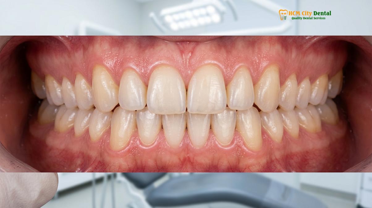
The structural diversity of these teeth is not accidental; it is a marvel of evolutionary biomechanics. Each tooth type is morphologically adapted to perform a specific phase of mastication (chewing), ensuring that food is properly incised, lacerated, and triturated before deglutition (swallowing)[4]. Preserving the integrity of each tooth type is paramount for maintaining overall occlusal harmony and digestive efficiency.
The Four Classifications of Permanent Teeth
The 32 permanent teeth are classified into four distinct morphological categories. A detailed clinical examination reveals the following distribution:
| Tooth Classification | Total Count (Adult) | Primary Biomechanical Function | Location in Dental Arch |
|---|---|---|---|
| Incisors | 8 | Shearing and cutting food during the initial bite. | Anterior (front) center of the mouth. |
| Canines (Cuspids) | 4 | Gripping, piercing, and tearing fibrous foods. | Corners of the dental arch, adjacent to incisors. |
| Premolars (Bicuspids) | 8 | Transitional tearing and initial crushing of food. | Between the canines and the molars. |
| Molars | 12 | Heavy trituration and fine grinding for digestion. | Posterior (back) of the oral cavity. |
Histological Layers of a Human Tooth
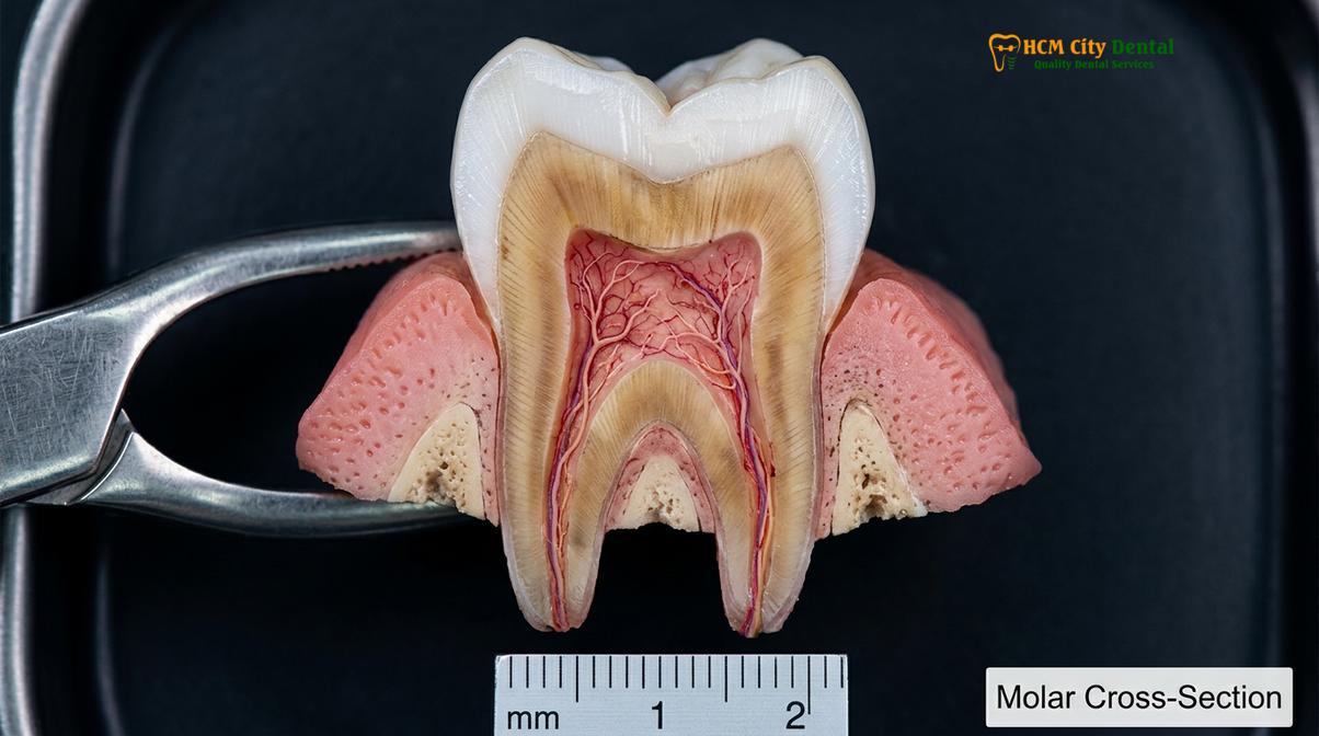
Beyond their macroscopic classification, understanding how many teeth adults have requires an appreciation of their microscopic architecture. A tooth is not a solid block of bone; rather, it is a complex, multi-layered organ consisting of both highly mineralized tissues and vital neurovascular bundles.
The outermost layer of the anatomical crown is the enamel. Composed of 96% inorganic hydroxyapatite crystals, enamel is the hardest substance in the human body, designed to withstand immense occlusal forces and resist bacterial acid attacks. However, enamel is acellular and cannot biologically regenerate once damaged by caries or trauma.
“The structural integrity of dental enamel is unparalleled in human biology. However, its inability to regenerate makes preventive dentistry and early intervention the absolute cornerstones of lifelong tooth preservation.”
Beneath the enamel lies the dentin, a slightly softer, yellowish tissue that forms the bulk of the tooth structure. Dentin is composed of microscopic tubules that radiate outward from the center of the tooth. Because these tubules contain fluid and cellular processes, exposed dentin is highly sensitive to thermal and tactile stimuli. If a patient undergoes procedures like charcoal teeth whitening using highly abrasive substances, the protective enamel can be irreversibly stripped away, exposing the vulnerable dentin and leading to severe hypersensitivity and accelerated decay.
At the very core of the tooth is the dental pulp. This soft tissue chamber houses the tooth’s life support system: the blood vessels, nerves, and connective tissues. The pulp extends down through the root canals and exits at the apical foramen, connecting the tooth to the body’s systemic circulation.
The Transition from Primary to Permanent Teeth
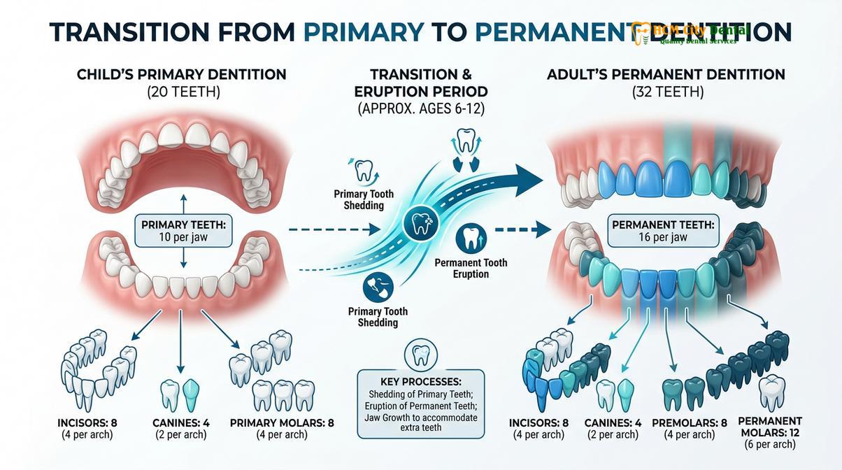
The transition from 20 deciduous baby teeth to 32 permanent adult teeth is a complex physiological process that typically spans from early childhood to the late teenage years.
The human species is diphyodont, meaning we develop exactly two successive sets of teeth during our lifespan. The initial set, known as the primary, deciduous, or “baby” dentition, consists of only 20 teeth: 8 incisors, 4 canines, and 8 molars. Noticeably absent from the primary dentition are the premolars and the third molars. The smaller number and size of primary teeth are perfectly proportioned to fit the smaller dimensions of a child’s developing maxillofacial skeleton.
As a child grows, the jawbones expand in all three dimensions, creating the necessary spatial volume to accommodate the larger and more numerous permanent teeth. The transition period, clinically referred to as the mixed dentition phase, begins when the first permanent teeth erupt. It is crucial to note that the first permanent molars erupt posterior to the primary molars; they do not replace any shedding baby teeth. This often leads to parental confusion, as these vital permanent teeth are sometimes mistaken for primary teeth and neglected.
During this mixed dentition phase, orthodontic anomalies frequently become apparent. Discrepancies between the size of the erupting permanent teeth and the available arch length can lead to severe crowding, ectopic eruptions, or an increased overjet. Early clinical evaluation is essential. For instance, interceptive orthodontics or clinical overjet correction can guide the developing jaws and erupting teeth into optimal functional positions, minimizing the need for complex orthognathic interventions later in adulthood. Patients undergoing such treatments often seek guidance on how to keep teeth white with braces to maintain optimal hygiene during the alignment process.
Wisdom Teeth: Why Many Adults Have 28 Instead of 32 Teeth
Due to evolutionary reductions in human jaw size, the four third molars often lack sufficient space to erupt properly, leading to their frequent surgical removal and a functional adult count of 28 teeth.
When discussing how many teeth adults have, the number 32 is the anatomical ideal. However, in modern clinical practice, a vast majority of adults function perfectly with only 28 teeth. The discrepancy of four teeth is almost exclusively attributed to the third molars, colloquially known as wisdom teeth. These are the final teeth to develop and typically attempt to erupt into the oral cavity during late adolescence or early adulthood.
The high prevalence of wisdom tooth complications is a fascinating subject of evolutionary anthropology. Millennia ago, the diet of early hominids consisted of coarse, abrasive, and uncooked foods such as raw roots, tough meats, and fibrous vegetation. Processing this diet required massive, robust jawbones and heavy masticatory forces. The third molars were essential for this heavy grinding[2].
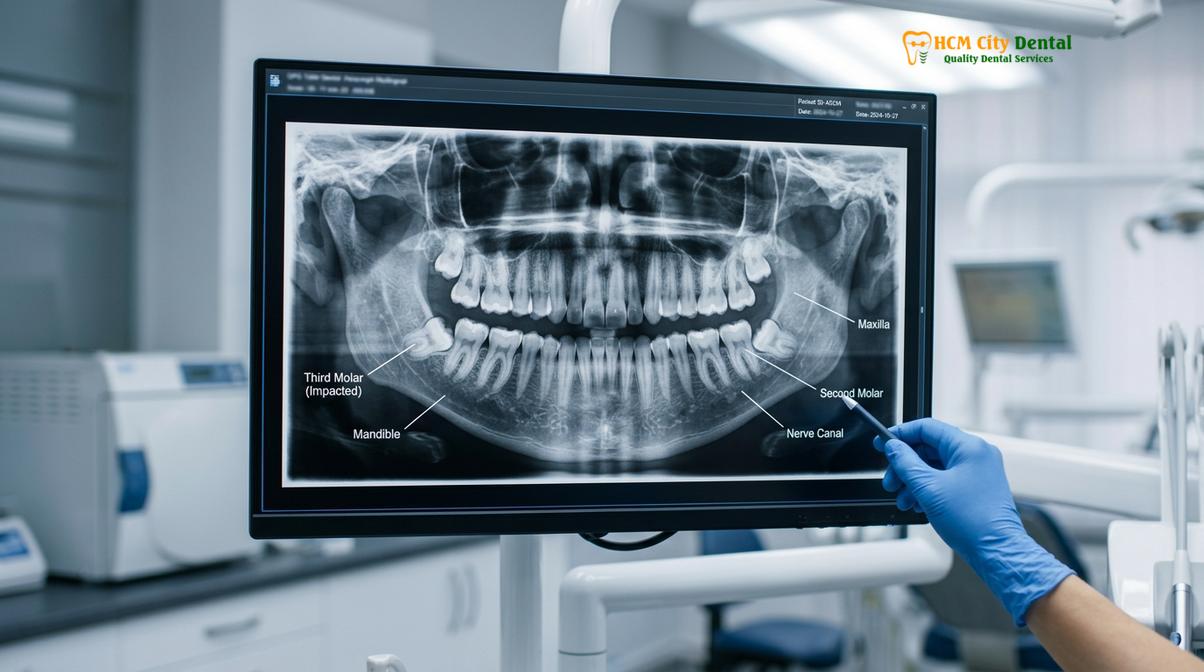
However, with the advent of agriculture, the mastery of fire for cooking, and modern food processing techniques, the human diet has become significantly softer. Consequently, the evolutionary demand for massive jaws has diminished. Over thousands of years, human jawbones have progressively decreased in size. Unfortunately, the genetic coding for the size and number of our teeth has not decreased at the same evolutionary rate. The result is a modern human jaw that is frequently too small to accommodate the full complement of 32 teeth.
Clinical Warning: Partially erupted wisdom teeth create deep periodontal pockets that are highly susceptible to bacterial accumulation. This can lead to pericoronitis, a severe and painful infection of the gum tissue that requires immediate clinical intervention to prevent systemic spread.
Clinical Complications of Impacted Wisdom Teeth
Because they are the last to erupt, third molars often find insufficient arch space available. This spatial deficiency leads to impaction—a condition where the tooth is physically blocked from fully erupting through the gingival tissue and bone. Impactions are clinically classified based on their angulation, such as mesioangular, vertical, horizontal, or distoangular.
Patients often inquire whether they should undergo wisdom teeth removal in a hospital or dentist setting. The decision depends entirely on the anatomical complexity of the impaction, the proximity of the tooth roots to the inferior alveolar nerve, and the patient’s medical history. A thorough radiographic assessment, often utilizing 3D Cone Beam Computed Tomography (CBCT), is mandatory to evaluate these risks and determine the appropriate wisdom teeth removal cost and surgical approach.
Common Causes of Tooth Loss in Adults
Despite the durability of permanent teeth, adults frequently experience tooth loss due to progressive periodontal disease, untreated dental caries, and acute maxillofacial trauma.
While the biological blueprint dictates that adults should have 32 (or 28) permanent teeth intended to last a lifetime, clinical reality paints a different picture. Edentulism (tooth loss) remains a significant global health challenge. Understanding the etiology of tooth loss is critical for implementing effective preventive strategies.
The two primary pathological culprits responsible for the vast majority of adult tooth loss are dental caries (tooth decay) and periodontal (gum) disease. Dental caries is a localized, biofilm-mediated disease driven by frequent exposure to fermentable carbohydrates. Cariogenic bacteria metabolize these sugars and excrete lactic acid as a byproduct. This acid rapidly drops the pH of the oral environment, leading to the demineralization of the inorganic enamel crystals. If the carious lesion is not intercepted and restored, the bacterial infection penetrates the dentin and eventually invades the vital pulp chamber, causing irreversible pulpitis.
However, in older adults, periodontal disease surpasses caries as the leading cause of tooth loss. Periodontitis is a chronic, destructive inflammatory disease initiated by the accumulation of bacterial plaque and calculus (tartar) below the gingival margin. The host’s immune system responds to these bacterial endotoxins by releasing inflammatory cytokines. While intended to fight the infection, this hyperactive immune response inadvertently destroys the tooth’s supporting structures—the periodontal ligament and the alveolar bone[3].
“Periodontal disease is often described as a silent epidemic. Because it is largely painless in its early and moderate stages, patients frequently do not seek care until severe alveolar bone loss has occurred and the teeth exhibit terminal mobility.”
When to See a Dentist: Important Clinical Notes
Routine dental examinations are essential for monitoring the health of all 32 permanent teeth, allowing clinicians to detect early signs of decay, gum disease, or wisdom tooth impaction before they cause irreversible damage.
Maintaining a full, functional adult dentition requires proactive professional care. Patients should not wait until they experience acute pain or visible swelling to visit a dental clinic. According to the Vietnam Ministry of Health (MOH) guidelines on adult oral health, regular biannual checkups and professional prophylaxis (scaling and root planing) are critical for preventing the progression of periodontal disease and dental caries[5].
You should schedule an immediate clinical evaluation if you experience persistent tooth sensitivity, bleeding gums during brushing, localized swelling behind your second molars, or any signs of occlusal shifting. Early intervention can often save a compromised tooth, whereas delayed treatment frequently results in complex extractions and the need for expensive prosthetic replacements.
Clinical Case Study: Restoring a Full Adult Dentition
Comprehensive restorative dentistry can successfully rehabilitate a compromised adult dentition, returning both function and aesthetics to patients suffering from severe tooth loss or structural damage.
Clinical Case Study: A 45-year-old patient visited HCMC Dental Clinic in Ho Chi Minh City presenting with multiple missing posterior teeth, severe occlusal wear on the remaining anterior incisors, and generalized periodontal inflammation. The patient had previously lost several molars due to untreated caries and was struggling with masticatory dysfunction and aesthetic concerns.
Following a comprehensive clinical and radiographic examination, the dental team developed a phased treatment plan. The initial phase focused on disease control, utilizing deep periodontal scaling to arrest the active gum disease. Once the foundational tissues were stabilized, the restorative phase began. The missing molars were replaced using strategically placed titanium dental implants, restoring the patient’s posterior grinding capacity. The worn anterior teeth were rehabilitated using custom-fabricated porcelain crowns, re-establishing the proper vertical dimension of occlusion. This dental reconstruction before and after transformation not only restored the patient’s functional adult tooth count but also significantly improved their overall quality of life and confidence.
Expert Insights on Adult Dental Anatomy
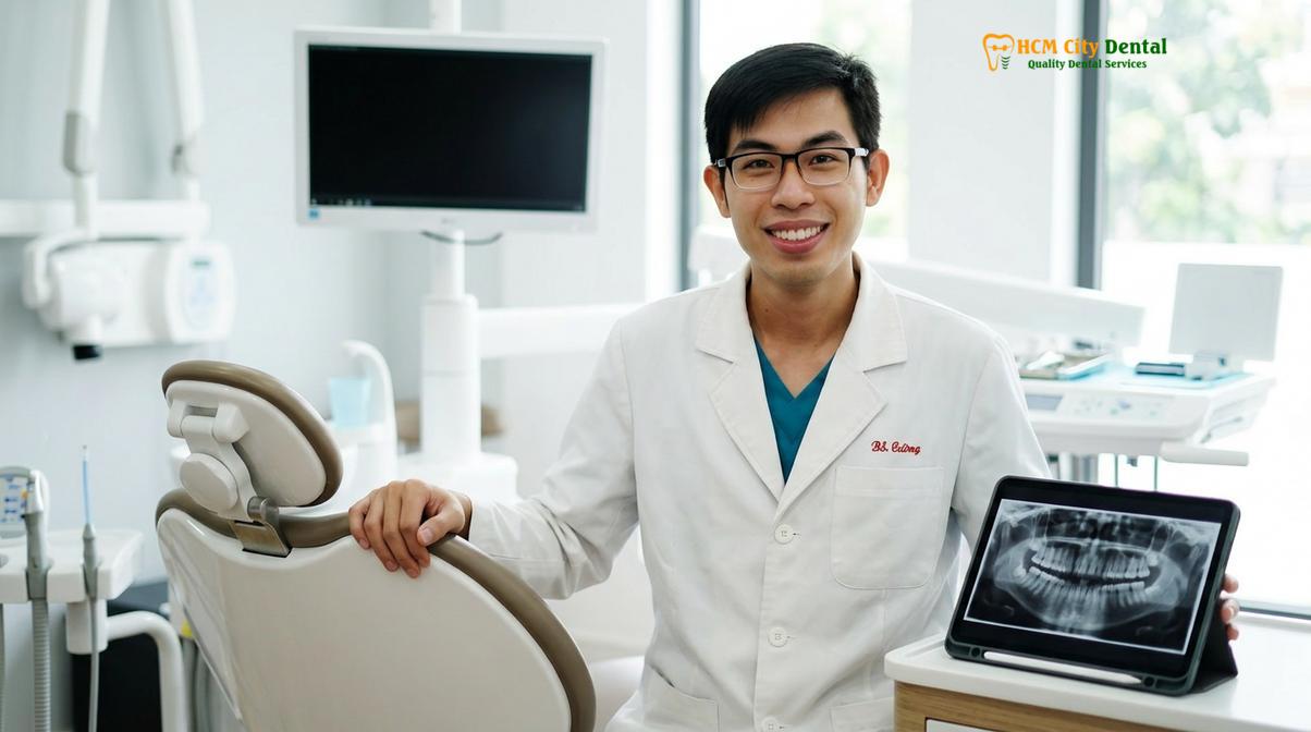
Dr. Nguyen Van Cuong, a leading specialist in restorative dentistry, emphasizes that understanding the precise anatomy of the adult dentition is the first step toward lifelong oral health. Dr. Cuong frequently educates patients on how preserving each of the 32 permanent teeth maintains occlusal harmony and prevents complex structural collapse. His clinical approach integrates advanced diagnostics with comprehensive General Dentistry tại HCMC Dental Clinic to ensure optimal patient outcomes, focusing on both the functional and aesthetic preservation of the natural smile.
Frequently Asked Questions (FAQ)
Do all adults naturally develop exactly 32 teeth?
No, while 32 is the standard anatomical number, genetic variations frequently occur. Many individuals experience hypodontia (congenitally missing teeth), most commonly affecting the third molars or lateral incisors, while others may develop supernumerary teeth (hyperdontia).
Why do most adults only have 28 functional teeth?
Most adults have 28 functional teeth because the four third molars (wisdom teeth) are routinely extracted. Due to evolutionary reductions in jaw size, these teeth often become impacted and are removed to prevent infection and crowding.
Can an adult naturally regrow a lost permanent tooth?
No, human adults are diphyodonts, meaning they only develop two sets of teeth during their lifetime. Once a permanent tooth is lost to trauma or decay, it cannot naturally regenerate and requires prosthetic replacement.
What happens if I have more than 32 teeth?
Having more than 32 teeth is a clinical condition known as hyperdontia. These extra (supernumerary) teeth can cause severe crowding, delayed eruption of adjacent teeth, and cyst formation, typically requiring surgical extraction and orthodontic management.
How does the adult tooth count differ from a child’s?
A complete adult dentition consists of 32 permanent teeth, whereas a child’s primary dentition contains only 20 teeth. Children lack premolars and have fewer molars, which are later accommodated as the jawbone grows and expands.
References
- Journal of the American Dental Association. Anatomical variations in human permanent dentition and clinical implications. (2021).
- International Journal of Oral and Maxillofacial Surgery. Evolutionary trends in human jaw size and third molar impaction rates. (2020).
- Journal of Periodontology. Pathogenesis of periodontal disease and its impact on adult tooth loss. (2019).
- British Dental Journal. Biomechanics of mastication and the functional roles of human teeth. (2022).
- Vietnam Ministry of Health (MOH). National Guidelines on Oral Health and Preventive Dentistry for Adults. (2022).
If you are experiencing discomfort with your wisdom teeth, missing teeth, or simply need a comprehensive evaluation of your adult dentition, professional care is essential. Contact HCMC Dental Clinic, Ho Chi Minh City today to schedule a thorough clinical assessment and ensure your smile remains healthy and fully functional for years to come.