Digital scans for sleep apnea utilize advanced intraoral 3D scanners to capture highly accurate optical impressions of the teeth and oral cavity. This modern workflow eliminates uncomfortable putty, allowing dentists to design precise, custom-fitted oral appliances that effectively manage snoring and mild-to-moderate obstructive sleep apnea.
Laser Whitening vs Zoom: Which Is More Effective?
\\
Clinical Summary:
\
The integration of digital scanning technology has revolutionized dental sleep medicine by replacing traditional, gag-inducing putty impressions with highly precise optical data. Dentists collaborate closely with sleep physicians to treat primary snoring and mild-to-moderate Obstructive Sleep Apnea (OSA) using custom Mandibular Advancement Devices (MADs). By capturing the exact topography of the dentition and the specific protrusive bite registration digitally, clinicians can fabricate devices like the Antisnoring Telescopic and Twinblock Snoring appliances with micron-level accuracy. This digital lab workflow ensures a comfortable fit, minimizes temporomandibular joint (TMJ) stress, and significantly improves patient compliance compared to traditional fabrication methods.
\
\\
Key Takeaways:
\
- \
- Diagnostic Protocol: Dentists do not diagnose sleep apnea; a formal sleep study (PSG or HSAT) evaluated by a sleep physician is strictly required.
- Enhanced Comfort: An intraoral 3D scanner completely eliminates the severe gag reflex associated with traditional alginate or PVS putty impressions.
- Precision Fit: Digital scans create a perfect fit dental model, preventing unwanted orthodontic shifting and reducing TMJ discomfort.
- Advanced Devices: Custom appliances like the Antisnoring Telescopic allow for lateral jaw movement, enhancing nighttime comfort.
- Rapid Turnaround: The digital lab workflow allows for instant file transmission and rapid CAD/CAM fabrication of the sleep appliance.
\
\
\
\
\
\
\\
The Pathophysiology of Sleep Apnea and the Multidisciplinary Diagnostic Approach
\\
Obstructive sleep apnea is a complex medical condition requiring a formal sleep study for diagnosis, followed by collaborative treatment planning between a sleep physician and a qualified dental provider.
\
\\
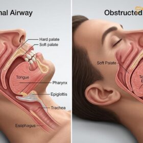
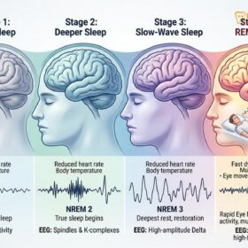
Obstructive Sleep Apnea (OSA) is a pervasive and potentially severe sleep disorder characterized by repeated episodes of partial or complete upper airway obstruction during sleep. When the muscles of the soft palate, tongue base, and pharynx relax excessively, they collapse into the airway. This mechanical blockage prevents normal breathing, leading to a drop in blood oxygen levels—a phenomenon known as oxygen desaturation. The brain senses this dangerous physiological state and triggers a micro-arousal, briefly waking the patient to restore muscle tone and reopen the airway, often accompanied by a loud gasp or snorting sound. These repetitive interruptions fragment the sleep cycle, preventing the patient from reaching the restorative stages of deep sleep and REM sleep.
\\
The severity of this condition is clinically measured using the Apnea-Hypopnea Index (AHI), which calculates the average number of breathing pauses (apneas) and periods of shallow breathing (hypopneas) per hour of sleep. An AHI of 5 to 15 indicates mild OSA, 15 to 30 indicates moderate OSA, and an AHI greater than 30 signifies severe OSA. Left untreated, the chronic intermittent hypoxia and sympathetic nervous system surges associated with OSA can lead to profound systemic health issues, including treatment-resistant hypertension, arrhythmias, stroke, metabolic syndrome, and severe daytime cognitive impairment.
\\
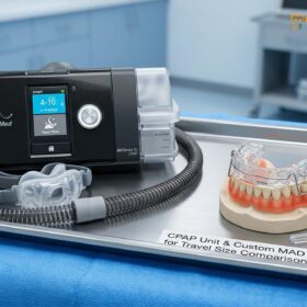
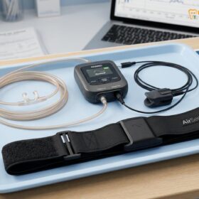
It is a critical tenet of dental sleep medicine that dentists do not diagnose sleep apnea. The diagnosis of OSA falls strictly under the purview of a board-certified sleep physician. Patients must undergo a comprehensive objective evaluation, typically through an overnight in-lab Polysomnography (PSG) or a medically supervised Home Sleep Apnea Test (HSAT). Once the sleep physician confirms the diagnosis and determines the AHI, they may prescribe Continuous Positive Airway Pressure (CPAP) therapy as the gold standard for severe cases. However, for patients with primary snoring, mild-to-moderate OSA, or those who are demonstrably intolerant to CPAP therapy, the sleep physician will refer the patient to a qualified dentist for the fabrication of custom sleep apnea appliances.
\\
\
“The successful management of obstructive sleep apnea relies heavily on a multidisciplinary paradigm. The sleep physician provides the definitive medical diagnosis and oversees the systemic health outcomes, while the dental sleep medicine practitioner utilizes advanced digital workflows to design an oral appliance that mechanically stabilizes the airway with maximum patient compliance.”
\
\\
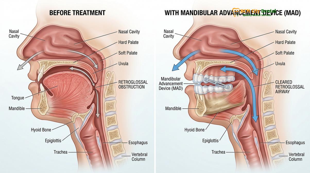
This collaborative approach ensures that the patient receives evidence-based, comprehensive care. The dentist’s role is to evaluate the patient’s dental and periodontal health, assess the temporomandibular joint (TMJ), and utilize advanced digital scans for sleep apnea to create a Mandibular Advancement Device (MAD) that safely and effectively holds the lower jaw in a forward position, thereby preventing the tongue base from collapsing into the pharyngeal space [1].
\\\
\\\
How 3D intraoral scanners replace traditional putty impressions
\\
Modern optical scanning technology eliminates the need for messy alginate or polyvinyl siloxane materials, capturing the oral topography digitally to ensure maximum patient comfort and clinical precision.
\
\\
For decades, the standard method for capturing the anatomical details of a patient’s teeth and gums involved filling a large metal or plastic tray with a viscous impression material—such as alginate or polyvinyl siloxane (PVS)—and pressing it firmly over the dental arches. The patient was required to hold this bulky tray in their mouth for several minutes while the material chemically set into a rubbery solid. For many patients, particularly those with a sensitive soft palate or underlying anxiety, this process triggered a severe and highly distressing gag reflex. Furthermore, traditional putty impressions are inherently susceptible to human error, material shrinkage, temperature distortions, and the introduction of air bubbles, all of which can compromise the accuracy of the final physical model.
\\
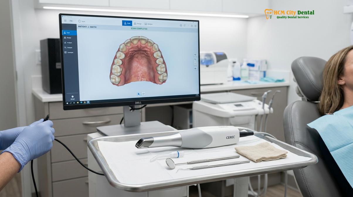
The advent of the intraoral 3D scanner has fundamentally transformed this clinical workflow. Instead of using physical putty, the dentist uses a sleek, handheld wand equipped with advanced optical sensors and structured light technology. As the wand glides smoothly over the surfaces of the teeth, it captures thousands of high-resolution images per second. Powerful software instantly stitches these images together to render a highly detailed, full-color, three-dimensional virtual model of the patient’s oral cavity on a computer monitor in real-time.
\\
The primary advantage of this technology in dental sleep medicine is the profound improvement in the patient experience. Because the wand is small and non-invasive, it completely bypasses the triggers that cause a gag reflex. Patients can breathe normally, swallow, and even take breaks if necessary during the scanning process. The digital scan captures the exact morphology of the clinical crowns, the gingival margins, and the complex occlusal anatomy without any physical distortion. This transition from analog to digital impressions represents a massive leap forward in ensuring that the foundational data used to build the sleep appliance is absolutely flawless.
\\\\
\\
The clinical benefit: Micron-level accuracy for a comfortable sleep guard fit
\\
High-resolution digital impressions create a flawless virtual model of the dentition, allowing for the fabrication of oral appliances that fit securely without causing orthodontic shifting or dental pain.
\
\\
When treating sleep apnea with an oral appliance, the physical fit of the device is arguably the most critical factor determining long-term patient compliance and therapeutic success. A Mandibular Advancement Device must anchor firmly to the upper and lower teeth to exert the necessary mechanical force to hold the heavy lower jaw and tongue forward against the pull of gravity and muscle relaxation during sleep. If the appliance is too loose, it will dislodge during the night, rendering the therapy useless and potentially creating a choking hazard. Conversely, if the appliance is too tight or applies uneven pressure, it acts like an active orthodontic aligner, causing severe tooth pain, unwanted shifting of the dentition, and significant morning soreness.
\\
By utilizing an intraoral 3D scanner, the dentist generates a perfect fit dental model that boasts micron-level accuracy. The digital file captures the exact undercuts and contours of every single tooth. When the dental laboratory designs the appliance using Computer-Aided Design (CAD) software, they can digitally block out specific undercuts and calibrate the exact amount of retention required. This ensures that the final appliance snaps into place with a satisfying, secure fit that distributes the biomechanical load evenly across the entire dental arch, rather than placing excessive stress on individual teeth [2].
\\
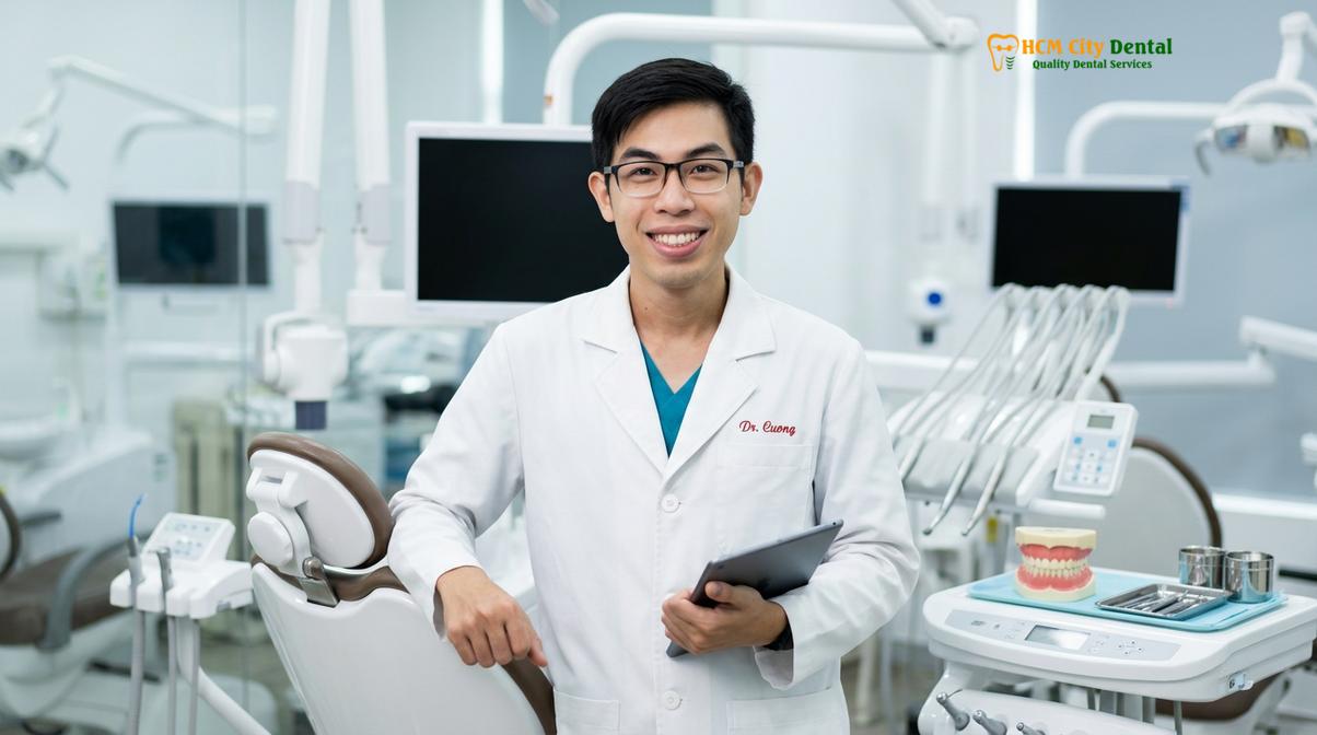
According to Dr. Nguyen Van Cuong’s clinical perspective on airway scanning, achieving a precise digital impression is the foundational step in mitigating TMJ stress during oral appliance therapy. When the appliance fits perfectly, the patient’s jaw is held in a stable, predictable position. There are no micro-movements or rocking of the device that could trigger muscle spasms or joint inflammation. Dr. Cuong emphasizes that comprehensive TMJ joint checks and meticulous titration calibration are essential when transitioning a patient from a CPAP machine to a custom mandibular advancement device, and this process relies entirely on the accuracy of the initial digital data.
\\\
\\\
Designing your customized bite registration in three dimensions
\\
Capturing the exact protrusive jaw position digitally ensures the resulting sleep appliance holds the airway open effectively while maintaining a safe, therapeutic relationship for the temporomandibular joint.
\
\\
While capturing the anatomy of the individual teeth is crucial, the true therapeutic mechanism of a sleep appliance lies in the spatial relationship between the upper and lower jaws. To open the retroglossal airway (the space behind the base of the tongue), the lower jaw must be advanced forward. However, the degree of advancement must be carefully calculated. Advancing the jaw too little will fail to resolve the apneas, while advancing it too far can cause severe, sometimes irreversible, damage to the temporomandibular joint and the associated masticatory muscles.
\\
To determine the optimal starting position, the dentist utilizes a specialized measuring tool, often a George Gauge or a similar bite registration fork. The patient is asked to bite down in their normal resting position (centric occlusion), and then asked to slide their lower jaw as far forward as possible (maximum protrusion). The dentist measures this total range of motion. Typically, the initial therapeutic position is set at approximately 50% to 70% of the patient’s maximum protrusion, combined with a specific amount of vertical opening to allow room for the appliance material and to optimize airway dynamics.
\\
Once this precise three-dimensional position is established, the dentist uses the intraoral 3D scanner to capture the bite registration digitally. The patient holds their jaw in the measured protrusive position, and the scanner records the exact interocclusal relationship from the buccal (cheek) side. The software then aligns the previously scanned upper and lower digital models into this exact therapeutic posture. This digital bite record is infinitely more accurate than traditional wax bites, which can easily warp, compress, or distort during transit to the laboratory [3].
\\
This initial position is just the starting point. Custom sleep appliances are designed to be titratable, meaning the dentist can make micro-adjustments (usually in 0.25mm to 0.5mm increments) over several weeks or months. This gradual titration allows the TMJ and muscles to adapt slowly to the new posture, maximizing airway clearance while minimizing clinical side effects.
\\\
\\\
Custom Sleep Appliances at HCMC Dental Clinic
\\
Based on the digital scans and clinical evaluation, patients can receive specialized mandibular advancement devices tailored to their specific anatomical needs and budget requirements.
\
\\
At HCMC Dental Clinic, the integration of digital scanning technology allows for the provision of highly sophisticated, custom-milled or 3D-printed oral appliances. Following a thorough clinical examination, joint assessment, and review of the sleep physician’s diagnostic report, the dental team will recommend a specific device architecture based on the patient’s anatomy, severity of OSA, and bruxism (teeth grinding) habits.
\\
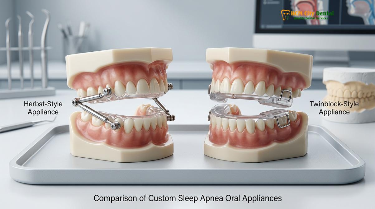
Two of the most highly regarded and frequently prescribed devices at the clinic are the Antisnoring Telescopic appliance and the Twinblock Snoring appliance. Both devices are fabricated using premium, biocompatible medical-grade materials, ensuring durability and safety for long-term intraoral use.
\\
The Antisnoring Telescopic Appliance
\
The Antisnoring Telescopic device utilizes a Herbst-style mechanism. It features custom-fitted upper and lower acrylic or milled nylon splints connected by lateral telescopic metal rods. The primary clinical advantage of this design is its allowance for significant lateral (side-to-side) and vertical jaw movement while strictly preventing the jaw from falling backward into the airway. This freedom of movement is crucial for patients who suffer from nocturnal bruxism or those who experience claustrophobia with rigid devices. By allowing the jaw to move naturally during sleep, the telescopic rods drastically minimize TMJ stiffness and muscle fatigue. Currently, this premium device is priced at 12,000,000 VND (approximately $480 USD when utilizing the clinic’s WhatsApp discount program).
\\
The Twinblock Snoring Appliance
\
The Twinblock Snoring appliance is a two-piece device characterized by interlocking acrylic bite blocks. The upper and lower splints feature precision-angled 70-degree ramps. When the patient closes their mouth, the lower blocks slide forward against the upper blocks, gently guiding the mandible into the predetermined protrusive position. Because the upper and lower pieces are not physically linked by metal hardware, the patient can easily open their mouth to speak, drink water, or yawn without removing the device. This design is highly effective for patients who breathe through their mouths or require maximum vertical clearance. This device is currently priced at 11,000,000 VND (approximately $440 USD with the WhatsApp discount).
\\
| Feature | Antisnoring Telescopic | Twinblock Snoring |
|---|---|---|
| Mechanism | Lateral telescopic metal rods (Herbst-style) | Interlocking 70-degree acrylic bite blocks |
| Jaw Movement | Allows lateral and vertical movement | Allows full mouth opening; guides jaw forward on closure |
| Best Suited For | Patients with bruxism, TMJ sensitivity | Mouth breathers, patients preferring no metal hardware |
| Titration Method | Adjustable rod lengths / shims | Modification of acrylic ramps / advancement screws |
| Estimated Cost | 12,000,000 VND (~$480 USD) | 11,000,000 VND (~$440 USD) |
\\
Clinical Case Review: Transitioning from CPAP to MAD
\
A 45-year-old male patient visited HCMC Dental Clinic in Ho Chi Minh City seeking an alternative to his CPAP machine, which he found intolerable due to mask leaks and claustrophobia. His sleep physician had diagnosed him with moderate OSA (AHI of 22). Dr. Cuong performed a comprehensive TMJ evaluation and utilized an intraoral 3D scanner to capture the dentition without triggering the patient’s severe gag reflex. The digital data was used to fabricate an Antisnoring Telescopic appliance. Following a 6-week titration protocol, a follow-up Home Sleep Apnea Test (HSAT) revealed his AHI had dropped to 4, well within the normal range, with complete resolution of his primary snoring and significant improvement in daytime energy levels.
\
\\
How the digital lab workflow speeds up fabrication turnaround in Saigon
\\
Transmitting digital scan files instantly to a dental laboratory allows for rapid CAD/CAM design and manufacturing, significantly reducing the waiting time for the final sleep appliance.
\
\\
In the traditional analog workflow, once the dentist captured the physical putty impressions and wax bite registration, these delicate items had to be carefully disinfected, packaged, and shipped via courier to a specialized dental laboratory. This physical transit introduced delays, risk of loss, and the potential for the materials to warp due to temperature changes during shipping. Upon arrival, the lab technician had to pour liquid stone into the impressions to create physical plaster models—a process that takes hours to set and introduces further potential for dimensional inaccuracy.
\\
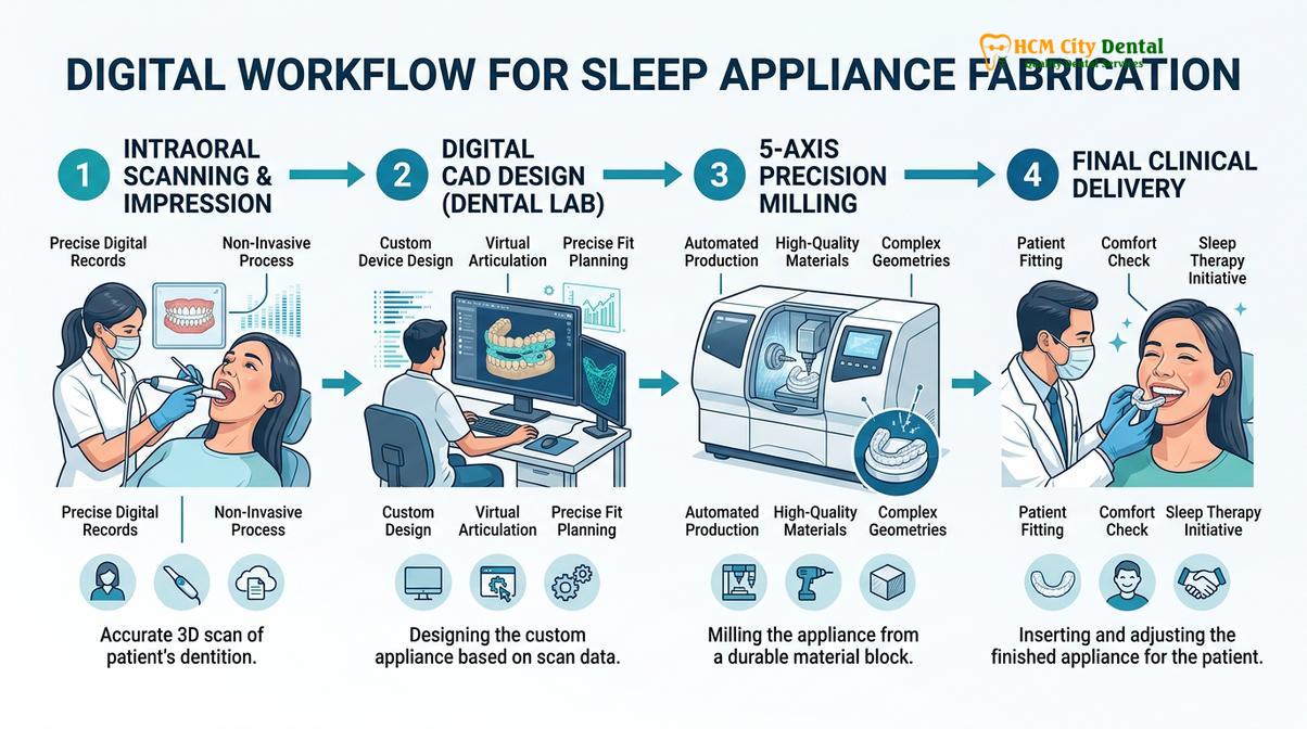
The implementation of a fully digital lab workflow eliminates these analog bottlenecks entirely. The moment the dentist completes the digital scans for sleep apnea, the high-resolution STL or PLY files are encrypted and transmitted instantly via a secure cloud portal to the dental laboratory. There is no physical shipping, no risk of damage, and zero transit time. The laboratory technician receives the perfect fit dental model immediately and imports it into specialized CAD software [4].
\\
Within the digital environment, the technician articulates the virtual models using the precise digital bite registration. They then design the custom sleep appliance, carefully defining the thickness of the material, the placement of the telescopic rods or twinblock ramps, and the exact degree of retention around the undercuts of the teeth. Once the digital design is approved, the file is sent to advanced CAM (Computer-Aided Manufacturing) equipment.
\\
Depending on the specific device, it may be manufactured using high-precision 3D printers that build the appliance layer by layer using biocompatible resins, or it may be milled from a solid puck of medical-grade PMMA (polymethyl methacrylate) or nylon using a 5-axis CNC milling machine. Milled appliances are particularly renowned for their exceptional strength, lack of porosity, and resistance to bacterial accumulation. This streamlined digital lab workflow allows clinics in Ho Chi Minh City to provide patients with their custom sleep appliances much faster than traditional methods, ensuring that critical airway therapy is not unnecessarily delayed.
\\\
\\\
When to See a Doctor
\\
Recognizing the severe systemic warning signs of sleep apnea is critical, as untreated airway obstruction can lead to life-threatening cardiovascular and metabolic complications.
\
\\
While snoring is often dismissed as a mere nuisance to a bed partner, it can be the primary audible symptom of a collapsing airway. It is imperative to understand that not everyone who snores has sleep apnea, but almost everyone with obstructive sleep apnea snores. If you or your partner notice specific warning signs, it is crucial to seek a formal medical evaluation from a sleep physician before pursuing any dental interventions.
\\
Important Clinical Considerations & Warning Signs
\
You should schedule an immediate consultation with a sleep specialist or your primary care physician if you experience any of the following symptoms:
\
- \
- Witnessed Apneas: Your bed partner observes you stopping breathing, gasping, or choking during sleep.
- Excessive Daytime Sleepiness: You feel overwhelmingly fatigued during the day, falling asleep during routine activities like reading, watching television, or dangerously, while driving.
- Morning Symptoms: Waking up with severe, unexplainable morning headaches, a profoundly dry mouth, or a sore throat.
- Systemic Indicators: Difficulty managing high blood pressure, sudden weight gain, or the onset of metabolic issues like type 2 diabetes.
\
\
\
\
\
Disclaimer: A dentist cannot diagnose OSA. If you present to a dental clinic with these symptoms, the dentist will mandate a referral for a Polysomnography (PSG) or Home Sleep Apnea Test (HSAT) prior to fabricating any oral appliance.
\
\\
Once a formal diagnosis is established, and if the sleep physician determines that an oral appliance is a clinically appropriate therapy for your specific AHI and anatomical profile, you can then proceed with the digital scanning workflow. Regular follow-up appointments with both the sleep physician (to monitor systemic health and verify AHI reduction via a follow-up sleep study) and the dentist (to evaluate the appliance fit, TMJ health, and perform necessary titrations) are essential for long-term success [5].
\\
\
“The transition to digital dentistry in sleep medicine is not merely a matter of convenience; it is a profound clinical upgrade. By eliminating the variables of analog impressions, we provide our patients with appliances that are safer for the jaw joint, more comfortable to wear, and ultimately more effective at maintaining a patent airway throughout the night.”
\
\\
If you have been diagnosed with mild-to-moderate sleep apnea, or if you are struggling to tolerate your prescribed CPAP machine, contact the team at HCMC Dental Clinic to schedule a comprehensive airway assessment. Utilizing state-of-the-art intraoral 3D scanners, the clinical team can determine your candidacy for a custom mandibular advancement device and guide you through the seamless digital fabrication process.
\\\
\\\
Frequently Asked Questions
\\
Is a digital dental scan uncomfortable?
\
No, a digital dental scan is completely painless and highly comfortable. The intraoral camera is small, non-invasive, and simply glides over your teeth to capture images, entirely eliminating the severe gag reflex often caused by traditional impression putty. Patients can breathe normally and swallow throughout the brief procedure, making it an ideal solution for those with dental anxiety or sensitive palates.
\\
How long does a 3D dental scan take in office?
\
A comprehensive 3D dental scan typically takes between three to five minutes to complete. The dentist captures the upper arch, lower arch, and the specific protrusive bite registration required for your sleep appliance in one brief, continuous session. Because the software stitches the images together in real-time, the clinician can immediately verify that all necessary anatomical data has been captured accurately.
\\
Why do 3D scans result in better fitting sleep mouthguards?
\
3D scans provide micron-level accuracy without the material shrinkage or distortion associated with traditional putty. This flawless digital replica ensures the resulting sleep mouthguard fits securely, preventing unwanted tooth movement and minimizing temporomandibular joint discomfort during the night. The CAD/CAM software allows the laboratory to precisely calibrate the retention, ensuring the device snaps into place perfectly without causing morning dental soreness.
\\
Can a dentist diagnose my sleep apnea using a 3D scan?
\
No, a dentist cannot diagnose sleep apnea using a 3D scan or any other dental tool. Diagnosis strictly requires a formal sleep study (polysomnography or HSAT) evaluated by a board-certified sleep physician, who then collaborates with the dentist for treatment. The 3D scan is solely used to capture the anatomical data necessary to fabricate the prescribed therapeutic oral appliance.
\\
How do I clean and maintain my custom sleep apnea appliance?
\
Clean your custom sleep appliance daily using a soft-bristled toothbrush and mild, non-abrasive antibacterial soap. Avoid using hot water, harsh toothpaste, or alcohol-based mouthwashes, as these can degrade the medical-grade materials and warp the precise fit of the device. When not in use, store the appliance in its protective case, away from direct sunlight or extreme heat, and bring it to your routine dental check-ups for professional ultrasonic cleaning and structural evaluation.
\
\\
References
\
- \
- Journal of Dental Sleep Medicine. Clinical accuracy of intraoral scanners in oral appliance therapy. (2021).
- American Academy of Sleep Medicine. Clinical practice guideline for the treatment of obstructive sleep apnea with oral appliance therapy. (2015).
- Sleep and Breathing. Biomechanical evaluation of telescopic Herbst appliances and TMJ safety. (2020).
- Journal of Clinical Sleep Medicine. The role of digital workflows in multidisciplinary dental sleep medicine. (2022).
- Journal of Oral Rehabilitation. Titration protocols and bite registration techniques for mandibular advancement devices. (2018).
\
\
\
\
\
\\