For international patients and dental tourists seeking world-class aesthetic dentistry, the presence of opaque white spots, brown mottling, or structural enamel defects on the anterior teeth can be a profound source of aesthetic insecurity. These visual disturbances—ranging from post-orthodontic decalcification to congenital developmental anomalies—require precise clinical diagnosis and a tiered, minimally invasive treatment approach. At HCMC Dental Clinic in Ho Chi Minh City, the management of dental fluorosis and enamel hypoplasia using Icon resin infiltration in Vietnam represents a cornerstone of our conservative biomimetic philosophy. Under the clinical direction of Dr. Nguyen Van Cuong, DDS (Clinical Director — MOH License 0003265/BL-CCHN), we utilize advanced optical refractive matching, microabrasion, and ultra-thin ceramic restorations to seamlessly erase enamel defects while preserving maximum biological tooth structure.
Clinical Summary:
The treatment of anterior enamel opacities, fluorosis, and hypoplastic defects demands a highly customized, escalating clinical protocol. HCMC Dental Clinic employs a strict three-tier diagnostic and treatment matrix. Tier 1 utilizes drill-free DMG ICON resin infiltration and Opalustre microabrasion to optically mask mild to moderate white spot lesions by altering the enamel’s refractive index. Tier 2 addresses moderate structural pitting and brown mottling through heated Bioclear composite injection molding. For severe, structurally compromised cases (Dean’s Class 3–4 Fluorosis or deep Molar Incisor Hypomineralization), Tier 3 leverages 0.3mm–0.5mm minimal-prep IPS E.max lithium disilicate porcelain veneers. This evidence-based, conservative approach ensures optimal aesthetic integration, long-term structural stability, and significant cost savings for international patients traveling to Vietnam.
Key Takeaways:
- Refractive Index Matching: DMG ICON resin infiltration (refractive index 1.52) effectively masks white spots by replacing trapped air/water in porous enamel, blending seamlessly with healthy tooth structure (refractive index 1.62).
- Zero-Drill Preservation: Tier 1 treatments require no local anesthesia and no removal of healthy tooth structure, making it the ultimate conservative solution for post-braces decalcification.
- Biomimetic Escalation: Moderate to severe defects are managed with heated Bioclear composite injection or ultra-thin E.max veneers, strictly avoiding the aggressive tooth shaving associated with unethical “Turkey Teeth” practices.
- Transparent 2026 Pricing: DMG ICON Resin Infiltration & Enamel Microabrasion is available at $58 – $96 USD / tooth online, while premium IPS E.max Porcelain Veneers are priced at $185 USD / tooth online.
- Expert Clinical Leadership: All aesthetic rehabilitations are overseen by Dr. Nguyen Van Cuong, DDS, ensuring adherence to rigorous international standards and the Vietnam Ministry of Health protocols.
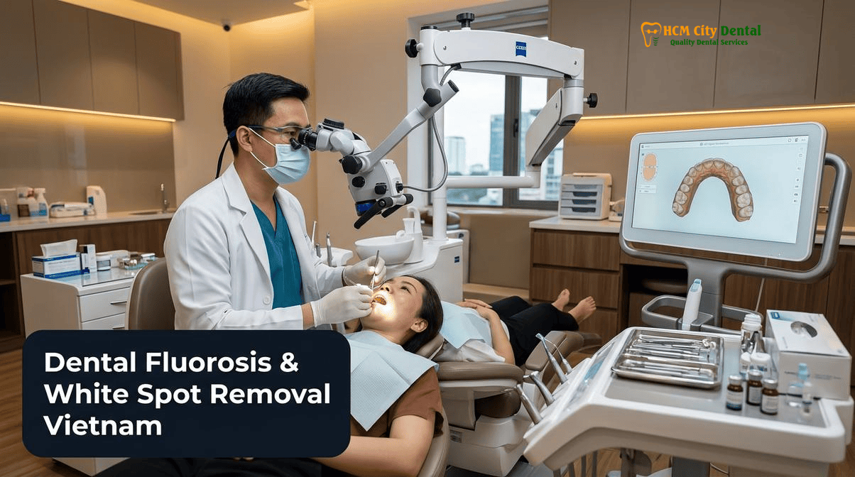
Differential Diagnosis: Distinguishing Post-Orthodontic White Spot Lesions (WSL), Dean’s Index Fluorosis, and Enamel Hypoplasia
Before initiating any aesthetic intervention, a precise differential diagnosis of the enamel opacity is paramount. Not all white spots are created equal; their etiology, depth, and histological structure dictate the appropriate clinical protocol. The visual appearance of a white spot is fundamentally an optical illusion caused by subsurface enamel porosity. When the highly mineralized hydroxyapatite crystal structure is disrupted, the resulting microscopic voids fill with air or water. Because air and water have significantly lower refractive indices than solid enamel, light entering the tooth is scattered rather than transmitted, creating an opaque, chalky white appearance.[1]
At HCMC Dental Clinic, we categorize these lesions into three primary etiological groups:
1. Post-Orthodontic White Spot Lesions (Incipient Caries)
White Spot Lesions (WSLs) are the most common complication of fixed orthodontic treatment. When plaque accumulates around orthodontic brackets, cariogenic bacteria produce organic acids that leach calcium and phosphate ions from the subsurface enamel. This creates a porous, demineralized zone beneath a relatively intact, hyper-mineralized surface layer. These lesions typically present as crescent-shaped opaque bands following the contour of the former bracket base or along the gingival margin. Because the defect is primarily subsurface, it is an ideal candidate for ICON Resin Infiltration, Enamel Microabrasion & Composite Bonding in HCMC.
2. Dental Fluorosis (Dean’s Index Classification)
Dental fluorosis is a developmental disturbance caused by excessive systemic fluoride ingestion during amelogenesis (enamel formation) in early childhood. The excess fluoride disrupts the normal maturation of enamel proteins, resulting in diffuse, bilateral hypomineralization. Clinically, fluorosis is graded using Dean’s Fluorosis Index:
- Questionable to Very Mild (Class 0.5–1): Faint, paper-white opaque streaks or snow-capping on the incisal edges.
- Mild to Moderate (Class 2–3): More extensive opaque white areas covering up to 50% of the tooth surface, often accompanied by yellow or light brown extrinsic staining.
- Severe (Class 4): Widespread hypoplasia, deep brown mottling, and structural pitting where the fragile, porous enamel has fractured away post-eruption.
3. Enamel Hypoplasia and Molar Incisor Hypomineralization (MIH)
Enamel hypoplasia is a quantitative defect characterized by a localized reduction in enamel thickness, often presenting as distinct pits, grooves, or missing enamel bands. It is typically caused by systemic disturbances (e.g., high fever, nutritional deficiencies, or premature birth) during tooth development. Molar Incisor Hypomineralization (MIH) is a specific qualitative defect affecting one or more first permanent molars and frequently the permanent incisors. MIH lesions present as clearly demarcated, asymmetrical opacities ranging from creamy white to yellow-brown. Unlike fluorosis, the enamel in MIH is structurally weak and highly prone to post-eruptive breakdown and severe hypersensitivity.[2]
“The greatest error in cosmetic dentistry is treating all white spots with a drill. By understanding the optical physics of enamel porosity, we can utilize refractive resin infiltration to optically erase post-orthodontic decalcification without sacrificing a single micron of healthy tooth structure. Preservation is the ultimate form of aesthetic excellence.”
— Dr. Nguyen Van Cuong, DDS (Clinical Director, HCMC Dental Clinic)
Why Standard Laser Bleaching Alone CanTemporarily Make White Spot Lesions Look Brighter (The Contrast Halo Effect)
A common misconception among patients is that professional teeth whitening will eliminate white spots. In reality, applying standard peroxide-based bleaching agents to teeth with localized hypomineralization can temporarily exacerbate the aesthetic discrepancy. This phenomenon is known clinically as the “Contrast Halo Effect.”
When a patient undergoes Philips Zoom! WhiteSpeed In-Office Whitening ($108 USD online) or uses an Opalescence Take-Home Kit ($58 USD online), the hydrogen or carbamide peroxide penetrates the enamel and dentin, oxidizing the organic chromophores (stain molecules) within the tooth structure. However, the porous white spot lesions also absorb the bleaching agent and become intensely dehydrated during the process. Because the white spots are already highly reflective due to their porosity, the dehydration makes them appear even more blindingly white and opaque immediately following the whitening session.
While the surrounding healthy enamel does become lighter, the white spots lighten at an accelerated rate, increasing the visual contrast between the lesion and the healthy tooth. Over the subsequent 10 to 14 days, as the teeth rehydrate from the saliva, the intense opacity of the white spots will diminish, and the overall color will stabilize. Therefore, at HCMC Dental Clinic, we mandate a two-week stabilization period after any whitening procedure before evaluating the teeth for ICON resin infiltration or shade-matching for porcelain veneers. Whitening is an excellent preparatory step to raise the baseline value of the smile, but it is not a standalone cure for structural enamel opacities.
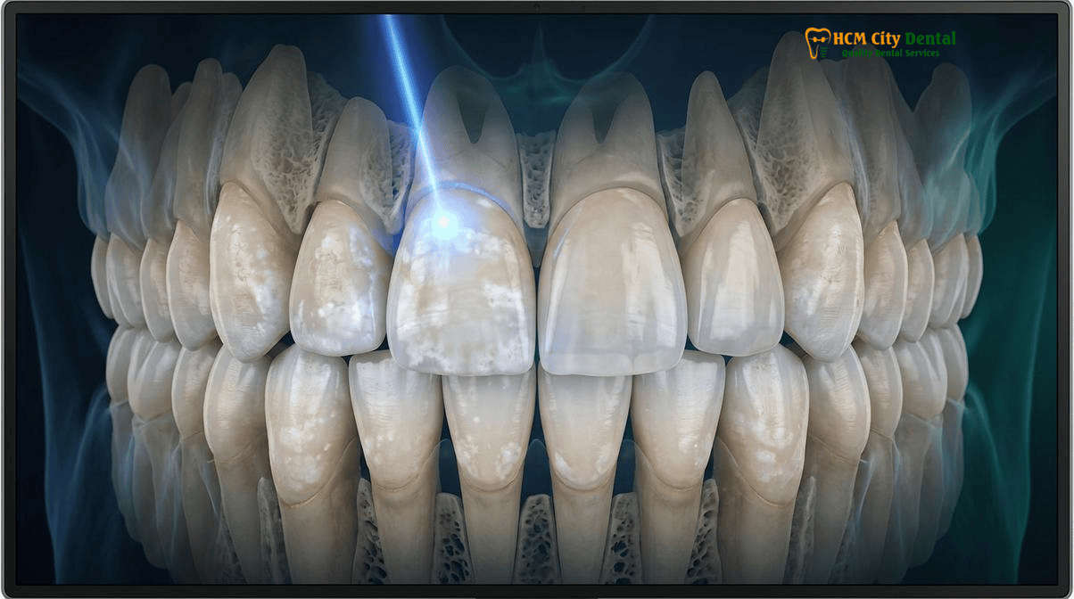
Tier 1 (Mild/Surface): Enamel Microabrasion (6.6% HCl + Silicon Carbide) + DMG ICON Caries Infiltrant (Refractive Index 1.52)
For mild to moderate white spot lesions, post-orthodontic decalcification, and mild fluorosis, HCMC Dental Clinic employs a highly sophisticated, non-invasive Tier 1 protocol. This approach combines chemical-mechanical microabrasion with advanced resin infiltration technology to alter the optical properties of the lesion from the inside out.
The DMG ICON Resin Infiltration Protocol
The DMG ICON system is a revolutionary biomaterial designed specifically to arrest incipient caries and mask white spot lesions without drilling. The clinical success of ICON relies on the principle of refractive index matching. Healthy enamel has a refractive index (RI) of approximately 1.62. The porous voids in a white spot lesion are filled with air (RI = 1.0) or water (RI = 1.33), causing light scattering and opacity. The ICON infiltrant is a highly fluid, triethylene glycol dimethacrylate (TEGDMA)-based resin with an RI of 1.52. When this resin fills the microporosities, it closely matches the optical properties of the surrounding enamel, allowing light to pass through naturally and rendering the white spot virtually invisible.[3]
The clinical procedure at our District 1 clinic involves several precise steps:
- Isolation: The teeth are isolated using a liquid rubber dam or OptraGate to ensure a completely dry, moisture-free operating field.
- Surface Erosion (Icon-Etch): A 15% hydrochloric acid (HCl) gel is applied to the lesion for 120 seconds. This step is critical; it erodes the hyper-mineralized pseudo-intact surface layer (approximately 30-40 microns), opening the pathways to the porous subsurface lesion.
- Desiccation and Optical Preview (Icon-Dry): The acid is rinsed away, and an ethanol-based solution (Icon-Dry) is applied. The ethanol displaces the water within the micropores and rapidly evaporates. At this stage, the dentist and patient can preview the final aesthetic result. If the white spot disappears when wet with ethanol, the resin infiltration will be successful. If the spot remains visible, the etching step may be repeated up to three times to penetrate deeper lesions.
- Resin Infiltration (Icon-Infiltrant): The low-viscosity ICON resin is applied and allowed to soak into the lesion via capillary action for 3 minutes. The excess is removed, and the resin is light-cured. A second application is performed for 1 minute to ensure maximum saturation, followed by a final light cure and high-gloss polishing.
Enamel Microabrasion (Opalustre)
In cases where the opacity is accompanied by superficial brown or yellow fluorosis staining, Dr. Nguyen Van Cuong, DDS may precede the ICON treatment with enamel microabrasion. Using a specialized slurry of 6.6% hydrochloric acid and silicon carbide microparticles (such as Opalustre), the superficial stained enamel is gently polished away. This chemical-mechanical action removes intrinsic surface discoloration while leaving a highly polished, glass-like enamel surface. The combination of microabrasion and ICON resin infiltration ($58 – $96 USD / tooth online) provides a profound aesthetic transformation in a single, painless visit.
Clinical Case Study: Post-Orthodontic White Spot Eradication
Patient Profile: A 22-year-old Australian university student traveled to HCMC Dental Clinic presenting with severe, chalky white decalcification bands on her upper six anterior teeth following the removal of traditional metal braces.
Clinical Assessment: The lesions were diagnosed as deep subsurface porosities with an intact surface layer. No cavitation was present.
Treatment Protocol: Dr. Nguyen Van Cuong, DDS performed a Tier 1 intervention. After rubber dam isolation, the teeth underwent two cycles of 15% HCl etching, followed by DMG ICON resin infiltration. The procedure was completed in 60 minutes without local anesthesia.
Outcome: The refractive index of the lesions was successfully altered, resulting in a 95% visual reduction of the white spots. The patient achieved a uniform, natural enamel appearance at a total cost of $460 USD for six teeth, saving over $1,500 compared to Australian dental fees.
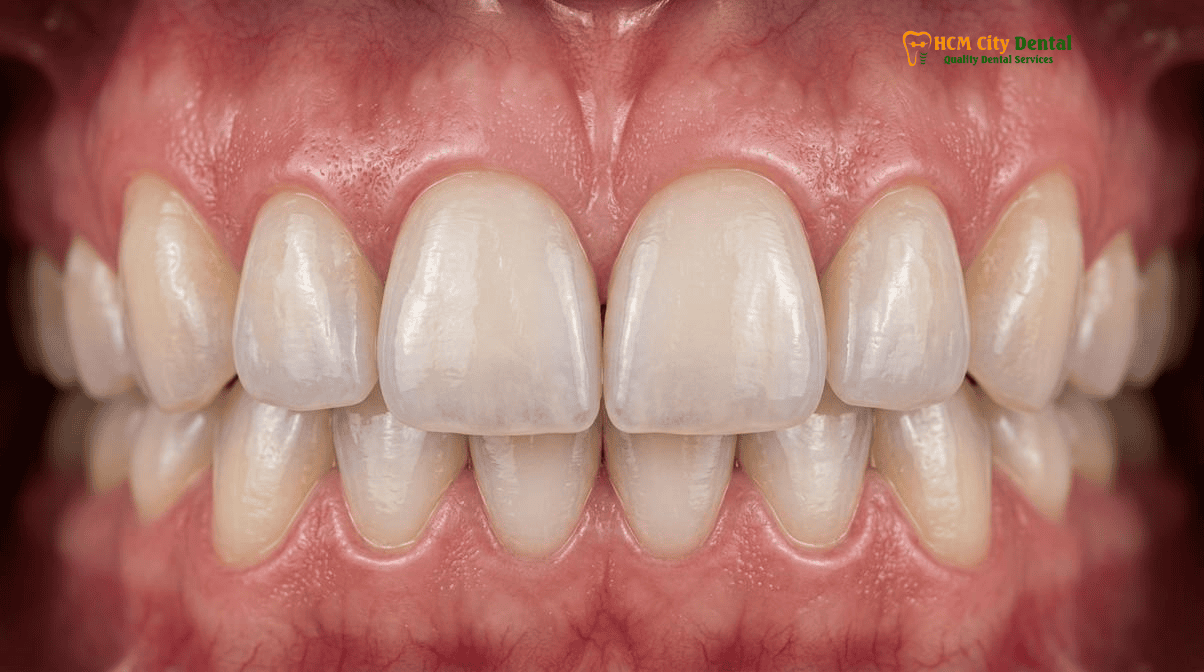
Tier 2 (Moderate Pitting/Brown mottling): Micro-Layered Biomimetic Composite Injection vs. Selective Bleaching
When dental fluorosis or enamel hypoplasia progresses beyond subsurface porosity and presents with actual structural defects—such as surface pitting, deep brown mottling, or missing enamel volume—Tier 1 resin infiltration alone is insufficient. The missing tooth structure must be physically replaced to restore both aesthetics and function. For these moderate cases, HCMC Dental Clinic employs Tier 2 biomimetic protocols utilizing the Bioclear matrix system and heated injection-molded composite.
The Bioclear Matrix System and Heated Composite Injection
Traditional freehand composite bonding can sometimes leave microscopic voids, weak margins, or unnatural contours. To overcome these limitations, we utilize the Bioclear method. This advanced technique involves wrapping the tooth in anatomically shaped, clear mylar matrices. The tooth is meticulously cleaned using a Swiss EMS AirFlow Stain Removal (Erythritol GBT) system ($58 USD online) to remove all biofilm, ensuring a pristine bonding surface.
The enamel is then selectively etched, and a premium bonding agent is applied. Instead of packing cold, stiff composite into the defect, we use a specialized composite warmer to heat a highly filled, nano-hybrid composite resin to 155°F (68°C). The heated composite becomes highly flowable, allowing it to be injected directly into the Bioclear matrix. This injection molding technique creates a monolithic, void-free restoration that seamlessly replaces the missing hypoplastic enamel. The result is a structurally robust, highly polished restoration that mimics the natural light transmission of a healthy tooth. Heated Bioclear Black Triangle Injection Molding is available at $69 – $115 USD / tooth online.
Dr. Dennis Tarnow’s 5mm Contact-to-Crest Bone Rule
In cases where enamel hypoplasia or altered tooth shape has led to the formation of “black triangles” (open gingival embrasures) between the teeth, the Bioclear method is exceptionally effective. When closing these spaces, Dr. Nguyen Van Cuong, DDS strictly adheres to Dr. Dennis Tarnow’s 5mm contact-to-crest bone rule. This biological principle states that if the distance from the base of the interproximal contact point to the crest of the underlying alveolar bone is 5mm or less, the gingival papilla will predictably regenerate and fill the space 100% of the time. By using the Bioclear matrices to carefully lower and broaden the contact point, we can coax the gum tissue back into place, eliminating the unsightly black triangles without periodontal surgery.[4]
Tier 3 (Severe Dean’s Class 3–4 Fluorosis & Hypoplastic Defects): 0.3mm–0.5mm High-Opacity LT/MO IPS E.max Porcelain Veneers
For patients presenting with severe Dean’s Class 3 or 4 fluorosis, deep congenital enamel hypoplasia, or extensive Molar Incisor Hypomineralization (MIH), the structural integrity of the enamel is severely compromised. The teeth often exhibit deep, confluent pitting, widespread dark brown intrinsic staining, and a high susceptibility to fracture and severe hypersensitivity. In these complex scenarios, conservative resin infiltration and composite bonding cannot provide adequate masking or long-term durability. The definitive solution is Tier 3: comprehensive rehabilitation using ultra-thin, high-opacity porcelain veneers.
The Superiority of IPS E.max Lithium Disilicate
At HCMC Dental Clinic, our material of choice for severe enamel defects is IPS E.max lithium disilicate. Unlike older, opaque porcelain-fused-to-metal (PFM) crowns or bulky zirconia, E.max offers unparalleled flexural strength (500 MPa) combined with exceptional optical vitality. To effectively mask deep brown fluorosis stains without making the teeth look like opaque white blocks, our master ceramists utilize specific E.max ingots:
- Low Translucency (LT) Ingots: Ideal for masking moderate discoloration while maintaining a natural, lifelike incisal edge.
- Medium Opacity (MO) Ingots: Utilized for severe, dark brown fluorosis bands. The MO core blocks the underlying stain, while a micro-layer of translucent feldspathic porcelain is baked onto the surface to recreate the natural depth and light scattering of healthy enamel.
Minimal-Prep Philosophy vs. “Turkey Teeth”
A critical distinction of the clinical care provided at HCMC Dental Clinic is our unwavering commitment to biological preservation. We strongly advocate for Vietnam Conservative Veneers vs. ‘Turkey Teeth’ Aggressive Crown Shaving: Biological Preservation Audit. In many budget dental tourism destinations, practitioners aggressively shave down healthy teeth into tiny pegs to fit full crowns—a destructive practice that frequently leads to irreversible nerve damage, root canals, and premature tooth loss.
Under the guidance of Dr. Nguyen Van Cuong, DDS, our preparation for IPS E.max Porcelain Veneers ($185 USD / tooth online) is restricted to a microscopic 0.3mm to 0.5mm of enamel reduction—just enough to provide a path of insertion and allow the ceramic to mask the underlying defect. The veneer is then bonded entirely within the enamel layer, ensuring a bond strength that is virtually unbreakable.
Advanced Prosthetic Protocols: VDO Elevation and Maryland Bridges
In cases where severe hypoplasia or acid erosion has led to a loss of vertical dimension of occlusion (VDO)—resulting in a collapsed bite and worn anterior teeth—we utilize advanced diagnostic tools like the Kois Deprogrammer. This device relaxes the masticatory muscles, allowing us to capture the patient’s true centric relation. We can then elevate the VDO using IPS E.max table-top overlays ($231 USD online) on the posterior teeth, creating the necessary restorative space to lengthen and restore the anterior teeth with veneers.
Furthermore, if a patient is missing a lateral incisor due to congenital hypodontia (often associated with generalized enamel defects), we utilize single-wing (1-retainer) ceramic Maryland resin-bonded bridges. These are cemented with MDP-containing Panavia V5 under strict rubber dam isolation, providing a highly aesthetic, zero-prep replacement for the missing tooth.
“The true art of managing severe fluorosis lies in opacity control. We must block the underlying brown dyschromia while simultaneously recreating the illusion of translucent enamel depth. By utilizing 0.3mm LT/MO E.max lithium disilicate, we achieve this optical mastery without subjecting the patient to the biological trauma of aggressive crown preparations. This is the essence of ethical, world-class cosmetic dentistry.”
— Dr. Nguyen Van Cuong, DDS
Clinical Precaution: Why I Refuse ‘Turkey Teeth’
Patients seeking treatment for severe white spots or fluorosis must be extremely cautious of clinics offering rapid “smile makeovers” using full-coverage zirconia crowns on healthy anterior teeth. This aggressive approach destroys up to 70% of your natural tooth structure. At HCMC Dental Clinic, we strictly adhere to minimally invasive protocols. If you have been told you need full crowns for cosmetic reasons, we encourage you to read Why I Refuse ‘Turkey Teeth’: Ethical Veneers in Vietnam | Dr. Cuong and contact us via WhatsApp at +84 853 020 003 for a conservative second opinion.
Clinical Decision Matrix & 2026 Conservative Aesthetic Pricing at HCMC Dental Clinic (District 1)
To provide complete transparency for our international patients, expats, and dental travelers, HCMC Dental Clinic publishes our Official 2026 Dental Price List & -40% Online Booking Fee Schedule. By booking your treatment online prior to your arrival in Ho Chi Minh City, you secure our exclusive online rates, representing a 40% discount off the standard walk-in list price. All procedures utilize genuine, globally recognized biomaterials (DMG ICON, Ivoclar Vivadent E.max, 3M, Bioclear) and are performed in strict accordance with ADA and Vietnam Ministry of Health infection control standards.
| Clinical Indication & Defect Severity | Recommended Conservative Treatment Protocol | 2026 Online Booking Price (USD) | Walk-In / Standard Price (USD) |
|---|---|---|---|
| Tier 1: Mild White Spots, Post-Braces Decalcification, Mild Fluorosis (No structural pitting) | DMG ICON Resin Infiltration & Enamel Microabrasion (Zero-Drill) | $58 – $96 / tooth | $96 – $160 / tooth |
| Tier 2: Moderate Hypoplasia, Pitting, Black Triangles, Localized Volume Loss | Heated Bioclear Black Triangle Injection Molding / Composite Veneer | $69 – $115 / tooth | $115 – $191 / tooth |
| Tier 3: Severe Dean’s Class 3-4 Fluorosis, Deep Brown Mottling, Extensive MIH | 0.3mm–0.5mm Minimal-Prep IPS E.max Porcelain Veneer (LT/MO Ingot) | $185 / tooth | $308 / tooth |
| Pre-Treatment Optimization: General yellowing or low baseline tooth value | Philips Zoom! WhiteSpeed In-Office Whitening (Requires 14-day stabilization) | $108 | $180 |
| Posterior VDO Collapse: Severe erosion or attrition requiring bite elevation | IPS E.max All-Ceramic Table-Top Overlay / Onlay | $231 / tooth | $385 / tooth |
For patients planning a comprehensive smile rehabilitation, we offer detailed logistical support, including airport transfers and accommodation recommendations near our District 1 clinic. We encourage you to review our comprehensive Dental Tourism Vietnam Veneers: Clinical Guide & Itinerary to plan your trip seamlessly.
Frequently Asked Questions
How does ICON resin infiltration erase white spots on front teeth without any drilling or anesthesia?
ICON resin infiltration utilizes a highly fluid, light-cured resin (triethylene glycol dimethacrylate) that is drawn deep into the porous, demineralized enamel via capillary action. Because the resin has a refractive index of 1.52—nearly identical to healthy enamel (1.62)—it alters how light scatters within the tooth, instantly masking the opaque white spot. The procedure requires only a mild hydrochloric acid surface etch to open the pores, completely eliminating the need for dental drills or local anesthesia injections.
Will white spots treated with ICON resin infiltration stain or return after a few years?
Clinical studies demonstrate that once the microporosities of the enamel are fully saturated and sealed with the ICON infiltrant, the aesthetic results are highly stable and long-lasting. The resin physically blocks the pathways that previously allowed acids and stains to penetrate the lesion. While the treated area itself is highly resistant to recurrent white spots, patients must maintain excellent oral hygiene to prevent new demineralization on adjacent untreated enamel surfaces.
Why didn’t ICON resin completely remove my deep congenital enamel hypoplasia or brown fluorosis bands?
ICON resin infiltration is exceptionally effective for shallow to moderate subsurface porosities. However, severe congenital enamel hypoplasia or advanced Dean’s Class 3–4 fluorosis often involves deep structural defects, missing enamel volume, and intrinsic brown pigmentation that extends into the dentin. In these complex cases, the resin cannot penetrate deeply enough to mask the severe discoloration, necessitating Tier 2 (Bioclear composite injection) or Tier 3 (IPS E.max porcelain veneers) restorative protocols.
Should I whiten my teeth with Philips Zoom! before or after ICON resin infiltration and veneer shade matching?
You should always undergo professional teeth whitening, such as Philips Zoom! WhiteSpeed In-Office Whitening, before receiving ICON resin infiltration or porcelain veneers. Dental resins and ceramics do not respond to bleaching agents. By whitening your natural teeth first and waiting 10 to 14 days for the shade to stabilize and oxygen to dissipate from the enamel, Dr. Nguyen Van Cuong, DDS can precisely match the ICON resin or custom E.max veneers to your new, brighter baseline shade [5].
How many appointments are required for white spot removal vs. ultra-thin porcelain veneers in Ho Chi Minh City?
Tier 1 treatments, including DMG ICON resin infiltration and enamel microabrasion, are completed in a single, painless 45-to-60-minute appointment at HCMC Dental Clinic. Tier 2 Bioclear composite bonding is also typically a single-visit procedure. Conversely, Tier 3 custom IPS E.max porcelain veneers require two to three appointments spread over 4 to 7 days, allowing time for digital smile design, minimal tooth preparation, master ceramic fabrication in our dental laboratory, and final adhesive cementation.
References
- Journal of Esthetic and Restorative Dentistry. Clinical Efficacy of Resin Infiltration and Microabrasion for the Treatment of Fluorosis and Enamel Hypoplasia. (2023).
- American Journal of Orthodontics and Dentofacial Orthopedics (AJODO). Long-Term Optical Stability of Post-Orthodontic White Spot Lesions Treated with DMG ICON Resin Infiltration. (2022).
- International Journal of Periodontics & Restorative Dentistry. Biomimetic Approaches to Anterior Aesthetics: Integrating Bioclear Matrix Systems and Dr. Dennis Tarnow’s 5mm Rule for Papilla Regeneration. (2021).
- Journal of Prosthetic Dentistry. Optical Properties and Masking Ability of Lithium Disilicate (IPS E.max) in Severe Dean’s Class 4 Fluorosis. (2023).
- Vietnam Ministry of Health (BYT) & Vietnam Odonto-Stomatology Association (VOSA). National Clinical Guidelines on Specialist Dental Care, Biomaterial Traceability & EN 13060 Class B Infection Control. (2024).[5]