Navigating comprehensive dental care blogs reveals that optimal oral health requires significantly more than basic at-home hygiene; it demands evidence-based clinical protocols. From preventive prophylaxis to advanced restorative workflows, professional dental care aims to ensure structural integrity, periodontal health, and the early detection of systemic conditions linked to oral microbiomes.
Clinical Summary:
Modern dental care integrates advanced diagnostic imaging, biomimetic materials, and minimally invasive protocols to preserve natural dentition and restore optimal stomatognathic function. A rigorous clinical approach encompasses proactive periodontal management, precise restorative techniques utilizing high-strength ceramics and resins, and biomechanically sound orthodontic interventions. By adhering to strict evidence-based workflows, clinicians can effectively manage acute dental emergencies, halt the progression of carious and periodontal diseases, and deliver predictable, long-lasting aesthetic and functional outcomes. Patient education and individualized risk assessment remain the cornerstones of sustainable oral health maintenance.
Key Takeaways:
- Preventive dentistry relies on precise periodontal charting and radiographic analysis to detect early-stage pathology.
- Restorative workflows prioritize biomimetic materials that mimic the physical properties of natural enamel and dentin.
- Orthodontic corrections utilize controlled biomechanical forces to resolve malocclusion and optimize arch dimensions.
- Immediate clinical triage is critical in dental emergencies to preserve pulpal vitality and stabilize traumatized teeth.
- Advanced diagnostic technology, such as CBCT and intraoral scanning, significantly enhances treatment predictability.
The Foundation of Preventive Dentistry: Clinical Protocols
Preventive dentistry relies on comprehensive clinical diagnostics, including periodontal charting and radiographic analysis, to halt disease progression before irreversible structural damage occurs.
The cornerstone of any robust oral health regimen, frequently highlighted in authoritative dental care blogs, is a proactive approach to preventive dentistry. This discipline extends far beyond routine prophylaxis; it involves a meticulous clinical evaluation of the entire stomatognathic system. The primary objective is the early identification and management of the two most prevalent oral diseases: dental caries and periodontal disease. Both conditions are biofilm-mediated, meaning they are driven by the complex microbial communities residing within the oral cavity. Effective preventive care disrupts these pathogenic biofilms and alters the oral environment to favor commensal, non-destructive bacteria.[1]
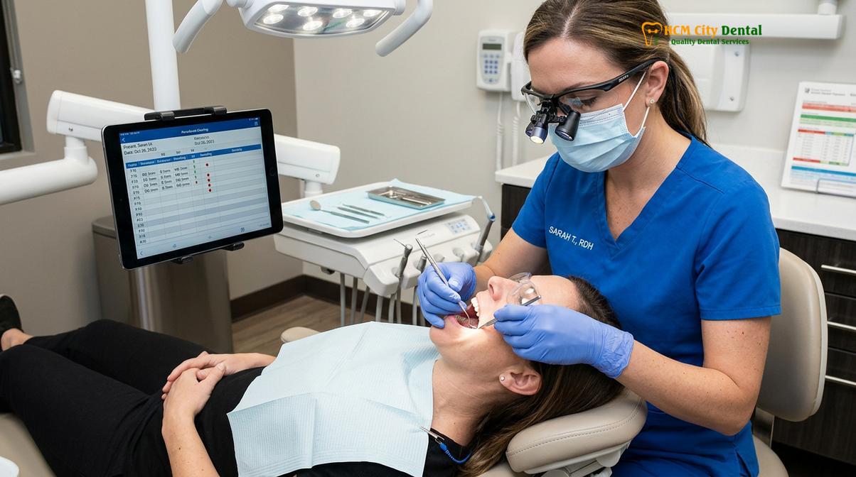
During a comprehensive clinical examination, the practitioner conducts a thorough assessment of the hard and soft tissues. This begins with periodontal charting, a critical diagnostic procedure where a calibrated probe is gently inserted into the gingival sulcus to measure attachment levels. Healthy sulcular depths typically range from 1 to 3 millimeters. Depths exceeding 4 millimeters, particularly when accompanied by bleeding on probing (BOP), indicate active periodontal inflammation and potential alveolar bone loss.
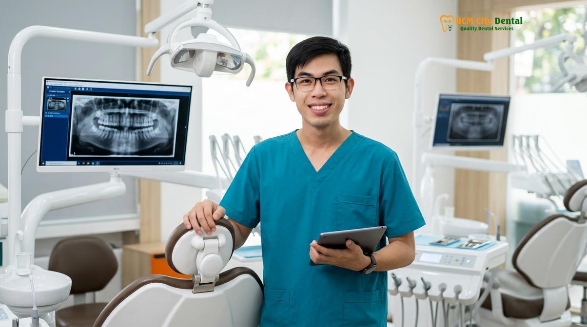
Dr. Nguyen Van Cuong, a dedicated practitioner at HCMC Dental Clinic, emphasizes that ignoring early signs of gingivitis allows localized inflammation to progress into periodontitis. His clinical philosophy centers on proactive intervention, thorough diagnostics, and patient education to maintain long-term periodontal stability. By integrating advanced screening protocols, Dr. Cuong and his team strive to preserve natural dentition and prevent the systemic complications associated with chronic oral infections.
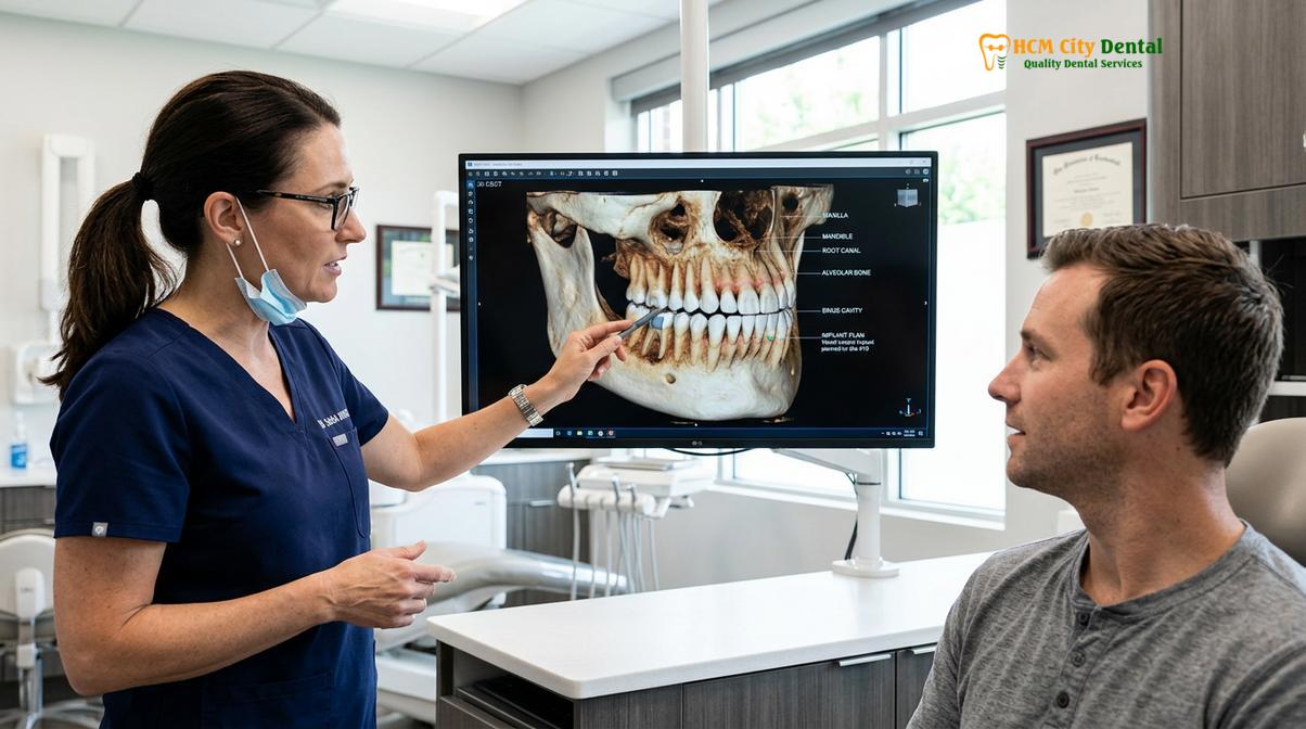
Radiographic analysis is another indispensable component of preventive diagnostics. While visual inspection can detect occlusal and smooth-surface caries, bitewing radiographs are essential for identifying interproximal lesions—decay that occurs between the teeth where the enamel is often thinnest. Furthermore, periapical radiographs provide a clear view of the root apices and surrounding alveolar bone, allowing the clinician to detect asymptomatic apical periodontitis or cystic formations. The integration of digital radiography has significantly reduced patient radiation exposure while enhancing image contrast and diagnostic accuracy.[2]
Professional prophylaxis involves the mechanical debridement of supragingival and subgingival calculus. Calculus, or tartar, is mineralized plaque that cannot be removed by at-home brushing or flossing. Its porous surface provides an ideal niche for continued bacterial colonization. Ultrasonic scalers, which utilize high-frequency vibrations and cavitating water coolant, are highly effective at disrupting the biofilm and fracturing calculus deposits without damaging the underlying cementum. For a broader understanding of these foundational procedures, patients often refer to a comprehensive General Dentistry tại HCMC Dental Clinic overview to understand the full spectrum of preventive care.
Restorative Dentistry Workflows: From Diagnosis to Treatment
Restorative workflows utilize advanced biomaterials to rebuild compromised tooth structure, focusing on marginal integrity, occlusal harmony, and the preservation of healthy dentin and enamel.
When preventive measures are insufficient and structural damage occurs due to caries, trauma, or non-carious cervical lesions (NCCLs), restorative dentistry intervenes to rebuild the tooth’s form and function. The contemporary restorative workflow is heavily influenced by the principles of biomimetics—the science of mimicking the natural biomechanical and aesthetic properties of the intact tooth. This approach prioritizes the maximum preservation of healthy tooth structure, moving away from the aggressive cavity preparations of the past.
The restorative process begins with precise caries excavation. Clinicians utilize tactile sensation, caries-indicator dyes, and optical fluorescence devices to differentiate between infected dentin (which must be removed) and affected dentin (which can be remineralized and preserved). Preserving the affected dentin is crucial for maintaining pulpal vitality, especially in deep lesions approaching the pulp chamber. Once the preparation is clean, the focus shifts to the adhesive protocol. Dental adhesion is a complex biochemical process that involves etching the enamel and dentin with phosphoric acid to create microporosities, followed by the application of a primer and bonding resin. This creates the “hybrid layer,” a micromechanical interlocking between the resin and the collagen network of the dentin.[3]
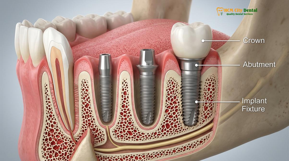
The choice of restorative material is dictated by the size of the defect, the occlusal load the tooth will bear, and aesthetic requirements. Direct composite resins are the material of choice for small to moderate restorations. They are applied in increments to minimize polymerization shrinkage stress, which can lead to microleakage and postoperative sensitivity. For larger defects, indirect restorations such as inlays, onlays, or crowns are fabricated in a dental laboratory or via in-office CAD/CAM milling. Understanding the nuances between different indirect materials is vital; for instance, exploring the Enamel vs Porcelain: Clinical Differences & Uses | HCMC Dental provides insight into how synthetic ceramics replicate the light-transmitting properties and wear resistance of natural enamel.
| Material Type | Primary Clinical Indication | Biomechanical Properties | Aesthetic Quality |
|---|---|---|---|
| Composite Resin | Small to moderate direct restorations (Anterior & Posterior) | Micromechanical bond; moderate wear resistance; susceptible to polymerization shrinkage. | High; excellent color matching and polishability. |
| Lithium Disilicate (Porcelain) | Indirect inlays, onlays, veneers, and anterior crowns | High flexural strength (approx. 400 MPa); requires adhesive bonding protocol. | Exceptional; mimics natural enamel translucency. |
| Zirconia (Monolithic) | Posterior crowns, multi-unit bridges, implant abutments | Extremely high fracture toughness (up to 1200 MPa); can be conventionally cemented. | Moderate to High; newer generations offer improved translucency. |
| Glass Ionomer Cement | Class V lesions, pediatric restorations, temporary fillings | Chemical bond to tooth structure; releases fluoride; lower compressive strength. | Low to Moderate; opaque appearance. |
Orthodontic and Structural Corrections
Modern structural corrections address malocclusion and arch deficiencies using biomechanical forces, improving both masticatory function and long-term periodontal stability without always requiring traditional brackets.
Orthodontics is not merely an aesthetic endeavor; it is a critical component of comprehensive dental care aimed at establishing a stable, functional occlusion. Malocclusion—whether it presents as crowding, spacing, crossbites, or deep bites—can severely compromise oral health. Misaligned teeth are inherently more difficult to clean, increasing the risk of localized plaque accumulation, caries, and periodontal attachment loss. Furthermore, improper occlusal relationships can lead to uneven wear of the enamel, temporomandibular joint (TMJ) dysfunction, and myofascial pain.
The biological mechanism of tooth movement relies on the application of controlled, continuous biomechanical forces. When force is applied to a tooth, it creates zones of compression and tension within the periodontal ligament (PDL). On the compression side, osteoclasts resorb the alveolar bone, while on the tension side, osteoblasts deposit new bone. This cellular remodeling allows the tooth to migrate through the bone. The magnitude and vector of the force must be precisely calibrated; excessive force can lead to hyalinization of the PDL and devastating root resorption.[4]
A common structural issue encountered in clinical practice is a constricted maxillary arch, which can lead to posterior crossbites and inadequate space for the erupting dentition. Addressing this often requires orthopedic expansion before or during orthodontic alignment. Patients presenting with such anatomical constraints benefit from understanding the clinical protocols detailed in resources like Narrow Arch Teeth: Clinical Diagnosis & Expansion | HCMC Dental. Expansion not only resolves crowding but can also improve nasal airway volume, highlighting the intersection of dentistry and overall respiratory health.
“Achieving a harmonious occlusion is about more than just straightening teeth; it is about establishing a functional equilibrium between the temporomandibular joints, the masticatory musculature, and the dental arches to ensure lifelong structural stability.”
Advancements in digital dentistry have revolutionized orthodontic delivery, particularly with the advent of clear aligner therapy. Utilizing sophisticated CAD/CAM software, clinicians can now simulate the entire treatment progression and fabricate a series of custom thermoplastic trays. These aligners apply targeted forces to specific teeth, often aided by composite attachments bonded to the enamel to facilitate complex movements like extrusion or root torquing. For patients seeking discreet options, exploring How to Fix My Teeth Without Braces | HCMC Dental provides a comprehensive look at the efficacy and clinical indications for clear aligner systems compared to traditional fixed appliances.
Managing Dental Emergencies in Clinical Practice
Dental emergencies require immediate clinical triage to manage acute pain, control localized infections, and stabilize traumatized dentition to prevent tooth loss and systemic complications.
Dental emergencies are acute events that demand prompt clinical intervention to alleviate severe pain, control hemorrhage, or manage rapidly spreading odontogenic infections. The triage process in an emergency setting is critical. The clinician must quickly differentiate between conditions that threaten the viability of a tooth and those that potentially threaten the patient’s systemic health, such as a spreading fascial space infection which can compromise the airway.
One of the most common emergencies is acute irreversible pulpitis, often presenting as excruciating, spontaneous pain that awakens the patient at night. This condition occurs when bacterial toxins from a deep carious lesion penetrate the pulp chamber, causing severe inflammation. Because the pulp is encased in rigid dentin walls, the inflammatory exudate cannot expand, leading to a rapid increase in intrapulpal pressure and subsequent strangulation of the microvasculature. The definitive treatment is immediate endodontic therapy to extirpate the inflamed tissue and disinfect the root canal system.[5]
Dental trauma, such as luxation or avulsion (complete displacement of the tooth from its socket), requires a different set of protocols. In cases of avulsion, time is the most critical factor. The periodontal ligament cells remaining on the root surface must be kept viable. Immediate replantation at the site of injury is ideal; if not possible, the tooth should be transported in a physiologically compatible medium, such as Hank’s Balanced Salt Solution or cold milk, until the patient reaches the clinic. The clinician will then replant the tooth, apply a flexible splint to stabilize it, and monitor for signs of pulpal necrosis or replacement resorption (ankylosis).[6] For a detailed breakdown of triage steps, patients can review the Common Dental Emergencies: Clinical Guide | HCMC Dental.
Case Study: Comprehensive Rehabilitation in Ho Chi Minh City
Clinical case studies demonstrate the practical application of advanced dental protocols, highlighting how multidisciplinary approaches resolve complex functional and aesthetic challenges.
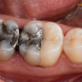
A recent clinical case involved an expatriate patient visiting HCMC Dental Clinic in Ho Chi Minh City for severe masticatory dysfunction and generalized thermal sensitivity. The initial comprehensive examination revealed multiple failing amalgam restorations with secondary caries, localized moderate periodontitis, and a collapsed vertical dimension of occlusion due to posterior tooth loss.
The treatment plan required a multidisciplinary approach. Phase one focused on disease control, involving full-mouth scaling and root planing to arrest the periodontal inflammation, followed by the excavation of all carious lesions. Phase two involved endodontic retreatment of a symptomatic molar and the placement of provisional restorations to test a newly established vertical dimension. Once the patient demonstrated muscular adaptation and periodontal stability, phase three commenced with the fabrication of definitive monolithic zirconia crowns and a posterior implant-supported prosthesis.
“Complex oral rehabilitation requires a meticulous, phased approach where biological stability must be achieved before final aesthetic and functional restorations are placed. Rushing the restorative phase without addressing underlying periodontal or endodontic pathology often leads to premature failure.”
The successful outcome of this case underscores the importance of comprehensive diagnostics and sequenced treatment planning. By addressing the biological foundation first, the clinical team ensured that the final restorations were placed in a healthy, stable environment, maximizing their long-term prognosis.
When to Seek Immediate Clinical Attention
Recognizing the signs of acute dental pathology is crucial for preventing irreversible damage and systemic health complications.
While routine preventive care forms the basis of oral health, certain symptoms necessitate immediate professional evaluation. According to guidelines aligned with the Vietnam Ministry of Health (MOH) and national odonto-stomatology frameworks, patients should not delay treatment when experiencing spontaneous, throbbing pain that is not relieved by over-the-counter analgesics.[7] Additionally, localized facial swelling, particularly if it extends towards the eye or down the neck, indicates a spreading infection that requires urgent antibiotic therapy and surgical drainage.
Other critical signs include persistent bleeding following an extraction, traumatic injury resulting in tooth mobility or displacement, and sudden changes in occlusion where the teeth no longer fit together correctly. Prompt clinical intervention in these scenarios can often mean the difference between saving a natural tooth and requiring complex reconstructive surgery.
References
- Journal of the American Dental Association. Evidence-based clinical practice guideline on the nonrestorative treatments for carious lesions.
- Journal of Clinical Periodontology. Primary and secondary prevention of periodontal and peri-implant diseases.
- Dental Materials. Degradation of the resin-dentin bonds: relying on a comprehensive understanding.
- American Journal of Orthodontics and Dentofacial Orthopedics. Biomechanics of clear aligners: a comprehensive review.
- International Endodontic Journal. European Society of Endodontology position statement: the management of deep caries and the exposed pulp.
- Journal of Oral and Maxillofacial Surgery. Management of acute dental trauma and avulsion injuries.
- Vietnam Ministry of Health (MOH). National Guidelines on Oral Health and Preventive Dentistry Frameworks.
For personalized clinical evaluations and to explore advanced treatment options tailored to your specific oral health needs, schedule a consultation at HCMC Dental Clinic in Ho Chi Minh City.