A regular cleaning removes plaque and tartar above the gumline to maintain healthy teeth, while a deep cleaning treats periodontal disease by removing bacterial buildup below the gumline and smoothing tooth roots. Your clinical diagnosis dictates which procedure is necessary to preserve your oral health.
Clinical Summary:
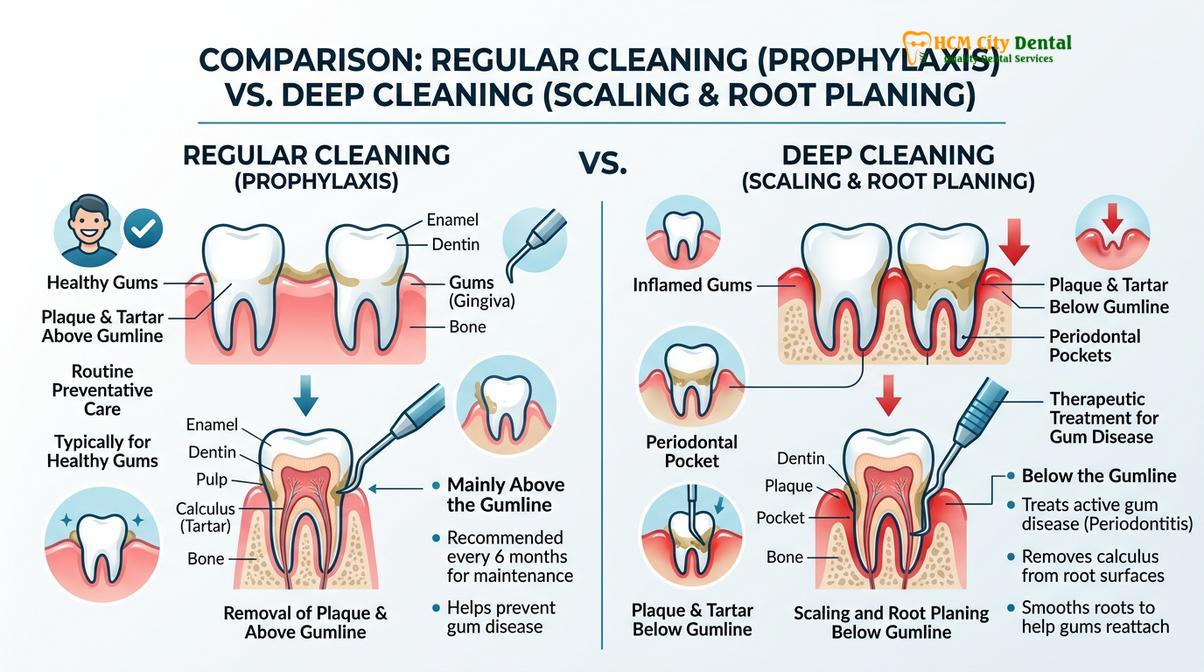
The distinction between a regular cleaning (prophylaxis) and a deep cleaning (scaling and root planing) lies in the clinical objective and anatomical focus. Prophylaxis is a preventive measure targeting supragingival calculus and plaque to maintain gingival health in patients without bone loss. Conversely, deep cleaning is a therapeutic intervention for periodontitis, requiring the removal of subgingival calculus and the smoothing of root surfaces to facilitate gingival reattachment. Accurate diagnosis relies on periodontal probing; pocket depths exceeding 3 millimeters with bleeding typically indicate the need for scaling and root planing. Local anesthesia is often utilized during deep cleaning to ensure patient comfort while the clinician meticulously debrides the periodontal pockets. Post-treatment maintenance is critical to prevent disease recurrence.
Key Takeaways:
- Regular cleanings are preventive, focusing on the visible crowns of the teeth above the gumline.
- Deep cleanings are therapeutic, treating active gum disease by cleaning beneath the gumline.
- Periodontal probing determines the need for deep cleaning; pockets over 3mm usually require intervention.
- Scaling and root planing (SRP) often requires local anesthesia for patient comfort.
- Strict post-treatment care and frequent periodontics maintenance visits are essential after SRP.
Defining Regular Cleaning: Prophylaxis and Supragingival Scaling
A regular cleaning, or prophylaxis, is a preventive dental procedure designed to remove plaque, calculus, and stains from the exposed surfaces of teeth above the gumline.
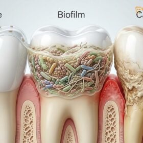
In the realm of preventive dentistry, a regular cleaning—clinically referred to as dental prophylaxis—is the cornerstone of maintaining optimal oral health for patients who do not exhibit signs of active periodontal disease. The primary objective of this procedure is to disrupt and remove the bacterial biofilm (plaque) before it can mineralize into hardened tartar. When plaque is left undisturbed on the tooth surfaces, minerals from the saliva, primarily calcium and phosphate, precipitate into the biofilm, creating a hard, porous substance known as supragingival calculus. Because supragingival calculus is firmly attached to the enamel, it cannot be removed by standard at-home brushing and flossing; it requires professional instrumentation [1].
During a standard prophylaxis appointment, the dental professional focuses entirely on the anatomical crown of the tooth—the portion visible above the gingival margin. The procedure typically begins with the use of an ultrasonic scaler. This sophisticated device utilizes high-frequency vibrations combined with a continuous stream of water to fracture and dislodge supragingival calculus deposits. The cavitation effect produced by the water spray also helps to flush away debris and disrupt the cell walls of harmful bacteria. Following the ultrasonic phase, the clinician will use specialized hand instruments, such as scalers and curettes, to meticulously remove any remaining fine calculus and plaque from the interproximal spaces (between the teeth) and along the gumline.
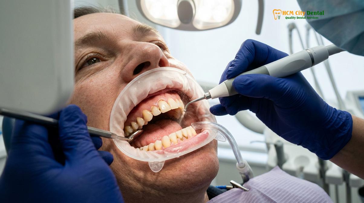
Once the tooth surfaces are entirely free of deposits, the final step of a regular cleaning involves polishing. Using a slow-speed handpiece equipped with a soft rubber cup and a mildly abrasive prophylaxis paste, the clinician polishes the enamel. This step not only removes extrinsic stains caused by coffee, tea, or tobacco but also creates a highly smooth surface that makes it more difficult for new plaque to adhere. For patients with healthy gums, this preventive routine general dentistry care is typically recommended every six months to prevent the onset of gingivitis and subsequent periodontitis.
“Prophylaxis is fundamentally a preventive measure. By consistently managing bacterial load and removing supragingival calculus, we maintain the integrity of the gingival attachment and prevent the initiation of destructive inflammatory cascades.”
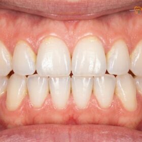
It is crucial to understand that a regular cleaning is strictly indicated for patients with a healthy periodontium. This means the gums are firm, pink, and do not bleed easily upon probing, and there is no evidence of alveolar bone loss on dental radiographs. If a patient presents with active gum disease, performing only a regular cleaning is clinically insufficient and can even be detrimental, as it leaves the underlying subgingival infection untreated. Dr. Nguyen Van Cuong frequently emphasizes to patients that while a regular cleaning makes the teeth look and feel clean, it is the comprehensive diagnostic assessment that ensures the correct level of care is provided.
What is Deep Cleaning? Scaling and Root Planing (SRP) Explained
Deep cleaning, clinically known as scaling and root planing, is a non-surgical periodontal therapy that removes subgingival bacterial deposits and smooths root surfaces to treat active gum disease.
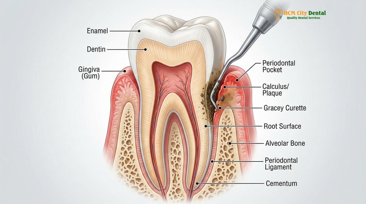
When gingivitis—the mildest form of gum disease—is left untreated, the bacterial infection can advance deeper into the periodontium, leading to periodontitis. This chronic inflammatory condition causes the gingival tissues to pull away from the teeth, creating spaces known as periodontal pockets. These pockets become an ideal anaerobic environment for highly pathogenic bacteria to thrive and multiply. As the body’s immune system responds to this bacterial invasion, the resulting inflammation inadvertently destroys the connective tissue and alveolar bone that anchor the teeth in the jaw. To halt this destructive process, a specialized procedure known as scaling and root planing is required [2].
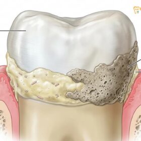
Scaling and root planing is the gold standard of non-surgical periodontal therapy. Unlike a regular cleaning, which stops at the gumline, this procedure delves deep into the periodontal pockets to eradicate the source of the infection. The process is divided into two distinct but continuous phases. The first phase, scaling, involves the meticulous removal of plaque, bacterial toxins, and hardened calculus from the root surfaces below the gumline. Subgingival calculus is often darker in color and much more tenacious than supragingival calculus because it derives its mineral content from inflammatory exudate (crevicular fluid) rather than saliva.
The second phase, root planing, addresses the structural alterations that occur on the root surface due to disease. The cementum—the calcified tissue covering the tooth root—can become porous and impregnated with bacterial endotoxins. Root planing involves smoothing the rough surfaces of the cementum and removing the necrotic, toxin-laden outer layer. By creating a hard, smooth, and biologically acceptable root surface, the procedure facilitates the reduction of inflammation and encourages the gingival tissues to heal and reattach to the tooth. This comprehensive approach is often referred to as gum inflammation therapy, as its primary goal is to resolve the acute immune response and stabilize the periodontium.
Because scaling and root planing is a physically demanding and technically precise procedure, it is rarely completed in a single visit for a patient with generalized periodontitis. The mouth is typically divided into quadrants (upper right, lower right, upper left, lower left), and the clinician will treat one or two quadrants per appointment. This phased approach allows for thorough debridement and minimizes patient fatigue. Furthermore, because the instruments must reach deep into inflamed and sensitive tissues, local anesthesia is almost always administered to ensure the patient remains completely comfortable throughout the process.
The success of scaling and root planing relies heavily on the clinician’s tactile sensitivity and anatomical knowledge. Since the subgingival environment is largely hidden from direct view, the dentist or hygienist must rely on the feel of the instruments against the root surface to detect and remove calculus. Dr. Nguyen Van Cuong notes that the meticulous nature of this gum inflammation therapy is what ultimately dictates the clinical outcome; incomplete removal of subgingival deposits will result in persistent inflammation and continued disease progression.
The Clinical Diagnostic: Pocket Depths and Gum Inflammation
Dentists determine the need for a deep cleaning by measuring the space between the tooth and gum tissue using a periodontal probe, alongside assessing radiographic bone levels.
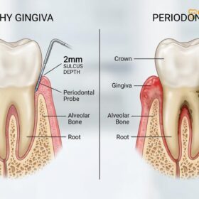
The decision to perform a regular cleaning versus a deep cleaning is never arbitrary; it is based on a rigorous clinical and radiographic evaluation. The most critical diagnostic tool in this process is the periodontal probe, a specialized dental instrument featuring millimeter markings. During a comprehensive examination, the clinician gently inserts the probe into the gingival sulcus—the natural space between the tooth and the surrounding gum tissue—to measure its depth. This measurement is taken at six specific points around every single tooth to create a detailed periodontal chart [3].
In a healthy periodontium, the sulcus depth typically ranges from 1 to 3 millimeters. At these depths, the space is easily accessible for daily cleaning with a toothbrush and dental floss, and a regular prophylaxis is sufficient to maintain health. However, when the subgingival pocket depth measures 4 millimeters or greater, it indicates that the gingival attachment has migrated apically (downward) due to disease. Pockets of this depth create an anaerobic environment where pathogenic bacteria flourish, completely shielded from the patient’s home care efforts. When a subgingival pocket depth reaches 5 millimeters or more, it is a definitive clinical indicator that scaling and root planing is necessary to access and clean the base of the pocket.
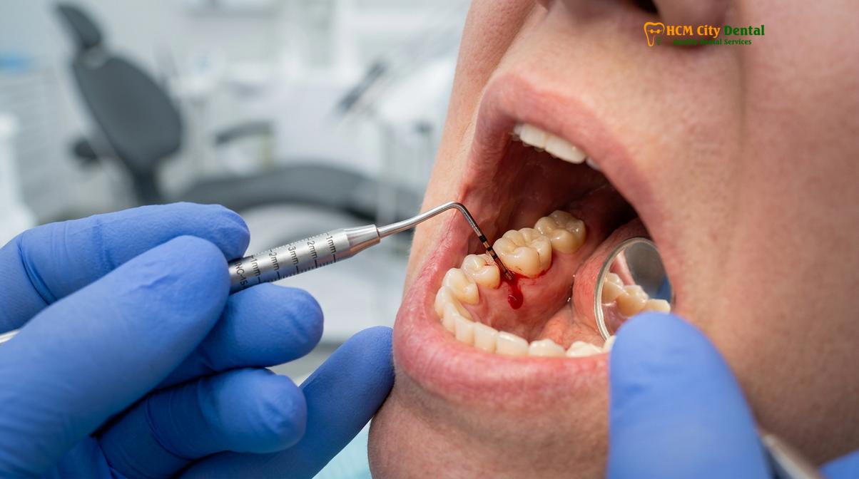
Alongside measuring pocket depths, the clinician closely monitors the tissue for bleeding on probing (BOP). Healthy gums do not bleed when gently probed. If bleeding occurs, it is a clear sign of active ulceration and inflammation within the pocket lining. The presence of deep pockets combined with BOP is the hallmark of active periodontitis. Furthermore, the clinical examination is always correlated with dental radiographs (X-rays). Radiographs allow the dentist to visualize the crestal alveolar bone levels. If bone loss is evident on the X-rays, it confirms the diagnosis of periodontitis and underscores the urgent need for deep cleaning to halt further destruction.
| Clinical Parameter | Regular Cleaning (Prophylaxis) | Deep Cleaning (Scaling & Root Planing) |
|---|---|---|
| Pocket Depths | 1 – 3 millimeters (Healthy) | 4+ millimeters (Diseased) |
| Bone Loss | None present on radiographs | Visible alveolar bone loss |
| Bleeding on Probing | Minimal to none | Moderate to severe (Active infection) |
| Primary Objective | Preventive maintenance | Therapeutic disease intervention |
| Anesthesia Requirement | Rarely needed | Local anesthesia typically required |
It is important to recognize that gum disease is often referred to as a “silent disease” because it can progress significantly without causing noticeable pain. A patient may feel that their teeth are perfectly fine, yet a clinical examination may reveal deep pockets and severe bone loss. This highlights the critical importance of routine professional dental cleanings and comprehensive evaluations. Relying solely on symptoms like pain or loose teeth means the disease has already reached an advanced, and potentially irreversible, stage.
Procedures and Anesthesia: What to Expect in the Chair
A deep cleaning is typically performed over two or more visits using local anesthesia to numb the gums, allowing the clinician to thoroughly clean deep pockets without causing discomfort.
Understanding the workflow of a scaling and root planing appointment can significantly alleviate patient anxiety. Because the procedure involves instrumentation deep within inflamed periodontal pockets, patient comfort is a primary concern. Therefore, the first step in a deep cleaning appointment is the administration of local anesthesia. By numbing the specific quadrant or half of the mouth being treated, the clinician can perform local anesthesia scaling. This ensures that the patient feels only pressure and vibration, rather than sharp pain, allowing the dentist or hygienist to work meticulously and effectively without causing distress [4].
Once profound anesthesia is achieved, the mechanical debridement begins. The clinician will typically utilize an ultrasonic scaler as the primary instrument. The ultrasonic tip vibrates at a high frequency (ranging from 25,000 to 42,000 cycles per second), effectively shattering the hard calculus deposits attached to the root surface. Simultaneously, the device emits a steady stream of water that serves multiple purposes: it cools the instrument tip, flushes away the dislodged debris and blood, and creates a cavitational effect that disrupts the bacterial cell walls within the biofilm. This local anesthesia scaling process is highly efficient at removing the bulk of the subgingival infection.
Following the ultrasonic phase, the clinician transitions to hand instrumentation. Using specialized tools called Gracey curettes, which are designed with specific angles to adapt to the complex anatomy of different tooth roots, the clinician performs the fine scaling and root planing. The curette is carefully inserted to the base of the periodontal pocket, and with precise, overlapping strokes, the clinician removes any residual fine calculus and smooths the cementum. This tactile phase is crucial, as the clinician relies on the “feel” of the instrument to ensure the root surface is completely free of necrotic tissue and endotoxins, creating an optimal environment for healing.
Depending on the severity of the infection, the dentist may also incorporate adjunctive therapies during the procedure. This can include the localized delivery of antimicrobial agents or antibiotics directly into the periodontal pockets to further suppress the pathogenic bacteria. Laser therapy is also increasingly used as an adjunct to traditional scaling and root planing, helping to decontaminate the pocket and stimulate tissue regeneration. After the designated quadrants are thoroughly treated, the patient is given specific post-operative instructions and scheduled for the completion of the remaining quadrants.
Post-Treatment Care: Preventing Infection After Deep Scaling
Successful recovery from a deep cleaning requires meticulous home oral hygiene, potential use of antimicrobial rinses, and adherence to a strict periodontal maintenance schedule.
The completion of the scaling and root planing procedure marks the beginning of the healing phase, not the end of the treatment. Because the gums have been thoroughly manipulated and the deep infection removed, it is normal to experience some mild side effects in the immediate post-operative period. Patients often report slight gingival tenderness, minor bleeding during brushing, and increased thermal sensitivity, particularly to cold. These symptoms are a natural part of the inflammatory resolution process and typically subside within a few days to a week. Using a desensitizing toothpaste containing potassium nitrate and taking over-the-counter anti-inflammatory medications can help manage these temporary discomforts [5].
During the initial healing phase, maintaining impeccable oral hygiene is paramount, though it must be done gently. Patients are usually advised to use a soft-bristled toothbrush and to avoid aggressive flossing in the treated areas for the first 24 to 48 hours. Warm saltwater rinses can be highly beneficial, acting as a mild natural antiseptic and soothing the inflamed tissues. In some cases, the dentist may prescribe a chlorhexidine gluconate mouthrinse to chemically control bacterial plaque while the gums are too tender for vigorous mechanical cleaning. Adhering strictly to these post-treatment protocols ensures that the newly cleaned root surfaces are not immediately recolonized by pathogenic bacteria.
“The clinical success of scaling and root planing is equally dependent on the clinician’s thoroughness in the chair and the patient’s dedication to daily plaque control at home. Without excellent oral hygiene, the bacterial biofilm will rapidly re-establish, and the disease will recur.”
Approximately four to six weeks after the completion of the deep cleaning, the patient must return for a comprehensive re-evaluation. During this critical appointment, the dentist will re-probe the gingival sulcus to assess the healing response. A successful outcome is characterized by a significant reduction in pocket depths, the cessation of bleeding on probing, and gums that appear firm and pink. The reduction in pocket depth occurs primarily due to the resolution of tissue edema (swelling) and the formation of a long junctional epithelium, where the gum tissue reattaches to the newly smoothed root surface.
Following a successful re-evaluation, the patient is placed on a strict periodontics maintenance schedule. Unlike a regular prophylaxis which occurs every six months, periodontics maintenance is typically required every three to four months. This accelerated schedule is scientifically based on the repopulation rate of periodontal pathogens; it takes approximately 90 to 120 days for the destructive bacteria to re-establish themselves in the pockets and trigger inflammation again. Regular periodontics maintenance visits are essential to disrupt this biofilm, monitor pocket depths, and prevent the relapse of periodontitis, ensuring the long-term stability of the teeth and supporting structures.
When to See a Doctor
Gum disease can progress silently, making professional evaluation crucial. You should schedule a clinical examination immediately if you experience persistent bad breath, gums that are red, swollen, or bleed easily when brushing or flossing, or if you notice your gums pulling away from your teeth. Additionally, increased tooth sensitivity, pain when chewing, or the sensation that your teeth are becoming loose or shifting are severe warning signs of advanced periodontitis that require urgent intervention.
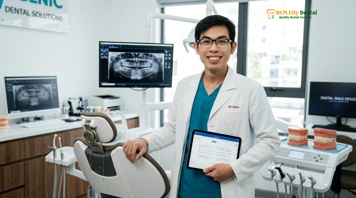
Only a qualified dental professional can accurately diagnose the extent of periodontal disease through comprehensive probing and radiographic analysis. If you suspect you may need more than a routine cleaning, seeking timely expert care is vital to prevent irreversible bone loss. We encourage patients to visit HCMC Dental Clinic in Ho Chi Minh City for a thorough periodontal assessment. Our clinical team will provide a personalized diagnosis and recommend the appropriate therapeutic approach to restore and maintain your oral health.
Frequently Asked Questions
How do I know if I need a deep cleaning instead of a regular one?
You need a deep cleaning if a clinical examination reveals periodontal pocket depths exceeding 3 millimeters, accompanied by bleeding and radiographic evidence of bone loss. A regular cleaning is only sufficient if your gums are healthy, pocket depths are 1-3 millimeters, and there is no active periodontal disease. Your dentist will use a specialized probe to measure these depths and determine the appropriate therapeutic intervention.
Is deep cleaning painful?
Deep cleaning is generally not painful because dentists utilize local anesthesia to thoroughly numb the gums and surrounding tissues before the procedure begins. While you may feel some pressure or vibration from the ultrasonic instruments, sharp pain is prevented. Post-treatment, patients may experience mild gingival tenderness and thermal sensitivity for a few days, which can be managed with over-the-counter analgesics and desensitizing toothpaste.
How much does scaling and root planing cost per quadrant?
The cost of scaling and root planing typically ranges from $150 to $300 per quadrant, depending on the geographic location, the severity of the periodontal disease, and the specific clinic. According to the latest clinic fee schedule, this fee generally includes the local anesthesia and the meticulous debridement required. It is highly recommended to consult directly with your dental provider for an accurate, personalized estimate based on your clinical needs.
Can teeth fall out after a deep cleaning?
Teeth do not fall out as a direct result of a deep cleaning; rather, the procedure is designed to save teeth by halting the progression of bone loss. However, if severe periodontitis has already destroyed significant supporting bone, removing the heavy calculus that was artificially splinting the teeth together might make them feel temporarily looser. As the gums heal and reattach, tooth mobility often stabilizes or improves.
How long does it take for gums to heal after scaling and root planing?
Initial gum healing after scaling and root planing takes approximately 5 to 7 days, during which time acute inflammation, bleeding, and tenderness subside. However, the complete biological process of gingival reattachment to the tooth root and the maturation of the connective tissue can take up to 6 to 8 weeks. A follow-up evaluation is typically scheduled at this time to assess the reduction in pocket depths.
References
- Journal of Periodontology. Clinical parameters of scaling and root planing in the treatment of chronic periodontitis. (2020).
- American Dental Association. Evidence-based clinical practice guideline on the nonsurgical treatment of chronic periodontitis. (2015).
- International Journal of Dental Hygiene. The efficacy of supragingival and subgingival biofilm control. (2019).
- Journal of Clinical Periodontology. Healing dynamics following non-surgical periodontal therapy. (2021).
- Periodontology 2000. The role of periodontal maintenance in long-term tooth survival. (2018).