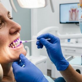
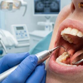
Ultrasonic scaling uses high-frequency vibrations and water irrigation to shatter hard calculus, while hand scaling relies on manual scraping with sharp, specialized instruments. Both methods effectively remove pathogenic plaque, but combining them offers the most comprehensive cleaning, ensuring optimal periodontal health and minimal tissue trauma during professional dental hygiene appointments.
Clinical Summary:
The debate between ultrasonic scaling vs hand scaling is a cornerstone of modern periodontal therapy. Dental calculus, a mineralized matrix of bacterial biofilm, cannot be removed by standard at-home brushing and requires professional mechanical debridement. Ultrasonic scalers utilize magnetostrictive or piezoelectric technology to generate high-frequency vibrations (20,000 to 45,000 Hz) that fracture calculus deposits, while simultaneously producing a cavitation effect that flushes out periodontal pockets. Conversely, hand scaling employs specialized periodontal curettes and scalers, offering clinicians unparalleled tactile sensitivity to detect and remove microscopic subgingival deposits. Clinical evidence strongly supports a blended protocol: utilizing ultrasonic devices for efficient gross debridement, followed by meticulous hand instrumentation for fine root planing. This combined approach maximizes calculus removal efficiency, minimizes clinical fatigue, and optimizes patient comfort during routine and deep cleaning procedures.
Key Takeaways:
- Ultrasonic scalers utilize high-frequency vibrations and water coolant to efficiently fracture heavy calculus deposits.
- Hand scaling provides superior tactile feedback, allowing clinicians to detect hidden subgingival irregularities.
- Cavitation and acoustic microstreaming from ultrasonic devices actively disrupt bacterial biofilms in deep pockets.
- A combined treatment protocol maximizes clinical outcomes and ensures thorough root surface debridement.
- Patient comfort varies; some prefer the speed of ultrasonics, while others prefer the quiet precision of manual tools.
The Science of Calculus Removal: Hardened Plaque
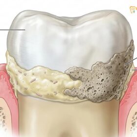
Dental calculus is mineralized bacterial plaque that bonds tightly to tooth enamel and root surfaces, requiring professional mechanical debridement for complete removal and disease prevention.
To fully understand the clinical necessity of professional scaling, one must first examine the pathophysiology of dental plaque and its transformation into calculus. The oral cavity is a complex ecosystem harboring hundreds of bacterial species. Within minutes of brushing, a glycoprotein pellicle derived from saliva coats the tooth surfaces. This pellicle acts as an adhesive substrate, allowing primary colonizing bacteria to attach and multiply, forming a structured biofilm known as dental plaque[1]. If this biofilm is not mechanically disrupted through daily oral hygiene practices, it undergoes a process of maturation and mineralization.
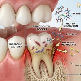
Mineralization occurs when calcium and phosphate ions from saliva (for supragingival calculus) or gingival crevicular fluid (for subgingival calculus) precipitate into the plaque matrix. This process transforms the soft, easily removable biofilm into a hardened, porous substance called calculus, commonly known as tartar. Once calculus forms, it creates a highly retentive surface that harbors even more pathogenic bacteria, initiating a localized inflammatory immune response. This chronic inflammation leads to gingivitis and, if left untreated, progresses to periodontitis—characterized by the destruction of the periodontal ligament and alveolar bone[2].
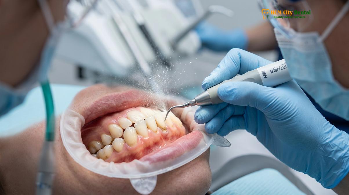
Because calculus is firmly bonded to the hydroxyapatite crystals of the tooth enamel and cementum, standard toothbrushes and dental floss are entirely ineffective at removing it. Professional intervention is mandatory. The primary objective of any professional dental cleaning is the meticulous removal of these calcified deposits to halt the inflammatory cascade and restore biocompatibility to the root surfaces. Achieving optimal Saigon dental hygiene standards requires clinicians to employ highly specialized instruments capable of fracturing this mineralized matrix without causing iatrogenic damage to the underlying tooth structure.
The clinical challenge lies in the topography of the teeth. Supragingival calculus, located above the gumline, is visible and relatively straightforward to access. However, subgingival calculus, hidden deep within periodontal pockets, presents a significant hurdle. It is often dark in color due to blood pigments and is tenaciously adhered to the root cementum. Removing these hidden deposits requires not just mechanical force, but profound clinical skill, an understanding of root anatomy, and the correct selection of dentist scaling tools.
Ultrasonic Scalers: High-Frequency Vibrations and Cavitation
Ultrasonic scalers operate between 20,000 and 45,000 hertz, utilizing micro-vibrations and acoustic microstreaming to fracture calculus and flush away periodontal pathogens.
The advent of power-driven instrumentation revolutionized periodontal therapy. Ultrasonic scalers are sophisticated electromechanical devices that convert electrical energy into high-frequency mechanical vibrations at the instrument tip. These devices are broadly categorized into two main technologies: magnetostrictive and piezoelectric. Magnetostrictive units utilize a stack of metal strips that expand and contract in a magnetic field, producing an elliptical tip motion where all surfaces of the tip are active. Piezoelectric units, on the other hand, use ceramic crystals that change dimension when an electrical charge is applied, resulting in a linear tip motion where primarily the lateral surfaces are active[3].
Regardless of the specific technology, the primary mechanism of action is mechanical chipping. As the vibrating tip contacts the calculus, the rapid micro-impacts shatter the mineralized matrix. However, the efficacy of a cavitron ultrasonic scaler extends far beyond mere mechanical force. These devices require a continuous flow of water for cooling, as the rapid vibrations generate significant frictional heat that could otherwise cause thermal necrosis to the dental pulp.
This water coolant introduces two critical secondary mechanisms of action: cavitation and acoustic microstreaming. Cavitation occurs when the high-frequency vibrations create microscopic bubbles in the water coolant. These bubbles rapidly expand and violently implode, releasing localized bursts of energy that rupture the cell walls of periodontal pathogens. Acoustic microstreaming refers to the forceful, swirling fluid dynamics generated around the vibrating tip, which effectively flushes unattached plaque, endotoxins, and debris out of the periodontal pocket.
“The hydrodynamic effects of ultrasonic instrumentation, specifically cavitation and acoustic microstreaming, provide a distinct antimicrobial advantage in deep periodontal pockets, disrupting the biofilm matrix in areas where mechanical tip contact is anatomically impossible.”
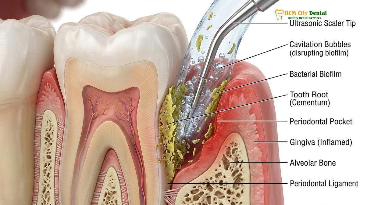
From a clinical perspective, ultrasonic scaling is highly efficient for gross debridement—the removal of large, tenacious calculus deposits. It requires a lighter touch compared to manual scaling, reducing hand fatigue for the clinician. Furthermore, the lavage effect of the water spray improves visibility by continuously clearing blood and debris from the operating field. However, the reliance on water spray also produces significant aerosols, requiring strict infection control protocols and high-volume evacuation to maintain a safe clinical environment[4].
Hand Instrumentation: Precision Scraping with Periodontal Scalers
Manual scaling utilizes specialized curettes and scalers to meticulously scrape away residual calculus, offering clinicians unparalleled tactile sensitivity to detect hidden subgingival deposits.
Despite the technological advancements of power-driven devices, hand instrumentation remains a fundamental and irreplaceable skill in comprehensive general dentistry services. Manual scaling relies on the clinician’s physical dexterity, utilizing precisely engineered instruments to mechanically scrape calculus from the tooth surfaces. The two primary categories of hand instruments are sickle scalers and periodontal curettes.
Sickle scalers feature a pointed toe and two cutting edges, making them ideal for removing heavy supragingival calculus, particularly in the tight interproximal spaces between anterior teeth. However, their sharp, pointed design makes them unsuitable for subgingival use, as they can easily traumatize the delicate gingival tissues. For subgingival debridement and root planing, clinicians rely on periodontal curettes. These instruments have a rounded toe and a rounded back, allowing them to be safely inserted beneath the gumline.
Curettes are further divided into universal curettes, which can be adapted to multiple tooth surfaces, and area-specific curettes, such as the Gracey series. Gracey curettes are meticulously designed with specific shank angles and a single cutting edge offset at 70 degrees, allowing for perfect adaptation to the complex anatomical curves of specific tooth roots. The mastery of these instruments requires a deep understanding of root morphology, precise angulation, and the use of a modified pen grasp coupled with a stable fulcrum to ensure controlled, overlapping strokes.
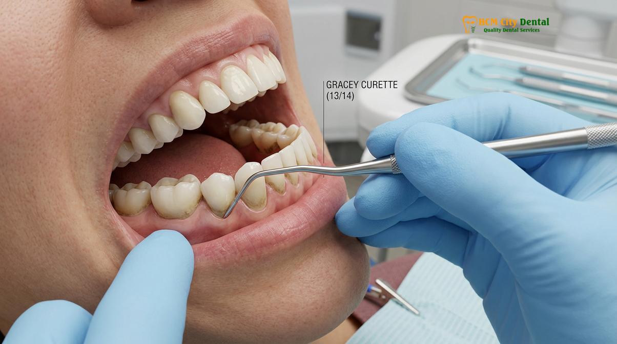
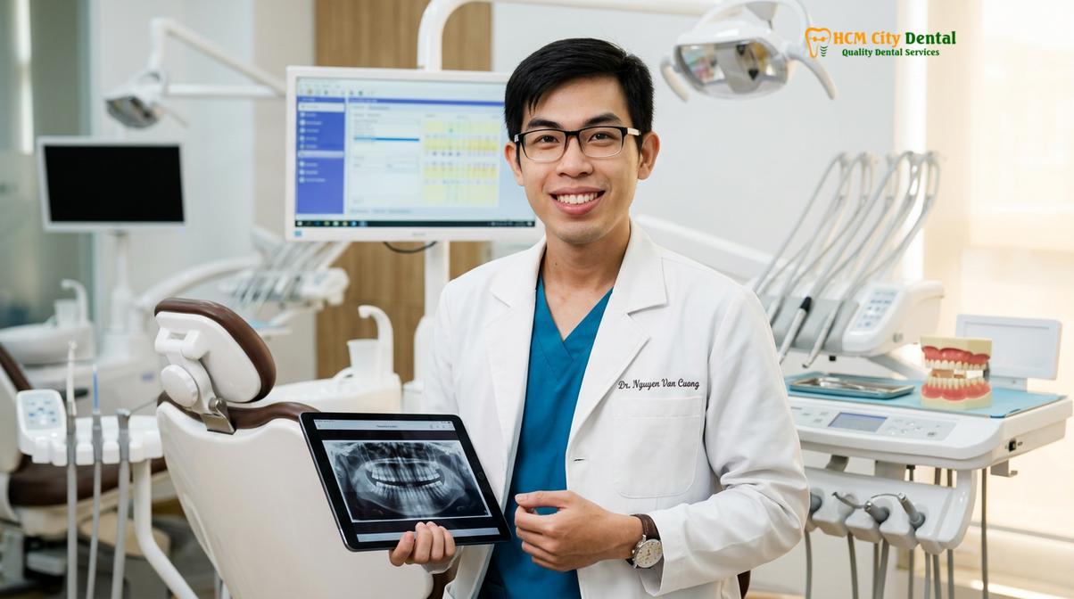
The most significant advantage of hand scaling is tactile sensitivity. As the clinician lightly guides the curette over the root surface, the metal shank transmits microscopic vibrations to their fingertips. This allows the practitioner to “feel” the difference between smooth, healthy cementum and rough, residual calculus hidden deep within a pocket. According to Dr. Nguyen Van Cuong, a leading specialist at HCMC Dental Clinic, this tactile feedback is crucial for verifying the endpoint of periodontal therapy, ensuring that no microscopic deposits are left behind to harbor future bacterial growth.
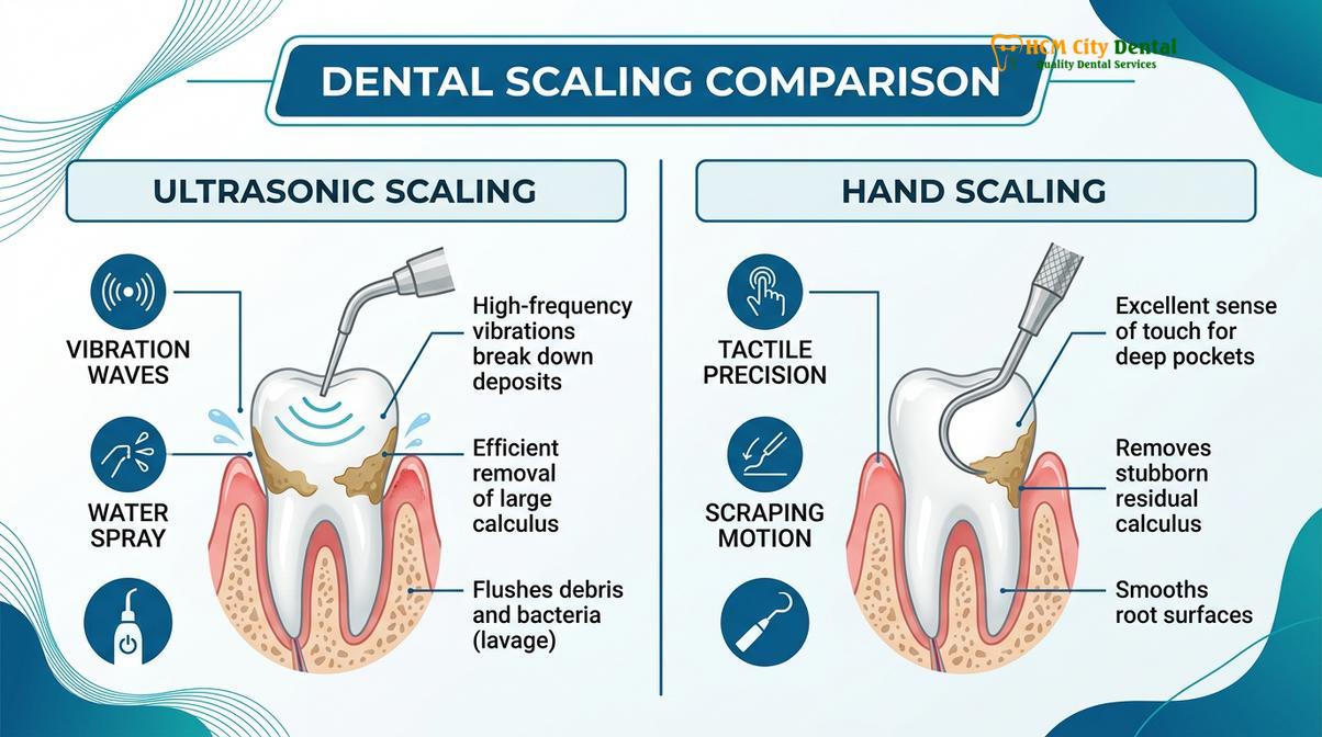
| Clinical Feature | Ultrasonic Scaling | Hand Scaling |
|---|---|---|
| Primary Mechanism | High-frequency vibration & cavitation | Mechanical scraping & root planing |
| Tactile Sensitivity | Low (vibration masks feedback) | High (direct transmission through shank) |
| Water Usage | Continuous irrigation required | None (dry field possible) |
| Aerosol Production | High (requires high-volume suction) | Minimal to None |
| Best Suited For | Heavy calculus, deep pocket flushing | Fine detailing, subgingival verification |
Comparing Patient Comfort, Tooth Sensitivity, and Cleaning Speed
While ultrasonic scaling is generally faster and requires less pressure, hand scaling avoids water spray and vibration, making the choice highly dependent on individual patient sensitivity and clinical needs.
The patient experience during periodontal therapy is a critical factor that influences compliance with recommended maintenance schedules. The choice between ultrasonic and hand scaling significantly impacts patient comfort, and there is no universal preference. Some patients find the high-frequency vibration and high-pitched sound of ultrasonic scalers distressing. Additionally, the continuous flow of cold water required for cooling can trigger sharp pain in patients suffering from dentin hypersensitivity. The hydrodynamic theory of tooth sensitivity explains that fluid movement within exposed dentinal tubules stimulates nerve endings in the pulp; the cold water spray from an ultrasonic device can easily provoke this response.
Conversely, hand scaling eliminates the water spray and the high-pitched noise, which many patients find more relaxing. However, manual instrumentation requires the clinician to apply lateral pressure against the tooth surface to effectively engage the cutting edge of the curette beneath the calculus deposit. Some patients perceive this scraping pressure and the accompanying sound as uncomfortable or invasive. Furthermore, hand scaling is generally a slower, more meticulous process, which can lead to jaw fatigue for the patient during extended appointments.
Clinical Contraindications: Ultrasonic scaling should be used with extreme caution or avoided entirely in patients with severe respiratory conditions (due to aerosol aspiration risks), unshielded cardiac pacemakers (specifically with older magnetostrictive units), and areas of severe demineralization where the vibration could compromise weakened enamel.
From a clinical efficiency standpoint, ultrasonic scaling is undeniably faster for the removal of heavy, tenacious calculus. The power-driven tip can fracture large deposits in a fraction of the time it would take to manually scrape them away. This efficiency reduces chair time and minimizes hand fatigue for the dental professional. However, speed does not always equate to thoroughness. Relying solely on ultrasonic devices can sometimes result in “burnished” calculus—where the outer layer of the deposit is smoothed over rather than completely removed, making it incredibly difficult to detect and remove subsequently[5].
Ensuring tooth enamel safety is paramount in both techniques. When used with the correct angulation and appropriate power settings, ultrasonic tips will not damage healthy enamel. Similarly, perfectly sharpened hand instruments used with proper technique will safely plane the root surface without gouging the cementum. The key variable in both methods is the skill and experience of the clinician performing the procedure.
The Ideal Combination Protocol for Maximum Plaque Removal
The gold standard in modern periodontics is a blended approach: utilizing ultrasonic instruments for bulk calculus debridement followed by hand scaling for fine root planing and tactile verification.
In contemporary clinical practice, the debate is rarely an absolute choice between ultrasonic scaling vs hand scaling. Instead, evidence-based dentistry advocates for a synergistic, combined protocol that leverages the unique strengths of both modalities while mitigating their respective weaknesses. This blended approach ensures the highest calculus removal efficiency and provides the most predictable clinical outcomes for periodontal healing.
The standard workflow for a comprehensive periodontal maintenance session begins with a thorough diagnostic probing to map pocket depths. The clinician then initiates the active therapy using an ultrasonic scaler. The power-driven tip is systematically guided around the dentition to rapidly fracture and remove the bulk of supragingival and easily accessible subgingival calculus. Simultaneously, the cavitational water spray flushes the periodontal pockets, reducing the bacterial load and washing away blood to improve visibility.
Once the gross debridement is complete, the clinician transitions to hand instrumentation. Using finely sharpened Gracey curettes, the practitioner meticulously explores the subgingival root surfaces. This step relies entirely on the superior tactile sensitivity of hand tools to detect microscopic, residual calculus or rough patches of cementum that the ultrasonic tip may have missed or burnished. The clinician then performs precise root planing strokes to create a glass-smooth surface, which is biologically acceptable for the gingival tissues to reattach.
“Optimal periodontal debridement is rarely achieved by a single modality. The integration of ultrasonic cavitation for biofilm disruption, followed by the tactile precision of manual curettage, represents the definitive standard of care in non-surgical periodontal therapy.”
Clinical Case Review: A 45-year-old patient presented to Dr. Nguyen Van Cuong at HCMC Dental Clinic in Ho Chi Minh City with generalized moderate periodontitis, heavy subgingival calculus, and localized bleeding on probing. Dr. Cuong utilized a combined protocol: initial full-mouth debridement with a piezoelectric ultrasonic scaler to efficiently remove bulk deposits and flush the 5mm pockets, followed by targeted manual root planing using area-specific curettes. At the 4-week re-evaluation, the patient exhibited a significant reduction in pocket depths, complete resolution of gingival bleeding, and reported minimal post-operative sensitivity due to the efficient, blended approach.
Following the combined scaling protocol, the teeth are typically polished to remove extrinsic stains and residual soft plaque, leaving a smooth surface that is less susceptible to immediate bacterial recolonization. This comprehensive approach, combining the power of technology with the finesse of manual skill, ensures that patients receive the highest standard of preventative dental care.
When to See a Doctor for Professional Periodontal Therapy
Maintaining optimal oral health requires more than just diligent home care; it necessitates regular professional evaluation. You should schedule a consultation for a comprehensive examination and potential scaling if you experience persistent symptoms of periodontal distress. The most common early warning sign is gingival bleeding—healthy gums should never bleed during normal brushing or flossing. If you notice pink in the sink, it is a clear indicator of localized inflammation and bacterial buildup.
Other critical signs that warrant immediate professional attention include chronic halitosis (bad breath) that does not improve with mouthwash, visible hard, yellowish or brown deposits along the gumline, or gums that appear red, swollen, and tender to the touch. As periodontal disease progresses, you may notice gingival recession (teeth appearing longer), increased tooth sensitivity to hot and cold, or even slight mobility in your teeth. These are advanced signs of attachment loss and require prompt intervention.
At HCMC Dental Clinic in Ho Chi Minh City, our clinical team emphasizes the importance of preventative diagnostics. We recommend that patients with a healthy periodontium undergo routine professional cleaning and evaluation every six months. However, individuals with a history of gum disease, rapid calculus accumulation, or systemic risk factors such as diabetes or smoking may require customized periodontal maintenance intervals every three to four months to ensure long-term stability and prevent irreversible bone loss.
Frequently Asked Questions
Is ultrasonic scaling bad for tooth enamel?
When performed correctly by a trained dental professional, ultrasonic scaling is completely safe for tooth enamel. The instruments are designed to fracture porous calculus rather than dense enamel. However, improper angulation or excessive pressure could theoretically cause micro-scratches, which is why professional technique is paramount. The tip is kept in constant motion to prevent localized heat buildup or surface damage.
Can patients with pacemakers have ultrasonic scaling?
Most modern cardiac pacemakers are shielded and generally safe around piezoelectric ultrasonic scalers. However, older magnetostrictive devices may cause electromagnetic interference. Patients must always disclose their specific pacemaker model to their dentist so the appropriate and safest scaling method can be selected. When in doubt, hand scaling is the universally safe alternative.
Why do my teeth feel sensitive after ultrasonic cleaning?
Post-scaling sensitivity occurs because the removal of heavy calculus exposes previously insulated root surfaces to temperature changes in the mouth. Additionally, the hydrodynamic theory suggests that fluid movement within exposed dentinal tubules triggers nerve endings, causing temporary discomfort that usually subsides within a few days. Using a desensitizing toothpaste containing potassium nitrate can help alleviate these symptoms rapidly.
How often should I get professional scaling done?
For individuals with healthy gums, professional scaling is typically recommended every six months. Patients with a history of periodontitis, rapid calculus buildup, or systemic conditions like diabetes may require periodontal maintenance therapy every three to four months to prevent disease progression. Your dentist will determine the optimal frequency based on your specific clinical presentation and risk factors.
Does hand scaling hurt more than ultrasonic scaling?
Pain perception varies widely among patients; some find the scraping pressure of hand scaling uncomfortable, while others dislike the high-frequency vibration and water spray of ultrasonic devices. Local anesthesia or desensitizing agents can be applied during either procedure to ensure maximum patient comfort. Open communication with your hygienist allows them to tailor the technique to your specific tolerance levels.
References
- Journal of Clinical Periodontology. Efficacy of machine-driven and manual subgingival debridement in periodontal therapy. (2020).
- Periodontology 2000. The biofilm concept: consequences for future prophylaxis of oral diseases. (2019).
- International Journal of Dental Hygiene. Patient perception and comfort during ultrasonic versus manual instrumentation. (2021).
- Journal of the American Dental Association. Aerosol mitigation and safety protocols in modern periodontal practice. (2022).
- Clinical Oral Investigations. Comparative analysis of root surface alterations following ultrasonic and curette instrumentation. (2018).