If you notice persistent bleeding, chronic bad breath, receding gums, deep periodontal pockets, or loose teeth, these are clear signs you need deep cleaning for tartar beneath the gums. This therapeutic procedure, known as scaling and root planing, halts periodontal disease progression and prevents irreversible bone loss.
Clinical Summary:
Periodontal disease is a progressive bacterial infection that destroys the supporting structures of the teeth. When plaque biofilm calcifies into hard calculus (tartar) below the gumline, standard brushing and routine dental cleanings are no longer sufficient. The presence of subgingival tartar triggers a chronic inflammatory response, leading to symptoms such as persistent bleeding, halitosis, gum recession, and increased tooth mobility. A deep cleaning, clinically termed scaling and root planing (SRP), is a non-surgical periodontal therapy designed to meticulously remove these bacterial deposits from the root surfaces. By eliminating the source of infection and smoothing the cementum, this procedure allows the gingival tissue to heal, reduces pocket depths, and prevents systemic health complications associated with untreated oral infections. Early recognition of these warning signs and prompt intervention by a dental professional are critical for preserving the natural dentition and maintaining overall systemic health.
Key Takeaways:
- Bleeding during routine oral hygiene is an abnormal sign indicating active gingival inflammation and ulceration.
- Chronic halitosis often stems from volatile sulfur compounds produced by anaerobic bacteria in deep gum pockets.
- Gum recession and the appearance of elongated teeth are direct results of underlying alveolar bone destruction.
- Periodontal pockets measuring 4 millimeters or deeper require professional scaling and root planing to debride.
- Untreated periodontal infections significantly elevate the risk of systemic conditions, including cardiovascular disease.
Gingivitis vs. Periodontitis: When Standard Scaling is Not Enough
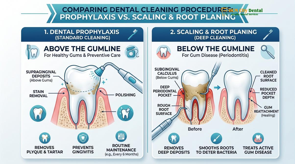
While gingivitis is reversible surface inflammation treated with standard scaling, periodontitis involves irreversible bone loss requiring deep cleaning to remove subgingival tartar.
To understand the clinical signs you need deep cleaning tartar gums, it is essential to differentiate between the stages of gum disease. The human mouth is a complex microbiome harboring hundreds of bacterial species. When oral hygiene is suboptimal, these bacteria colonize the tooth surfaces, forming a sticky, colorless film known as dental plaque biofilm. If this biofilm is not mechanically disrupted through daily brushing and flossing, minerals from saliva—primarily calcium and phosphate—precipitate into the plaque matrix, causing it to calcify into a hardened substance called calculus, or tartar[1].
In the initial stage, known as gingivitis, the bacterial accumulation is primarily supragingival (above the gumline). The body’s immune system responds to the bacterial endotoxins by initiating an inflammatory cascade. The gums become erythematous (red), edematous (swollen), and prone to bleeding. However, at this stage, the underlying alveolar bone and the periodontal ligament remain intact. Gingivitis is entirely reversible with a standard dental prophylaxis (routine cleaning) and improved home care.
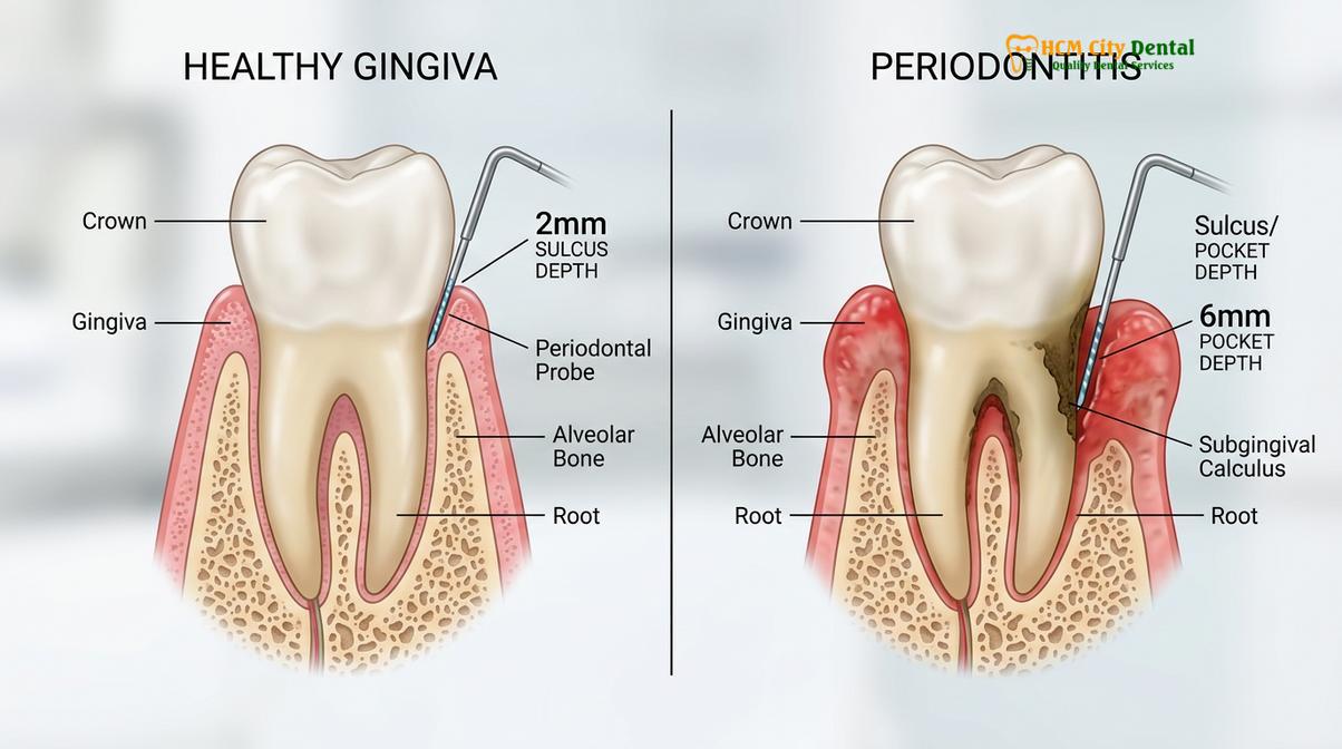
Conversely, if gingivitis is left untreated, the bacterial biofilm matures and migrates apically (downward) beneath the gumline, transitioning into periodontitis. The subgingival environment is highly anaerobic, favoring the proliferation of virulent, tissue-destroying pathogens such as Porphyromonas gingivalis and Treponema denticola. As these bacteria multiply, they release potent toxins that provoke a hyperactive host immune response. The immune system, in its attempt to eradicate the infection, inadvertently releases enzymes and pro-inflammatory cytokines (like Interleukin-1β and TNF-α) that break down the connective tissue and resorb the alveolar bone supporting the teeth[2].
Once tartar has formed below the gumline, a standard cleaning is clinically inadequate. The instruments used in a routine prophylaxis are not designed to reach deep into periodontal pockets. This is where scaling and root planing becomes a medical necessity. Dr. Nguyen Van Cuong frequently emphasizes in clinical practice that attempting to treat periodontitis with a standard cleaning is akin to washing the outside of a window when the dirt is on the inside; the root cause of the disease remains untouched, and the destruction continues silently.
Sign 1: Gums that Bleed Consistently During Brushing and Flossing
Healthy gums do not bleed; persistent bleeding during oral hygiene indicates active bacterial infection and ulceration of the pocket lining that requires professional intervention.
One of the most universally recognized, yet frequently ignored, indicators of periodontal distress is bleeding gums. Many patients mistakenly attribute bleeding during brushing to using a toothbrush with bristles that are too hard or applying excessive mechanical force. While traumatic brushing can cause localized abrasions, generalized and consistent bleeding is a hallmark sign of active periodontal inflammation.
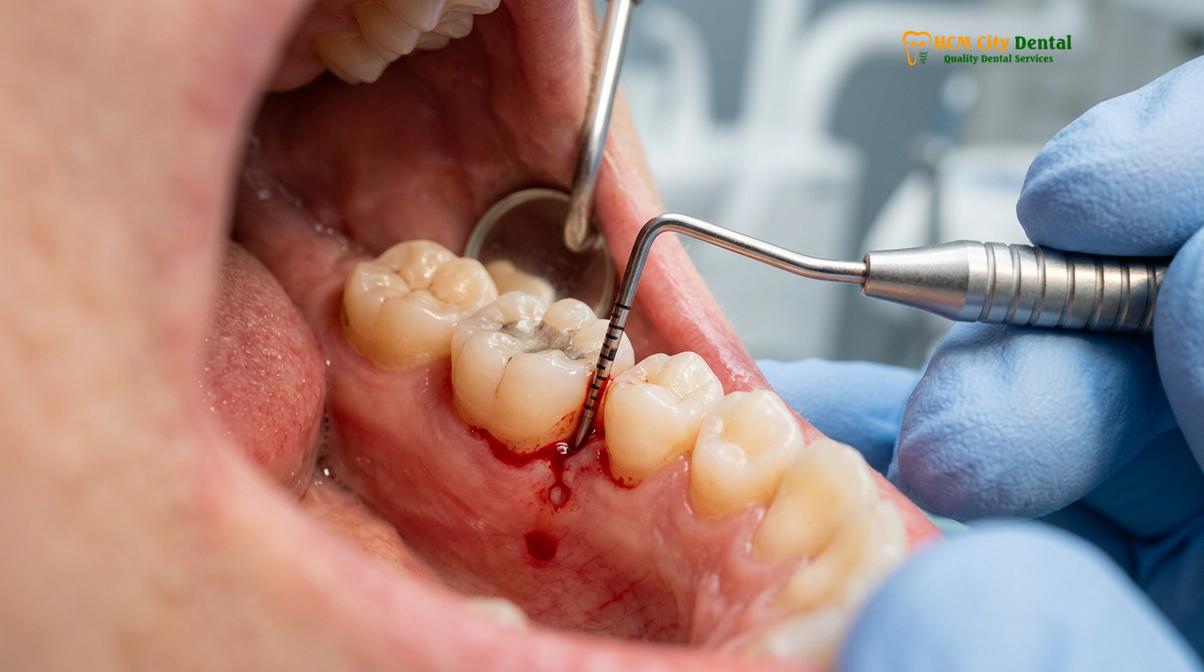
From a histological perspective, healthy gingival tissue is characterized by a pale pink color, a firm and stippled texture (resembling an orange peel), and a tightly adapted knife-edge margin around the neck of the tooth. The sulcular epithelium—the tissue lining the shallow crevice between the tooth and the gum—is intact and resilient. When subgingival plaque and tartar accumulate, the resulting inflammatory response causes profound vascular changes. The capillaries within the gingival connective tissue dilate and engorge with blood to deliver immune cells (neutrophils and macrophages) to the site of infection[3].
“Bleeding on probing is widely considered the most reliable clinical indicator of active periodontal inflammation and a predictor of future clinical attachment loss if left untreated.”
This vasodilation, combined with the thinning and micro-ulceration of the sulcular epithelium caused by bacterial enzymes, makes the tissue incredibly fragile. Consequently, the minor mechanical friction introduced by a toothbrush or dental floss easily ruptures these engorged microvessels, resulting in visible bleeding. If you experience bleeding during brushing on a daily or weekly basis, your body is signaling a chronic infection.
During a comprehensive general dental care examination, a clinician will assess the Bleeding on Probing (BOP) index. A high BOP percentage strongly correlates with the presence of subgingival calculus and active tissue destruction. A deep cleaning addresses this by meticulously debriding the root surfaces, removing the calcified irritants, and allowing the ulcerated sulcular epithelium to heal and re-epithelialize, thereby resolving the bleeding.
Sign 2: Persistent Bad Breath (Halitosis) and Metallic Taste
Chronic bad breath and a metallic taste often result from volatile sulfur compounds produced by anaerobic bacteria thriving in deep, oxygen-deprived periodontal pockets.
Occasional bad breath after consuming pungent foods like garlic or onions is a normal physiological occurrence. However, chronic bad breath that persists despite rigorous brushing, flossing, and the use of cosmetic mouthwashes is a significant clinical warning sign. In the context of periodontal disease, this persistent malodor is not a digestive issue, but rather a direct byproduct of bacterial metabolism occurring beneath the gumline.
The deep periodontal pockets created by advancing gum disease provide an ideal, oxygen-deprived (anaerobic) incubator for specific strains of Gram-negative bacteria. As these pathogens break down proteins derived from food debris, saliva, and sloughed epithelial cells, they release Volatile Sulfur Compounds (VSCs) as metabolic waste. The primary culprits include hydrogen sulfide (which smells like rotten eggs), methyl mercaptan (which smells like feces), and dimethyl sulfide. These gases seep out of the periodontal pockets and mix with the breath, creating a distinct and highly offensive odor[4].
Furthermore, patients with severe subgingival calculus often report a persistent metallic or sour taste in their mouth. This dysgeusia is typically caused by the continuous microscopic oozing of blood and purulent exudate (pus) from the infected gum pockets into the oral cavity. Blood contains iron, which imparts a distinct metallic flavor, while the purulent discharge contributes to the foul taste.
Cosmetic mouthwashes and breath mints only mask these odors temporarily; they cannot penetrate the deep periodontal pockets to eradicate the source of the VSCs. A deep cleaning physically dismantles the bacterial biofilm and removes the porous calculus that harbors these odor-causing colonies. By reducing the pocket depths and eliminating the anaerobic environment, scaling and root planing effectively cures periodontitis-induced halitosis at its source.
Sign 3: Gum Recession and the Appearance of ‘Long Teeth’
As periodontal disease destroys underlying alveolar bone, the overlying gum tissue recedes, exposing sensitive tooth roots and making teeth appear abnormally long.
Gum recession is a physical manifestation of underlying structural loss. Patients often look in the mirror and notice that their teeth appear longer than they used to, or they may observe dark triangular spaces (black triangles) forming between their teeth near the gumline. This phenomenon is rarely due to the teeth actually erupting further; rather, it is the result of the gingival margin migrating apically (towards the root) as the supporting bone is destroyed.
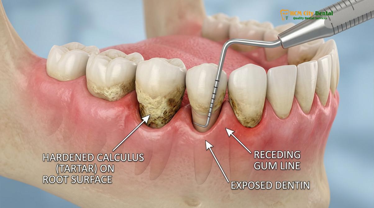
The architecture of the periodontium dictates that the gum tissue follows the contour of the underlying alveolar bone. When subgingival tartar triggers chronic inflammation, the osteoclasts (bone-resorbing cells) break down the bone crest to retreat from the advancing bacterial front. As the bone level drops, the overlying gum tissue loses its structural support and subsequently recedes. This receding gum line exposes the cementum—the relatively soft, porous tissue covering the tooth root.
| Stage of Recession | Clinical Presentation | Patient Symptoms | Recommended Intervention |
|---|---|---|---|
| Mild (Class I) | Slight root exposure, no interdental bone loss. | Mild thermal sensitivity, aesthetic concerns. | Scaling and root planing, brushing technique modification. |
| Moderate (Class II) | Recession extends to the mucogingival junction. | Increased dentinal hypersensitivity, visible root yellowing. | Deep cleaning, possible desensitizing agents. |
| Severe (Class III/IV) | Extensive root exposure with interdental bone loss. | Severe pain to cold/sweets, food impaction, tooth mobility. | Advanced periodontal therapy, potential soft tissue grafting. |
Unlike the crown of the tooth, which is protected by highly mineralized enamel, the root surface is covered by cementum and underlying dentin, both of which contain thousands of microscopic dentinal tubules leading directly to the tooth’s nerve center (the pulp). When these tubules are exposed to the oral environment, patients experience acute dentinal hypersensitivity. Consuming hot, cold, sweet, or acidic foods and beverages triggers a sharp, sudden pain.
Furthermore, exposed root surfaces are highly susceptible to root caries (decay) because cementum demineralizes at a much higher pH (around 6.7) compared to enamel (5.5). If you notice your teeth appearing longer or experience new, localized sensitivity near the gumline, it is imperative to undergo a thorough periodontal evaluation. While a deep cleaning cannot reverse the recession that has already occurred, it is the critical first step in halting the active bone loss and preventing further exposure of the roots.
Sign 4: Deep Periodontal Pockets and Tartar Build-Up Below the Gums
Pockets measuring 4 millimeters or deeper indicate that the gum tissue has detached from the tooth, creating a space where subgingival tartar rapidly accumulates.
In a healthy state, the gingival tissue attaches firmly to the tooth just above the crest of the alveolar bone, creating a shallow crevice called a sulcus. A healthy sulcus typically measures between 1 to 3 millimeters in depth. This shallow space can be effectively cleaned by the bristles of a toothbrush and standard dental floss. However, when periodontal disease takes hold, the inflammatory process destroys the periodontal ligament fibers that anchor the gum to the tooth, causing the tissue to detach.
This detachment creates a pathological space known as a periodontal pocket. As the pocket deepens, it becomes a highly protected environment for bacteria. The deeper the pocket, the more anaerobic the environment becomes, fostering the growth of the most aggressive periodontal pathogens. Because oxygen cannot reach these depths, and home care tools are physically incapable of cleaning beyond 3 millimeters subgingivally, plaque below gumline rapidly calcifies into dark, tenacious calculus.
Clinical Case Review: Managing Deep Periodontal Pockets
A 45-year-old patient presented to HCMC Dental Clinic in Ho Chi Minh City complaining of dull, aching gums and chronic bad breath. The patient had not received a professional cleaning in over three years. During the clinical examination, periodontal probing revealed generalized pocket depths of 5 to 7 millimeters, accompanied by heavy subgingival calculus visible on digital radiographs. The diagnosis was moderate generalized periodontitis. The treatment protocol involved two sessions of scaling and root planing under local anesthesia, utilizing ultrasonic instrumentation to shatter the calculus and hand curettes to smooth the root surfaces. At the 6-week re-evaluation, tissue inflammation had resolved, and pocket depths had reduced to a maintainable 3 to 4 millimeters, successfully avoiding the need for invasive flap surgery.
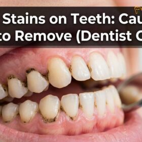
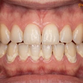
Subgingival calculus is distinct from supragingival calculus. It is often dark brown, green, or black due to the incorporation of blood pigments (hemosiderin) from the chronically bleeding tissues. It is also much harder and more tenaciously adhered to the root surface. This calculus acts as a porous reservoir, retaining bacterial endotoxins directly against the ulcerated pocket wall, perpetuating the cycle of inflammation and bone destruction.
During a comprehensive dental examination, the clinician uses a calibrated periodontal probe to measure the depth of these pockets around every tooth. Measurements of 4 millimeters indicate early periodontitis, 5 to 6 millimeters indicate moderate disease, and 7 millimeters or greater signify severe, advanced periodontitis. Any pocket measuring 4 millimeters or deeper with bleeding on probing is a definitive clinical indication for scaling and root planing.
Sign 5: Loose or Shifting Teeth and Gum Tenderness
Tooth mobility and shifting are late-stage indicators of severe bone destruction, requiring immediate deep cleaning and possible splinting to prevent tooth loss.
Teeth are not rigidly fused to the jawbone; they are suspended in the bony socket by a complex network of collagen fibers known as the periodontal ligament (PDL). This ligament acts as a shock absorber, allowing for microscopic physiological movement during chewing. However, when you can visibly see or feel your teeth moving, or if they have shifted out of their normal alignment, you are experiencing pathologic tooth mobility—a severe, late-stage sign of advanced periodontal disease.
As subgingival tartar drives chronic inflammation, the osteoclasts relentlessly resorb the alveolar bone. When a significant percentage of the supporting bone is lost (often exceeding 50% of the root length), the tooth loses its mechanical foundation. The remaining periodontal ligament fibers become overwhelmed by normal occlusal (biting) forces, leading to secondary occlusal trauma. This trauma further accelerates the widening of the PDL space and exacerbates the bone loss, creating a rapid downward spiral toward tooth exfoliation (loss)[5].
Clinical Warning: Pathologic Migration
If you notice new gaps forming between your front teeth, or if your teeth appear to be flaring outward, this is known as pathologic migration. It indicates that the structural support of the teeth has been severely compromised. Delaying treatment at this stage dramatically increases the risk of spontaneous tooth loss. Immediate periodontal intervention is required to stabilize the dentition.
Patients with loose teeth signs often report a dull, throbbing pain or tenderness in the gums, particularly when chewing fibrous or hard foods. The gums may feel “spongy” or fluctuant upon palpation, sometimes accompanied by the expression of purulent exudate from the gingival margins. The bite may feel “off” or uncomfortable because the shifting teeth no longer interlock correctly with their opposing counterparts.
When tooth mobility is present, a deep cleaning is urgently required to arrest the active infection and halt further bone destruction. Following the scaling and root planing procedure, as the inflammation subsides, the gingival tissues tighten around the teeth, which can slightly reduce mobility. However, in cases of severe bone loss, the dentist may also need to perform periodontal splinting—bonding the loose teeth to adjacent stable teeth—to distribute occlusal forces and provide mechanical stability while the periodontium heals.
The Risk of Ignoring Periodontal Disease: Systemic Health Connections
Untreated periodontal infections allow oral bacteria to enter the bloodstream, significantly increasing the risk of cardiovascular disease, diabetic complications, and adverse pregnancy outcomes.
The implications of ignoring the signs you need deep cleaning tartar gums extend far beyond the oral cavity. The mouth is the gateway to the body, and the highly vascularized nature of inflamed periodontal tissues provides a direct portal for oral pathogens to enter the systemic circulation. This phenomenon, known as bacteremia, occurs daily in patients with severe periodontitis, even during simple activities like chewing or brushing.
Extensive medical research has established a profound bidirectional relationship between periodontal disease and systemic health. When Gram-negative bacteria and their endotoxins enter the bloodstream, they trigger a systemic inflammatory response, elevating acute-phase proteins such as C-reactive protein (CRP). High levels of systemic inflammation are a known catalyst for endothelial dysfunction, contributing to the pathogenesis of atherosclerosis (hardening of the arteries). Consequently, patients with untreated periodontitis have a statistically higher risk of suffering from myocardial infarctions (heart attacks) and ischemic strokes.
“Periodontal medicine has demonstrated that managing oral inflammation through non-surgical therapies like scaling and root planing can significantly reduce systemic inflammatory markers, thereby lowering the overall cardiovascular risk profile of the patient.”
Furthermore, there is a well-documented synergistic relationship between periodontitis and diabetes mellitus. Chronic systemic inflammation impairs insulin signaling, exacerbating insulin resistance and making it exceedingly difficult for diabetic patients to maintain glycemic control. Conversely, poorly controlled diabetes impairs wound healing and immune function, accelerating the destruction of periodontal tissues. Deep cleaning has been shown to improve HbA1c levels in diabetic patients by reducing the systemic inflammatory burden.
Dr. Cuong strongly advocates for a holistic approach to dental care, emphasizing that periodontal therapy is not just about saving teeth; it is a critical component of comprehensive medical management. Pregnant women with severe gum disease are also at a higher risk for adverse outcomes, including pre-eclampsia, premature birth, and low birth weight infants, due to the systemic circulation of inflammatory cytokines triggering early uterine contractions.
When to See a Doctor for Periodontal Evaluation
Recognizing the early warning signs of gum disease is the first step in preserving your oral health. You should schedule a comprehensive periodontal evaluation if you experience any of the following symptoms:
- Gums that bleed regularly when brushing, flossing, or eating hard foods.
- Gums that appear bright red, swollen, or feel tender to the touch.
- Persistent bad breath or a metallic taste that does not resolve with oral hygiene.
- Visible recession of the gumline, exposing the yellowish root surfaces.
- Teeth that feel loose, have shifted position, or changes in the way your teeth fit together when you bite.
- A history of cardiovascular disease, diabetes, or if you are planning a pregnancy, as periodontal health is crucial in these scenarios.
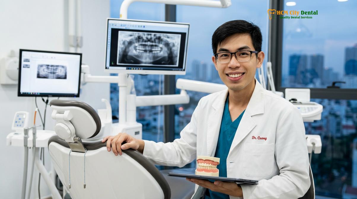
During a Saigon dental center exam, the clinical team will utilize advanced diagnostic tools, including digital radiography to assess alveolar bone levels and periodontal probing to measure pocket depths accurately. Based on these clinical diagnostics, a personalized treatment plan will be formulated. If subgingival tartar and attachment loss are identified, scaling and root planing will be recommended as the primary therapeutic intervention.
Do not wait for pain to dictate your dental visits; periodontal disease is often entirely painless until it reaches its most advanced, destructive stages. Early intervention through professional dental cleaning and targeted periodontal therapy at HCMC Dental Clinic can effectively arrest the disease process, restore gingival health, and safeguard your smile for a lifetime.
Frequently Asked Questions
Can receding gums grow back after deep cleaning?
No, receding gums cannot grow back naturally after a deep cleaning procedure. However, scaling and root planing halts the progression of periodontal disease, preventing further tissue loss. Once the infection is cleared and the gums are healthy, your dentist may recommend surgical interventions, such as gum grafting, to restore the lost tissue and cover exposed roots.
Why do my gums bleed when I use dental floss?
Gums bleed during flossing primarily due to underlying inflammation caused by plaque and tartar accumulation beneath the gumline. When bacterial biofilm is not adequately removed, it triggers an immune response, making the sulcular tissue fragile, ulcerated, and highly vascularized. Frequent bleeding is a primary indicator of gingivitis or periodontitis requiring professional evaluation.
How long does it take for gums to heal after deep cleaning?
Initial gum healing after a deep cleaning typically takes about one to two weeks. During this period, inflammation subsides, bleeding stops, and the gum tissue begins to reattach to the smoothed root surfaces. Complete structural healing and pocket depth reduction may take up to six to eight weeks, which is when a follow-up periodontal evaluation is usually scheduled.
Is scaling and root planing a painful procedure?
Scaling and root planing is generally not painful because local anesthesia is administered to numb the gums and tooth roots before the procedure begins. Patients may experience mild pressure or vibration from the ultrasonic instruments. Post-operative sensitivity to temperature and slight gum tenderness are common but typically resolve within a few days using over-the-counter analgesics.
How often should I get a deep cleaning if I have periodontitis?
A full deep cleaning is usually a one-time therapeutic intervention, provided the patient maintains strict oral hygiene and attends regular periodontal maintenance visits. After the initial scaling and root planing, patients with a history of periodontitis are typically placed on a 3-to-4-month recall schedule for targeted maintenance cleanings to prevent bacterial recolonization and disease relapse.
References
- Journal of Periodontology. Pathogenesis of periodontal diseases and the host immune response. (2021).
- American Dental Association (ADA). Clinical practice guidelines for the non-surgical treatment of chronic periodontitis. (2020).
- International Journal of Dental Hygiene. The efficacy of scaling and root planing in pocket depth reduction. (2019).
- Journal of Clinical Periodontology. Systemic implications of severe periodontitis and cardiovascular risk. (2022).
- World Health Organization (WHO). Global oral health status report: comprehensive periodontal care. (2023).