Jawbone cavitations, or Neuralgia-Inducing Cavitational Osteonecrosis (NICO), are hollow, infected areas of dead bone in the jaw, typically forming after incomplete healing from tooth extractions. These silent lesions harbor toxic bacteria and inflammatory cytokines, often causing chronic facial pain and systemic health issues.
Clinical Summary:
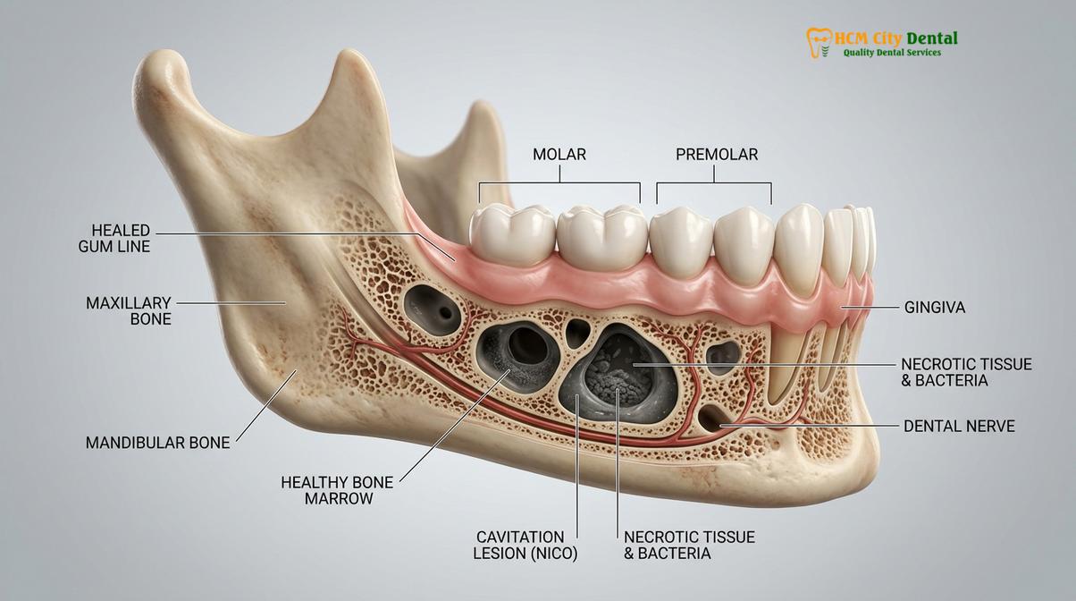
Neuralgia-Inducing Cavitational Osteonecrosis (NICO), commonly referred to as jawbone cavitations, represents a complex localized pathology where ischemic bone death occurs within the medullary spaces of the maxilla or mandible. These lesions frequently develop following routine dental extractions—particularly wisdom teeth—when the periodontal ligament is not fully removed, or when localized ischemia prevents proper angiogenesis and osteogenesis. Because the overlying cortical bone and gingival tissues often heal normally, these necrotic voids remain undetected by standard 2D radiography. Within these anaerobic environments, highly toxic metabolic byproducts and inflammatory chemokines, specifically RANTES/CCL5, accumulate and enter the systemic circulation. This focal infection can trigger profound systemic immune responses and severe, referred facial pain, such as trigeminal neuralgia. The biological dentistry approach to resolving NICO involves advanced 3D Cone Beam Computed Tomography (CBCT) for accurate diagnosis, followed by meticulous surgical debridement using piezoelectric instrumentation. The surgical site is subsequently sterilized with medical-grade ozone and grafted with autologous Platelet-Rich Fibrin (PRF) to ensure robust, healthy bone regeneration and the elimination of systemic toxic burden.
Key Takeaways:
- NICO (Neuralgia-Inducing Cavitational Osteonecrosis) refers to hidden areas of dead, toxic bone marrow in the jaw.
- These lesions most commonly originate from poorly healed tooth extraction sites, especially wisdom teeth.
- Standard 2D dental X-rays miss the vast majority of cavitations; 3D CBCT imaging is clinically required for diagnosis.
- Cavitations produce high levels of inflammatory chemokines (RANTES/CCL5), linking them to systemic autoimmune and fatigue symptoms.
- Effective treatment requires surgical debridement, ozone disinfection, and PRF bone grafting, rather than antibiotics alone.
What Are Jawbone Cavitations (NICO)?
Jawbone cavitations are areas of ischemic, necrotic bone marrow that fail to heal, creating hollow spaces filled with toxic metabolic waste and chronic inflammation.
The term “cavitation” in dentistry does not refer to a cavity in a tooth, but rather a cavity within the bone itself. Medically classified as Neuralgia-Inducing Cavitational Osteonecrosis (NICO), this condition describes a disease process where a lack of adequate blood flow (ischemia) leads to the death of bone cells (osteonecrosis) within the medullary spaces of the jaw. Over time, the dead bone marrow liquefies or turns into a toxic, fatty substance, leaving a hollow void inside the jawbone. Because the outer cortical layer of the bone and the overlying gum tissue typically heal and appear completely normal, these lesions remain entirely hidden from visual inspection.
The concept of jawbone cavitations is not new. In the early 20th century, Dr. G.V. Black, widely considered the father of modern dentistry, documented this phenomenon. He described a progressive disease process that destroyed jawbone tissue without causing the classic signs of infection, such as redness, swelling, or fever. He noted that these areas of dead bone could be easily scooped out during surgery, revealing a hollow space. Today, modern biological dentistry recognizes that a nico jaw bone infection is a significant source of chronic, low-grade systemic inflammation [1].
The Pathophysiology of Ischemic Osteonecrosis
To understand how a cavitation functions, one must look at the microvascular environment of the jawbone. Bone is living tissue that requires a constant supply of oxygen and nutrients delivered via microscopic blood vessels. When an event—such as physical trauma, infection, or the use of potent vasoconstrictors—disrupts this blood supply, the bone cells in that localized area begin to die. This is known as avascular or ischemic osteonecrosis.
Unlike a typical acute infection that triggers a massive immune response (resulting in an abscess, swelling, and acute pain), a cavitation is a chronic, silent process. The immune system walls off the dead tissue, creating a localized pocket of necrosis. Within this pocket, the environment is strictly anaerobic (lacking oxygen). This creates an ideal breeding ground for highly pathogenic anaerobic bacteria, fungi, and even viruses. As these microorganisms thrive in the dead bone, they produce highly toxic metabolic waste products, including thioethers and mercaptans, which are incredibly disruptive to human cellular function.
“NICO lesions represent a unique form of ischemic osteonecrosis where the absence of acute inflammatory signs masks a profound, chronic focal infection capable of altering systemic immune pathways.”
How Cavitations Form (Often from Standard Extractions)
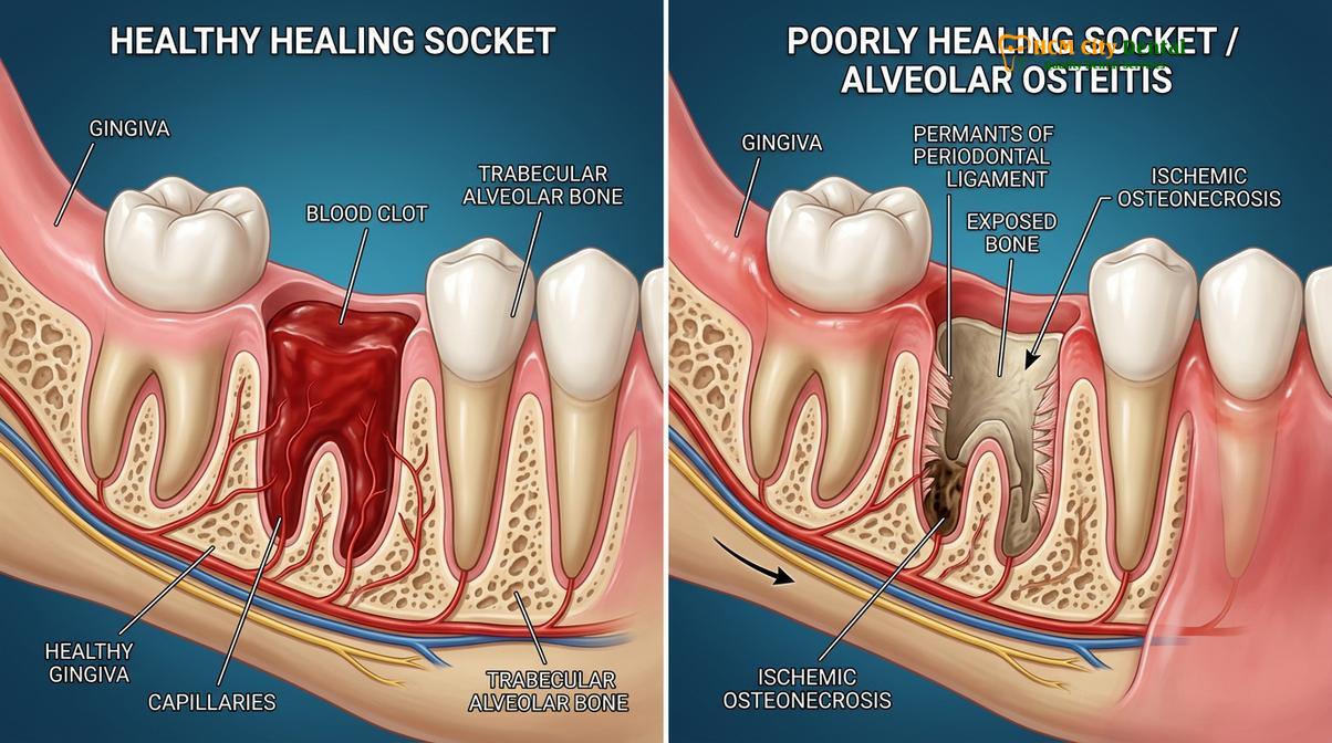
Cavitations typically develop when the periodontal ligament is left behind during a tooth extraction, preventing proper blood flow and bone regeneration in the socket.
The vast majority of jawbone cavitations originate from previous tooth extraction sites, particularly the extraction of third molars (wisdom teeth). When a tooth is removed using standard, conventional techniques, the primary goal is often simply the physical removal of the tooth structure. However, biological dentistry emphasizes that the biological management of the remaining socket is equally, if not more, important for long-term health.
Every tooth is suspended in its bony socket by a fibrous hammock known as the periodontal ligament (PDL). This ligament acts as a shock absorber and contains specialized nerve endings. When a tooth is extracted, if this ligament is not meticulously removed (a process called decortication or curettage), the body’s healing mechanism is severely compromised. The bone essentially “thinks” the tooth is still present because the ligament remains attached to the socket wall. Consequently, the bone does not receive the biological signal to fill the void with new osteoblasts (bone-forming cells) [2].
The Role of Vasoconstrictors and Local Ischemia
Another significant contributing factor to the formation of cavitations is the use of local anesthetics containing high concentrations of epinephrine (adrenaline). Epinephrine is a potent vasoconstrictor used to keep the anesthetic localized and reduce bleeding during surgery. However, in dense bone like the mandible, this severe restriction of blood flow can lead to localized ischemia. If the blood supply does not return promptly and robustly after the extraction, the bone cells lining the socket may die, initiating the cavitation process.
Furthermore, the occurrence of a “dry socket” (alveolar osteitis) significantly increases the risk of future cavitation development. A dry socket occurs when the initial blood clot, which is essential for healing, is dislodged or fails to form. This leaves the bone exposed to the oral environment, leading to localized necrosis and a high likelihood of incomplete bone fill over time. Patients often report jawbone cavitations symptoms years or even decades after a traumatic extraction.
| Clinical Factor | Healthy Bone Regeneration | Cavitation (NICO) Formation |
|---|---|---|
| Periodontal Ligament | Completely removed via surgical curettage | Left intact, preventing osteoblast migration |
| Blood Supply | Robust angiogenesis and clot formation | Ischemic environment; restricted blood flow |
| Bacterial Presence | Sterilized by immune system and blood flow | Anaerobic bacteria thrive in necrotic voids |
| Long-term Outcome | Dense, healthy trabecular bone fill | Hollow, fatty, toxic medullary space |
Systemic Symptoms of Silent Jawbone Osteonecrosis
While often painless locally, jawbone cavitations act as focal infections that release inflammatory cytokines into the bloodstream, triggering systemic autoimmune and neurological symptoms.
One of the most perplexing aspects of NICO lesions is their ability to cause profound systemic illness while remaining completely asymptomatic at the local site in the mouth. A patient may have a large, toxic cavitation in their jaw but feel absolutely no pain when chewing or pressing on the gums. This silent nature makes cavitations one of the most overlooked sources of chronic disease in modern medicine.
The systemic impact of cavitations is primarily driven by the concept of focal infection. The toxic byproducts produced by the anaerobic bacteria within the cavitation do not remain confined to the jaw. Because the jawbone is highly vascularized (despite the localized ischemia within the lesion), these toxins slowly seep into the systemic circulation. The immune system recognizes these toxins and mounts a continuous, low-grade inflammatory response.
The RANTES/CCL5 Chemokine Connection
Recent advancements in immunological research have identified a specific chemokine, known as RANTES (Regulated on Activation, Normal T Cell Expressed and Secreted) or CCL5, as a primary biomarker for jawbone cavitations. RANTES is a pro-inflammatory cytokine that plays a crucial role in the immune system’s response to infection. However, in the presence of a NICO lesion, the necrotic bone tissue produces massive, unregulated amounts of RANTES [3].
Elevated levels of RANTES in the bloodstream have been strongly linked to a variety of systemic conditions, including chronic fatigue syndrome, rheumatoid arthritis, multiple sclerosis, and even certain types of breast cancer. By acting as a constant source of RANTES, a jawbone cavitation keeps the body in a perpetual state of systemic inflammation, exhausting the immune system and exacerbating autoimmune conditions.
Clinical Warning: Jawbone cavitations should not be viewed merely as localized dental issues. The continuous release of RANTES/CCL5 and anaerobic toxins into the bloodstream can severely disrupt systemic immune function, making the resolution of these lesions a critical component of comprehensive biological medical care.
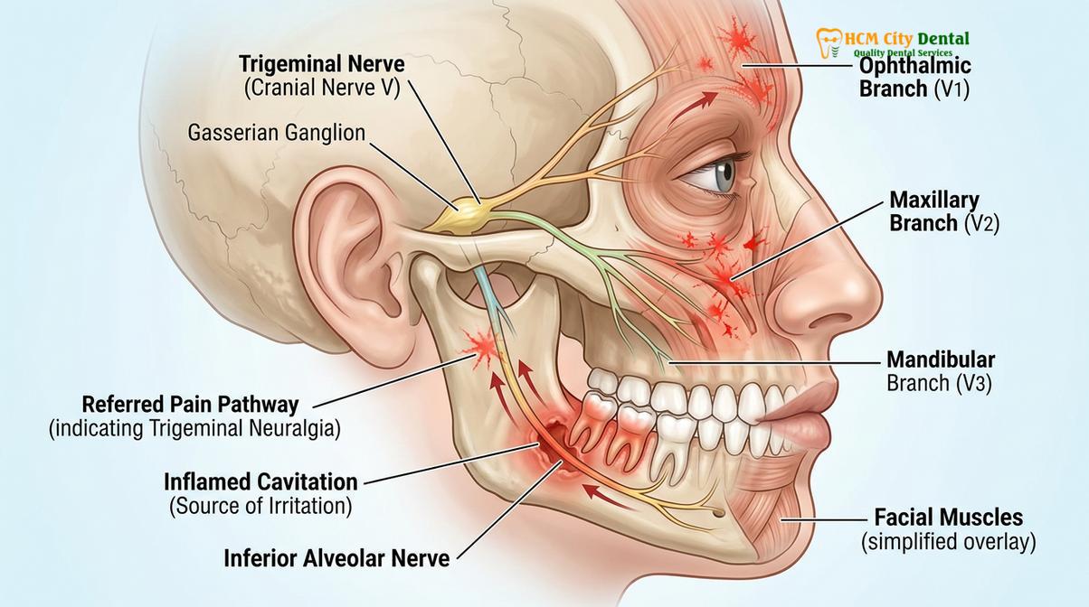
Trigeminal Neuralgia and Referred Pain
While many cavitations are painless, they are also the primary cause of a severe neurological condition known as Neuralgia-Inducing Cavitational Osteonecrosis. The necrotic lesions can irritate or compress the microscopic branches of the trigeminal nerve that run through the jawbone. This irritation can cause excruciating, sharp, shooting pain that radiates across the face, head, and neck. Often, patients suffering from trigeminal neuralgia undergo numerous neurological treatments or even brain surgeries without relief, because the root cause—a hidden jawbone cavitation—remains undiagnosed. Patients seeking relief from neuralgia inducing cavitational osteonecrosis HCMC often find that once the jawbone lesion is surgically cleared, their facial pain resolves completely.
Diagnostic Challenges: 3D CBCT vs. Traditional 2D X-Rays
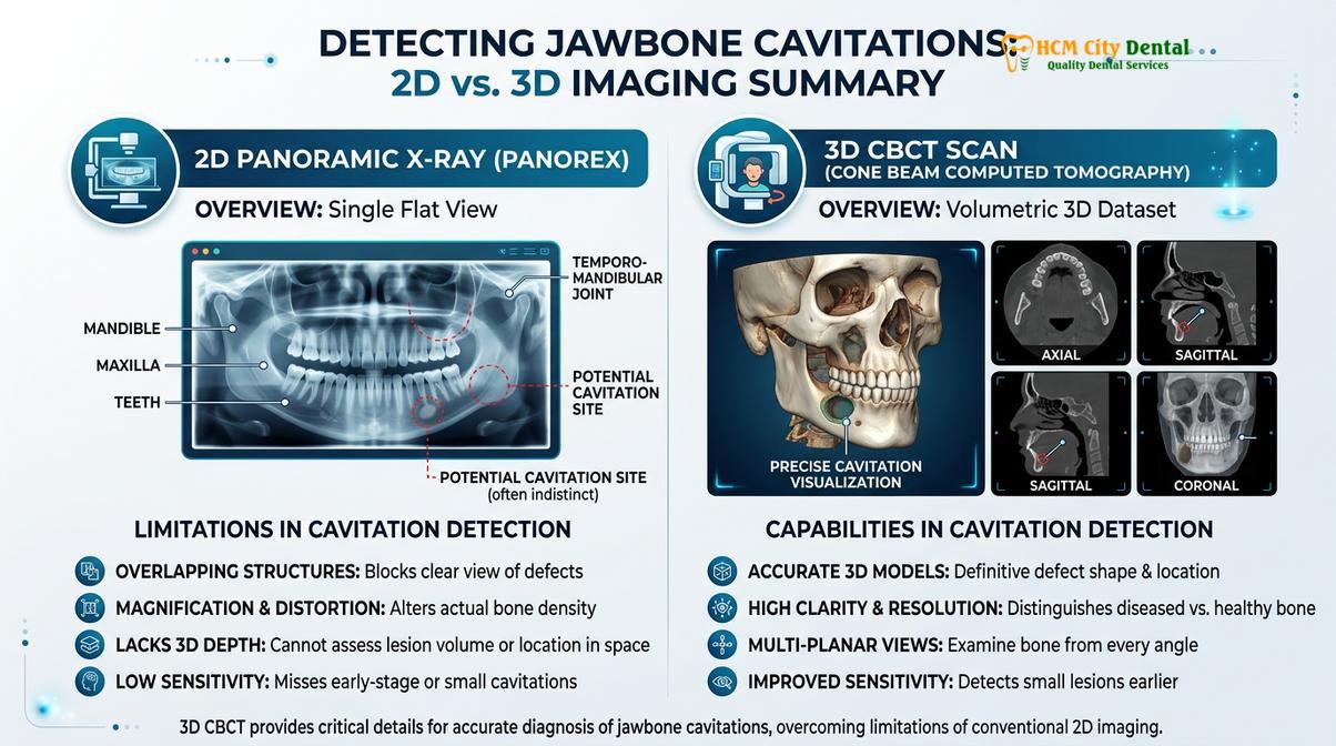
Traditional 2D panoramic X-rays cannot reliably detect cavitations due to overlapping structures; a 3D Cone Beam Computed Tomography (CBCT) scan is the gold standard for diagnosis.
The diagnosis of jawbone cavitations presents a significant challenge in conventional dentistry. The primary diagnostic tool used in most dental offices is the 2D panoramic X-ray. While excellent for detecting gross decay, impacted teeth, and large cysts, 2D radiography is fundamentally inadequate for diagnosing NICO lesions.
A 2D X-ray compresses a three-dimensional structure (the jaw) into a flat image. Because a cavitation is a void within the spongy marrow (cancellous bone) surrounded by dense outer cortical bone, the dense outer bone superimposes over the hollow lesion on a 2D image. Studies indicate that a bone must lose between 30% to 50% of its mineral density before a lesion becomes visible on a standard 2D X-ray. Consequently, up to 80% of jawbone cavitations are entirely missed by conventional panoramic imaging.
Cone Beam Computed Tomography (CBCT) Protocols
To accurately diagnose a cavitation, biological dentists utilize 3D Cone Beam Computed Tomography (CBCT). A CBCT scan captures hundreds of high-resolution images from different angles, allowing the dentist to view the jawbone in three dimensions and slice through the tissue millimeter by millimeter. This technology eliminates the problem of superimposition [4].
When analyzing a CBCT scan for cavitations, the clinician looks for specific radiographic markers. These include a lack of normal trabecular bone pattern, areas of extreme radiolucency (darkness indicating empty space), and thinning or expansion of the cortical plates. Advanced CBCT software can also measure bone density in Hounsfield Units (HU). A healthy jawbone typically registers a specific HU range; a cavitation will show a dramatically lower density, confirming the presence of a necrotic void.
In addition to CBCT, biological dentists may use meridian charting—based on traditional Chinese medicine principles linking specific teeth to specific organ systems—and applied kinesiology to correlate systemic symptoms with potential cavitation sites. This comprehensive diagnostic approach ensures that no hidden infections are overlooked before proceeding with General Dentistry or surgical interventions.
Biological Cavitation Surgery: Cleaning and Disinfecting with Ozone
Biological treatment involves surgically opening the site, meticulously removing necrotic tissue with piezoelectric instruments, and sterilizing the cavity with medical-grade ozone.
Once a cavitation is definitively diagnosed, the only effective treatment is surgical intervention. Antibiotics cannot resolve a NICO lesion because the area lacks a functional blood supply; medications simply cannot reach the dead bone. The goal of biological cavitation surgery is to physically remove the necrotic tissue, sterilize the bony defect, and stimulate the body’s natural regenerative processes to fill the void with healthy bone.
The procedure begins with profound local anesthesia, carefully selected to minimize vasoconstriction and maintain local blood flow. The surgeon creates a small, precise incision in the gum tissue and gently reflects a full-thickness mucoperiosteal flap to expose the underlying bone. A small window is then created in the cortical bone to access the medullary space where the cavitation resides.
Access and Piezosurgical Debridement
In biological dentistry, the removal of the necrotic bone is typically performed using piezosurgery rather than traditional high-speed dental drills. Piezosurgery utilizes ultrasonic micro-vibrations to cut bone with extreme precision. Unlike a drill, which generates significant heat and can damage adjacent healthy tissue, the piezotome cuts only mineralized bone and soft tissue is left completely unharmed. This protects delicate structures like the trigeminal nerve and blood vessels, minimizing surgical trauma and post-operative swelling.
The surgeon meticulously curettes (scrapes) the inside of the cavitation, removing all fatty, necrotic marrow, bacterial biofilms, and any remaining periodontal ligament fibers. The walls of the bony defect are scraped until healthy, bleeding bone is reached. This bleeding is crucial, as it brings fresh blood supply, oxygen, and immune cells back into the previously ischemic area.
Medical Ozone Application
Following thorough physical debridement, the surgical site must be sterilized. Because cavitations harbor highly resistant anaerobic bacteria and toxins, standard saline irrigation is insufficient. Biological dentists utilize medical-grade ozone therapy for this critical step [5].
Ozone (O3) is a highly reactive form of oxygen that possesses profound antimicrobial properties. The surgeon flushes the bony defect with ozonated water to physically wash away debris and instantly neutralize bacteria, fungi, and viruses. Subsequently, ozone gas is infused directly into the bone. The gas penetrates the microscopic dentinal tubules and porous bone structure, destroying pathogens on contact and oxidizing the toxic metabolic waste products. Furthermore, ozone stimulates local blood flow and upregulates the immune system, creating an optimal environment for rapid bone healing.
Restoring Bone Health Post-Surgery at HCMC Dental
Following thorough debridement, the surgical site is grafted with the patient’s own Platelet-Rich Fibrin (PRF) to stimulate rapid, healthy bone regeneration and prevent recurrence.
The final, and arguably most critical, step in cavitation surgery is ensuring that the newly cleaned bony defect heals properly and does not simply form another cavitation. To achieve this, biological dentists utilize advanced regenerative techniques, primarily Platelet-Rich Fibrin (PRF).
Prior to the surgery, a small amount of the patient’s blood is drawn and spun in a specialized centrifuge. This process separates the red blood cells from the plasma, concentrating the patient’s own platelets, white blood cells, and mesenchymal stem cells into a dense, gelatinous fibrin matrix. This PRF clot is entirely autologous (derived from the patient), meaning there is zero risk of rejection or allergic reaction.
Platelet-Rich Fibrin (PRF) Integration
The PRF clot is packed tightly into the cleaned cavitation site before the gum tissue is sutured closed. PRF acts as a biological scaffold, slowly releasing high concentrations of growth factors (such as PDGF, TGF-beta, and VEGF) over a period of 7 to 14 days. These growth factors signal the body to rapidly build new blood vessels (angiogenesis) and stimulate osteoblasts to lay down new, dense bone. By utilizing PRF, the healing time is significantly accelerated, post-operative pain is minimized, and the risk of the cavitation recurring is drastically reduced.
Clinical Case Review: A 45-year-old patient visited HCMC Dental Clinic in Ho Chi Minh City complaining of chronic fatigue and unexplained facial pain on the lower right side. A 3D CBCT scan revealed a large NICO lesion at the site of a wisdom tooth extracted 15 years prior. Dr. Nguyen Van Cuong performed a biological cavitation surgery utilizing piezosurgery, ozone gas disinfection, and PRF grafting. Within four weeks post-surgery, the patient reported a complete cessation of facial pain and a significant improvement in systemic energy levels, demonstrating the profound impact of clearing hidden focal infections.
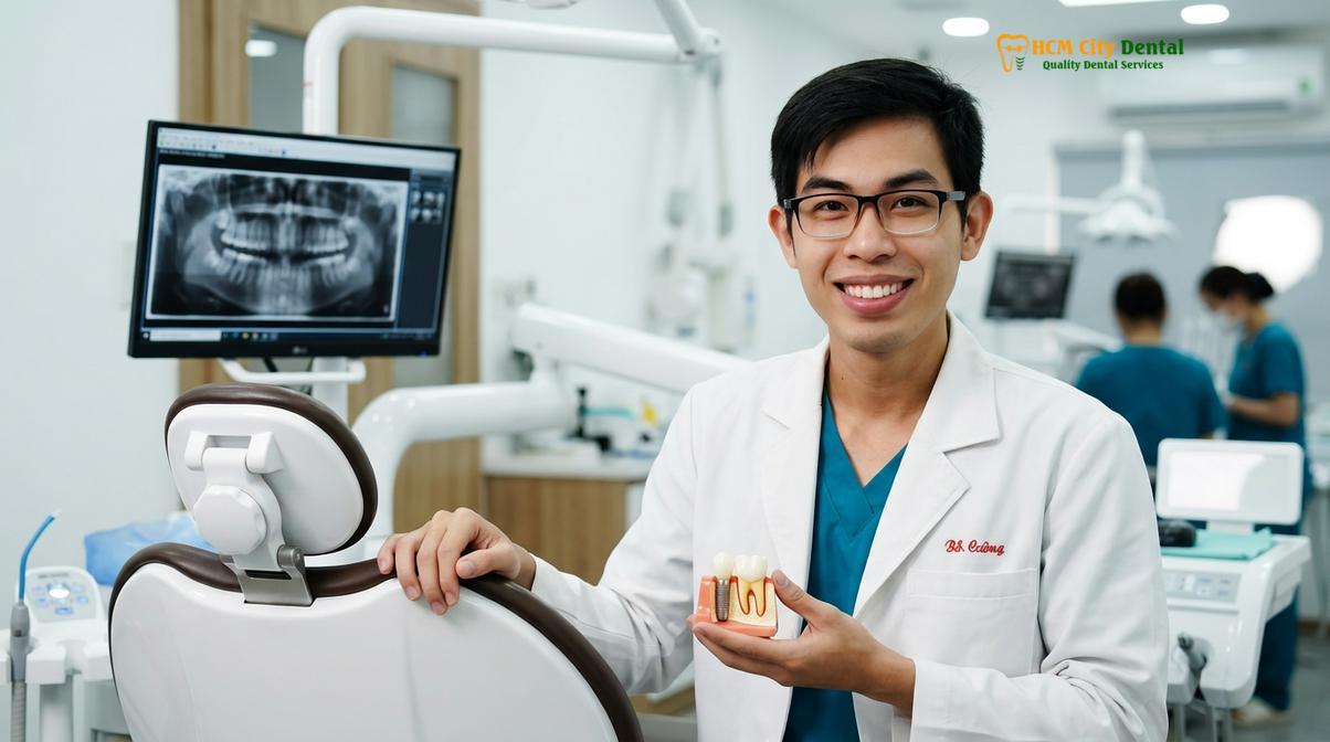
At HCMC Dental Clinic, the surgical protocols are strictly aligned with international biological dentistry standards. Dr. Nguyen Van Cuong emphasizes that the success of cavitation surgery relies not just on the physical removal of dead bone, but on the meticulous preparation of the site to support true biological regeneration. Following the procedure, patients are provided with comprehensive post-operative nutritional support protocols, including high-dose Vitamin C and specific minerals, to further enhance osteogenesis and systemic recovery. For patients requiring further restorative work after bone healing, seamless integration with dental implant planning or other biological dentistry services is carefully coordinated.
When to See a Doctor
Identifying a jawbone cavitation early can prevent years of chronic pain and systemic health degradation. Because these lesions are often silent, proactive screening is essential, particularly for individuals with a history of dental extractions. You should schedule a consultation with a biological dentist if you experience any of the following clinical indicators:
- History of Wisdom Tooth Extractions: Especially if the extractions were traumatic, involved dry sockets, or were performed many years ago without biological protocols.
- Unexplained Facial Pain: Chronic, shooting, or aching pain in the jaw, face, or neck (trigeminal neuralgia) that has not been resolved by standard dental or neurological treatments.
- Systemic Inflammatory Symptoms: Unexplained chronic fatigue, joint pain, or autoimmune flare-ups that seem resistant to conventional medical therapies.
- Phantom Tooth Pain: Experiencing pain in an area where a tooth has already been extracted or where a root canal has been previously performed.
“Patients suffering from idiopathic facial pain or chronic systemic inflammation should always be evaluated with a 3D CBCT scan to rule out hidden jawbone osteonecrosis. The mouth is intimately connected to the body, and silent infections here can dictate systemic health.”
If you suspect you may be suffering from a hidden jawbone infection, it is critical to seek care from a practitioner trained in biological diagnostics. Dr. Cuong and the team at HCMC Dental Clinic in Ho Chi Minh City utilize advanced 3D imaging to uncover these hidden lesions. Early diagnosis and precise surgical intervention can eliminate the toxic burden and restore both oral and systemic vitality. Contact our clinic to schedule a comprehensive biological dental evaluation.
Frequently Asked Questions
What is a jawbone cavitation (NICO)?
A jawbone cavitation, or NICO, is a hollow space of dead, infected bone marrow in the jaw that fails to heal properly. These hidden lesions harbor anaerobic bacteria and toxins, often causing chronic facial pain and contributing to systemic inflammation without showing obvious outward signs of infection. They act as silent focal infections that can continuously drain toxic byproducts into the bloodstream.
What causes a dental cavitation?
Dental cavitations are primarily caused by incomplete healing after a tooth extraction, particularly when the periodontal ligament is left behind. Other contributing factors include restricted blood flow from local anesthetics containing high levels of epinephrine, chronic low-grade infections from failing root canals, and localized trauma that leads to ischemic osteonecrosis in the jawbone.
How is a jawbone cavitation diagnosed?
A jawbone cavitation is definitively diagnosed using a 3D Cone Beam Computed Tomography (CBCT) scan, as traditional 2D X-rays miss up to 80% of these lesions. Biological dentists also rely on clinical symptom history, meridian charting, and sometimes specialized ultrasound technology to confirm the presence of necrotic bone. The CBCT allows the dentist to visualize the exact size and location of the hollow void within the marrow.
How do biological dentists treat cavitations?
Biological dentists treat cavitations through a specialized surgical procedure that involves opening the gum, thoroughly cleaning out the necrotic bone using piezosurgery, and disinfecting the area with medical ozone. The cleaned site is then grafted with the patient’s own Platelet-Rich Fibrin (PRF) to stimulate healthy bone regeneration. This comprehensive approach ensures the infection is eradicated and the bone heals densely.
Is cavitation surgery painful?
Cavitation surgery is performed under profound local anesthesia or conscious sedation, ensuring the procedure itself is virtually painless. Post-operative discomfort is typically mild to moderate and is effectively managed with biological healing protocols, PRF therapy, and standard pain management techniques prescribed by the dental surgeon. Most patients report that the recovery is smoother than the original tooth extraction.
References
- Journal of Oral Pathology & Medicine. Neuralgia-inducing cavitational osteonecrosis (NICO): Osteonecrosis of the jawbones. (2019).
- International Journal of Dentistry. The role of the periodontal ligament in socket healing and cavitation formation. (2020).
- Clinical Oral Investigations. Elevated RANTES/CCL5 levels in jawbone cavitations and systemic implications. (2021).
- Dentomaxillofacial Radiology. Diagnostic efficacy of CBCT versus panoramic radiography in detecting jawbone osteonecrosis. (2022).
- Journal of Cranio-Maxillofacial Surgery. Application of ozone therapy and PRF in the surgical management of jawbone cavitations. (2023).