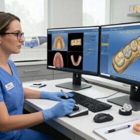
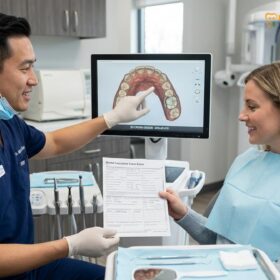
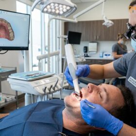
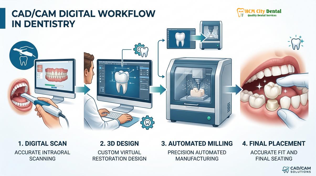
CAD/CAM digital crowns in Vietnam utilize advanced computer-aided design and milling to fabricate highly precise, durable, and aesthetic dental restorations. By replacing traditional silicone impressions with 3D intraoral scanning, patients experience faster turnaround times, superior marginal fit, and enhanced comfort during their restorative treatment.
Clinical Summary:
The integration of CAD/CAM (Computer-Aided Design and Computer-Aided Manufacturing) technology has fundamentally transformed restorative dentistry. This digital workflow encompasses intraoral optical scanning, virtual 3D design, and automated milling of high-strength ceramics such as monolithic zirconia and lithium disilicate. Clinical evidence demonstrates that digitally fabricated restorations exhibit exceptional marginal integrity, reducing the risk of microleakage and secondary caries. Furthermore, the elimination of traditional impression materials significantly improves patient comfort, particularly for those with a strong gag reflex. With direct laboratory partnerships, clinics can offer express turnaround times, making advanced prosthodontic rehabilitation highly accessible and efficient.
Key Takeaways:
- CAD/CAM technology enables the precise fabrication of custom dental crowns and bridges using digital scans and automated milling.
- Intraoral scanners (like iTero and Medit i700) eliminate the need for uncomfortable traditional putty impressions.
- Premium materials, including Zirconia HT and E.max, offer an optimal balance of mechanical strength and lifelike aesthetics.
- Direct laboratory partnerships facilitate express 3-5 day turnaround times for single crowns and full-arch rehabilitations.
- Digital workflows allow for highly accurate virtual bite adjustments and predictable clinical outcomes.
What is CAD/CAM Digital Dentistry?
CAD/CAM dentistry integrates 3D optical scanning, specialized design software, and automated milling machines to create custom dental prosthetics with sub-millimeter accuracy.
The evolution of restorative dentistry has been heavily influenced by the adoption of digital technologies. CAD/CAM systems represent a paradigm shift from manual, analog fabrication techniques to highly controlled, automated digital workflows. This technology allows prosthodontists to design and manufacture dental crowns and bridges with unprecedented precision, ensuring that the final restoration harmonizes perfectly with the patient’s natural dentition and dynamic occlusion.
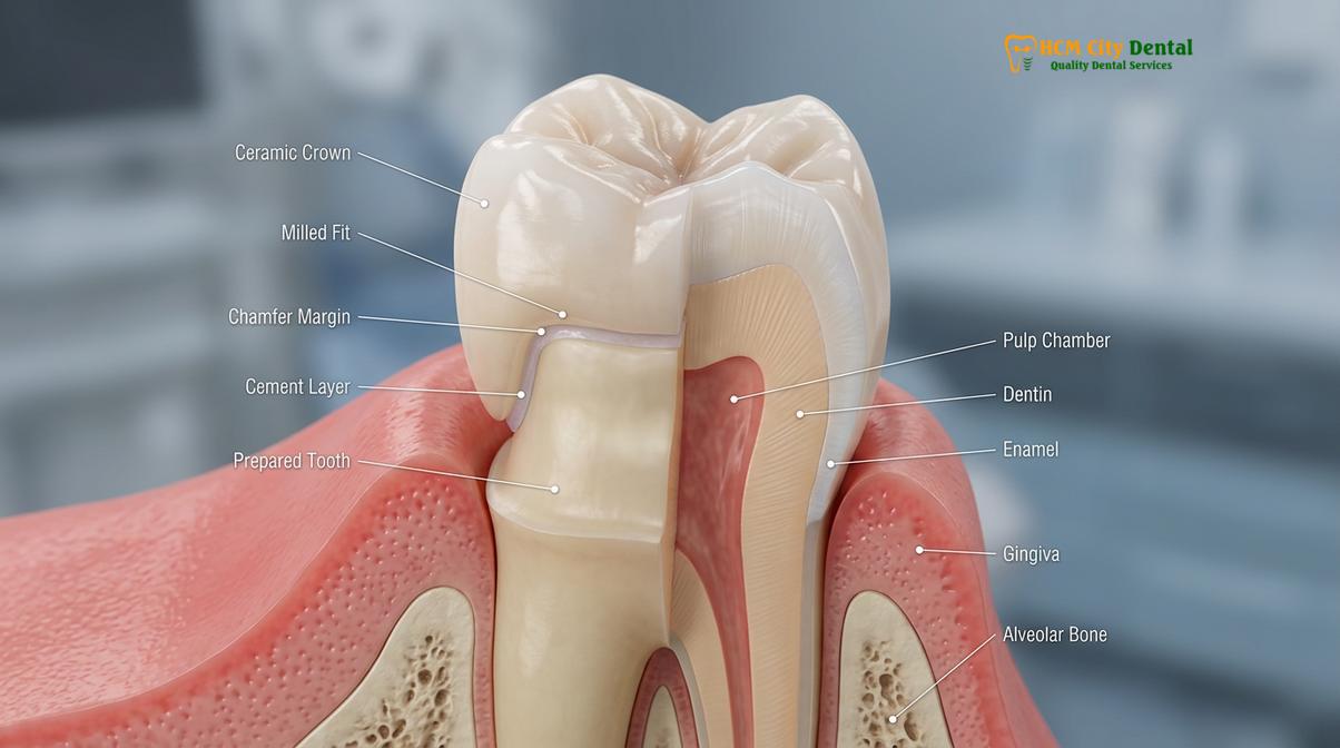
The foundation of a successful CAD/CAM restoration lies in meticulous clinical preparation. The tooth must be carefully reduced to create adequate restorative space while preserving as much healthy tooth structure as possible. The design of the preparation margin—typically a heavy chamfer or a rounded shoulder—is critical. These margin designs provide a distinct finish line that the optical scanner can easily capture and the milling machine can accurately reproduce, ensuring a seamless transition between the ceramic restoration and the natural root surface [1].
Furthermore, clinicians must respect the biologic width—the natural dimension of the gingival tissues attaching to the tooth above the alveolar bone. If a cavity or fracture extends deep below the gumline, a functional crown lengthening procedure may be necessary before scanning. This minor surgical intervention gently recontours the bone and gum tissue to expose more sound tooth structure, ensuring that the margins of the new crown do not impinge on the biologic width, which could otherwise lead to chronic gingival inflammation and bone loss [5].
“The precision of computer aided dental milling relies entirely on the quality of the initial clinical data. A flawless digital scan requires meticulous soft tissue management, absolute moisture control, and a clearly defined preparation margin that respects the periodontal architecture.”
Dr. Nguyen Van Cuong emphasizes that while the technology is highly advanced, the clinical fundamentals of tooth preparation remain paramount. “The software can design a beautiful restoration, but it cannot compensate for inadequate tooth reduction or poorly defined margins. Our goal is always to combine conservative preparation techniques with the precision of digital manufacturing to ensure long-term biomechanical success,” notes Dr. Cuong.
Intraoral Scanning vs. Traditional Goop Molds
Digital intraoral scanners replace uncomfortable putty impressions by capturing thousands of optical images per second to create a flawless 3D model of the dentition.
For decades, taking a dental impression involved filling a bulky tray with a viscous silicone or alginate material and holding it in the patient’s mouth until it set. This process was often uncomfortable, messy, and known to trigger severe gag reflexes in many patients. Moreover, traditional impression materials are susceptible to dimensional changes, tearing, and distortion, which can compromise the fit of the final crown.
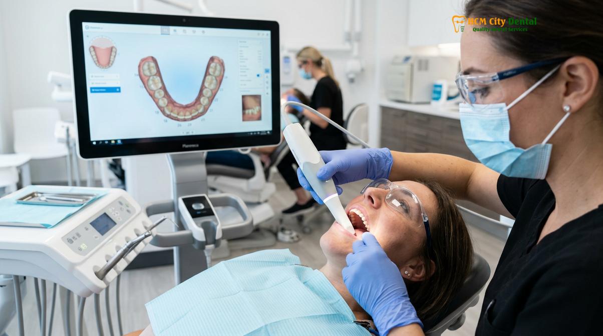
The introduction of advanced optical technology has revolutionized this step. Utilizing state-of-the-art devices like the iTero and Medit i700 Intraoral Scanners, clinicians can now capture the exact geometry of the prepared tooth and surrounding tissues digitally. These scanners use confocal microscopy and optical triangulation to project a light source onto the teeth, capturing thousands of data points per second. The result is a highly accurate, full-color 3D virtual model of the patient’s mouth, generated in real-time on a computer screen.
The benefits of utilizing an intraoral scanner saigon are multifaceted. Primarily, it significantly enhances patient comfort. The scanning wand is small, maneuverable, and requires no unpleasant materials, making the experience virtually stress-free. Clinically, digital impressions offer superior accuracy. The digital file can be magnified and inspected immediately; if a margin is unclear, the clinician can simply rescan that specific area rather than retaking the entire impression [3].
Effective digital scanning requires rigorous soft tissue management. Clinicians often use gingival retraction cords or specialized hemostatic agents to gently push the gum tissue away from the preparation margin and control any minor bleeding. This ensures that the optical sensor has an unobstructed view of the finish line, which is essential for the design software to calculate a precise marginal fit.
3D Milling and Material Precision
Automated milling units carve restorations from solid blocks of certified ceramics like Zirconia and Lithium Disilicate, ensuring exceptional structural integrity and lifelike aesthetics.
Once the digital impression is captured, the data is imported into specialized CAD software. Here, the restoration is virtually designed, taking into account the morphology of adjacent teeth, the opposing bite, and the desired aesthetic outcome. After the design is finalized, the data is sent to a CAM milling machine. This 5-axis CNC (Computer Numerical Control) unit uses diamond-coated burs to carve the crown out of a solid, industrially manufactured block of ceramic material.
The choice of material is a critical clinical decision based on the location of the tooth, the patient’s occlusal forces, and aesthetic requirements. Modern CAD/CAM systems primarily utilize two classes of all-ceramic materials: Zirconia and Lithium Disilicate.
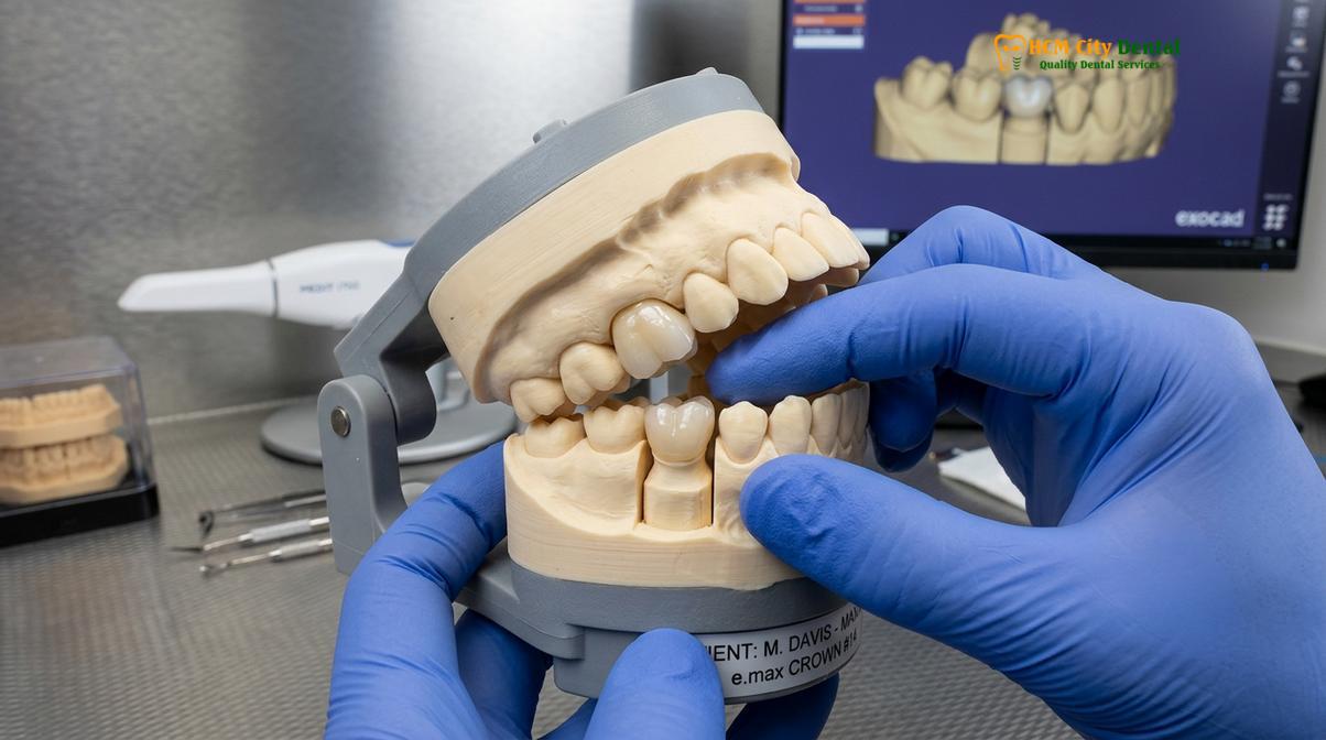
Zirconia HT (Cercon HT, Germany): Zirconia is a polycrystalline ceramic known for its extraordinary mechanical strength and fracture toughness. It undergoes a process called phase transformation toughening, which allows it to resist crack propagation under heavy chewing forces. Zirconia HT (High Translucency) offers an excellent balance of ultimate strength and improved aesthetics, making it ideal for posterior crowns, implant-supported restorations, and multi-unit bridges [1].
E.max (Ivoclar, Liechtenstein): IPS e.max is a lithium disilicate glass-ceramic renowned for its outstanding lifelike aesthetics. It possesses natural opalescence and fluorescence, allowing it to mimic the light-transmitting properties of natural enamel perfectly. While not as strong as zirconia, its flexural strength is more than sufficient for single crowns, particularly on front teeth where cosmetic demands are highest [2].
While 3d printed dental crowns are emerging as a technology for temporary restorations and diagnostic models, subtractive milling remains the gold standard for final, definitive ceramic restorations due to the superior density and structural integrity of the pre-manufactured ceramic blocks.
| Material Type | Primary Indication | Flexural Strength | Aesthetic Quality | Clinical Notes |
|---|---|---|---|---|
| Zirconia HT (Cercon HT) | Posterior crowns, Bridges, Implants | Very High (900-1200 MPa) | Good to Excellent | Requires high-temperature sintering; excellent for bruxism patients. |
| Lithium Disilicate (E.max) | Anterior crowns, Veneers, Premolars | High (400-500 MPa) | Superior (Lifelike) | Requires crystallization firing; bonds exceptionally well to tooth structure. |
| Porcelain-Fused-to-Metal (PFM) | Budget restorations, specific bridge designs | Moderate to High | Acceptable | Traditional option; may show a dark line at the gum margin over time. |
In cases where a tooth has undergone root canal therapy and lost significant structural volume, a post and core buildup is often required before scanning. A fiber post is cemented into the root canal, and composite resin is used to rebuild the core of the tooth, providing a stable foundation for the CAD/CAM crown. For dental implants, the digital workflow allows for the precise design of either screw-retained or cement-retained implant crowns, optimizing the emergence profile through the gingival tissues.
Same-Day and Express Turnaround Times
Through direct partnerships with in-house laboratories, digital workflows enable express fabrication of single crowns or full-arch bridges within three to five days.
One of the most significant advantages of CAD/CAM technology is the dramatic reduction in treatment time. Traditional crown fabrication often requires the patient to wear a fragile temporary crown for two to three weeks while the physical impression is shipped to an external laboratory. With digital dentistry, the data is transmitted instantly.
At HCMC Dental Clinic in Ho Chi Minh City, a Direct Lab Partnership facilitates an Express 3-5 Day Turnaround. This streamlined process is particularly tailored for dental tourists and expatriates, allowing complete smile makeovers or the fabrication of multiple crowns and 3-unit / 4-unit bridges to be completed within a single week. The direct collaboration between the prosthodontist and the master ceramist ensures complete quality control, allowing for immediate adjustments to shade matching and contouring.
The pricing structure for these premium restorations is highly competitive, especially when utilizing digital communication channels for pre-arrival planning. The clinic offers a transparent fee schedule, incorporating a 40% pre-arrival discount for WhatsApp bookings, making world-class dentistry highly accessible:
- PFM Crown: Walk-in: ~$140 (3.5M VND) | WhatsApp pre-booking discount: From $84 (2.1M VND) (-40%).
- Zirconia Crown (Cercon): Walk-in: ~$200 (5.0M VND) | WhatsApp pre-booking discount: From $120 (3.0M VND) (-40%).
- Zirconia HT Crown (Cercon HT): Walk-in: ~$260 (6.5M VND) | WhatsApp pre-booking discount: From $156 (3.9M VND) (-40%).
- E.max Crown (Ivoclar): Walk-in: ~$280 (7.0M VND) | WhatsApp pre-booking discount: From $168 (4.2M VND) (-40%).
- 3-Unit Bridge (Zirconia HT): Walk-in: ~$480 (12.0M VND) | WhatsApp pre-booking discount: From $288 (7.2M VND) (-40%).
- 4-Unit Bridge (Zirconia HT): Walk-in: ~$640 (16.0M VND) | WhatsApp pre-booking discount: From $384 (9.6M VND) (-40%).
- Inlay / Onlay (Sứ Lab): Walk-in: ~$180 (4.5M VND) | WhatsApp pre-booking discount: From $108 (2.7M VND) (-40%).
Clinical Case Example: Express Full-Arch Rehabilitation
A 55-year-old international patient presented to HCMC Dental Clinic with severe occlusal wear and multiple failing amalgam fillings. Utilizing the Medit i700 scanner, a complete digital impression was captured. The clinical team designed a comprehensive restorative plan involving eight Zirconia HT crowns on the posterior teeth for maximum durability and four E.max crowns on the anterior incisors for optimal aesthetics. Thanks to the direct lab partnership, the entire fabrication process was completed, and the final restorations were permanently cemented within a 5-day window, restoring both function and a youthful appearance.
Improving Patient Comfort and Accuracy
Digital workflows minimize chair time, eliminate gag reflexes, and allow for precise virtual bite adjustments before the final restoration is even milled.
The integration of digital smile design vietnam protocols allows patients to actively participate in their restorative journey. Before any teeth are prepared, the clinician can use digital software to simulate the proposed outcome, overlaying the new tooth shapes onto photographs of the patient’s face. This visual communication ensures that the final crowns will meet the patient’s aesthetic expectations regarding tooth length, shape, and shade matching.
Accuracy is further enhanced during the design phase through virtual articulation. The software analyzes the patient’s dynamic bite, calculating the exact contact points between the upper and lower teeth. This allows the technician to perform a virtual bite adjustment, ensuring that the milled crown requires minimal to no manual grinding during the final cementation appointment. This precision reduces chair time and prevents the introduction of micro-fractures into the ceramic surface that can occur with excessive manual adjustment [4].
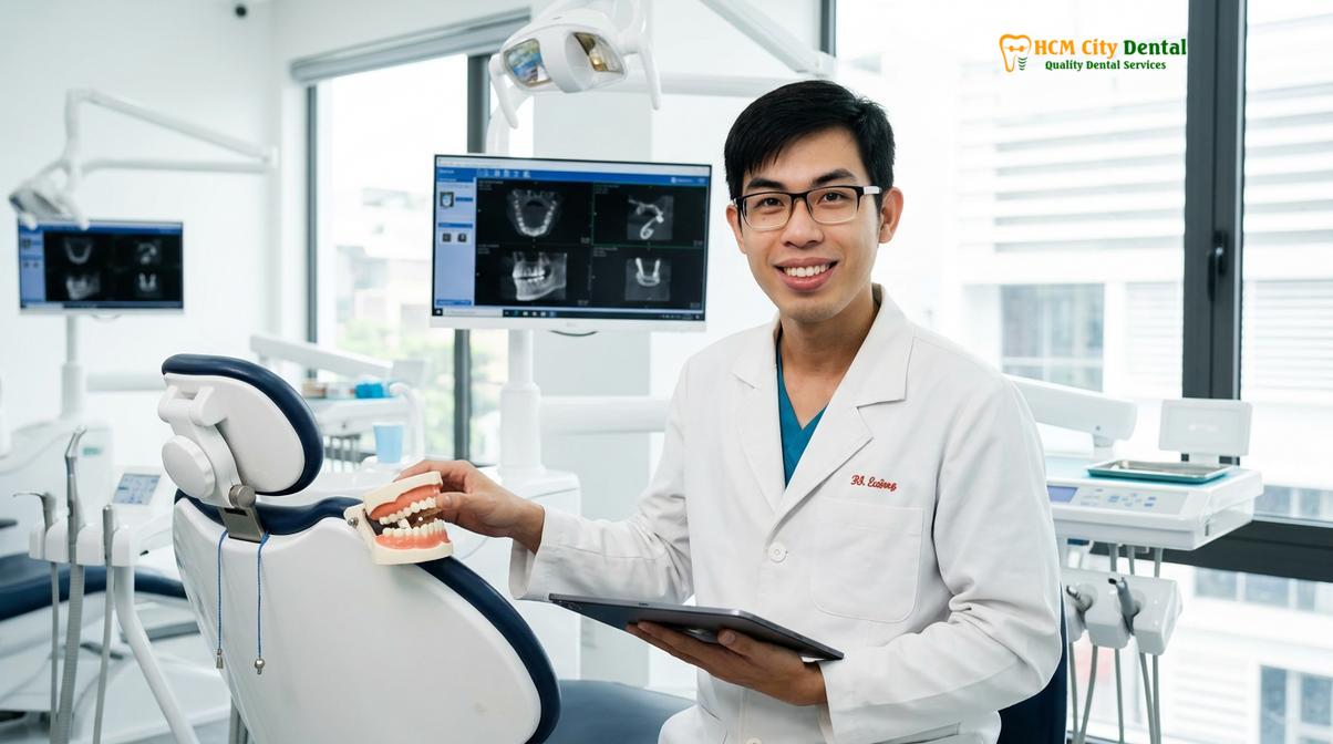
Dr. Cuong highlights the importance of comprehensive aftercare in the digital era. “We utilize only authentic, certified materials from Germany and Liechtenstein, which allows us to provide a robust global warranty policy. Even after our international patients return home, we maintain remote follow-up support via WhatsApp, utilizing photo and video assessment protocols to monitor tissue healing and occlusal stability,” explains Dr. Cuong. To further assist patients, the clinic offers flexible financials, including 0% interest monthly credit card installment plans.
Clinical Advisory: Emergency Loose Crown Management
If a temporary crown or an older permanent crown becomes loose or dislodged, it is considered a dental emergency. Do not attempt to reattach the crown with household glues, as this can cause severe chemical burns to the pulp and permanently damage the tooth structure. Keep the crown safe and seek professional emergency loose crown recementation or replacement immediately to prevent the adjacent teeth from shifting and to protect the exposed dentin from bacterial contamination.
“The true value of CAD/CAM technology is not just in the speed of fabrication, but in the predictable, reproducible accuracy it brings to complex prosthodontic rehabilitations, ensuring long-term biomechanical stability.”
When to See a Doctor
While CAD/CAM crowns are highly durable, regular clinical monitoring is essential. You should schedule a professional dental examination if you experience any of the following symptoms:
- Persistent Pain or Sensitivity: Sharp pain when biting down or lingering sensitivity to hot or cold temperatures may indicate underlying pulpal inflammation or a compromised cement margin.
- Gingival Inflammation: Localized redness, swelling, or bleeding around the margin of a crown can be a sign of plaque accumulation, biologic width violation, or an open margin requiring assessment.
- Structural Damage: Any visible chipping, cracking, or feeling of roughness on the ceramic surface should be evaluated to prevent further fracture propagation.
- Occlusal Discomfort: If a crown feels “high” or interferes with your natural bite, it requires professional occlusal adjustment to prevent trauma to the underlying periodontal ligament or the opposing teeth.
A comprehensive clinical evaluation, often involving digital radiographs and an occlusal analysis, is necessary to determine the appropriate individualized treatment plan.
Frequently Asked Questions
What does CAD/CAM stand for in dentistry?
CAD/CAM stands for Computer-Aided Design and Computer-Aided Manufacturing. In dentistry, this technology allows clinicians to digitally scan a prepared tooth, design a custom restoration on a computer, and mill the final crown from a solid block of ceramic with sub-millimeter precision. This digital workflow replaces many of the manual steps involved in traditional dental laboratory processes.
Are digital dental impressions more accurate than traditional molds?
Yes, digital impressions are highly accurate and eliminate the dimensional distortion associated with traditional impression materials. Intraoral scanners capture thousands of data points per second, creating a flawless 3D model that ensures a superior marginal fit for the final restoration. They also eliminate the risk of air bubbles or tears that can ruin a physical silicone impression.
How long does a CAD/CAM crown take to make?
With an integrated digital workflow and direct lab partnership, a CAD/CAM crown can typically be fabricated in three to five days. This express turnaround is highly beneficial for international patients seeking comprehensive restorative care within a limited timeframe, allowing for complete treatment without the need for multiple extended visits.
Is the tooth preparation process different for digital crowns?
The fundamental principles of tooth preparation remain the same, requiring precise chamfer or shoulder margins. However, digital scanning requires excellent soft tissue management and moisture control to ensure the optical sensors can clearly capture the preparation margins without interference from saliva, blood, or overlapping gingival tissue.
Can CAD/CAM technology be used for dental bridges?
Absolutely. CAD/CAM technology is routinely used to design and mill multi-unit restorations, including 3-unit and 4-unit bridges. The software calculates the optimal pontic design and connector dimensions to ensure the bridge can withstand occlusal forces while maintaining aesthetic harmony with the surrounding natural dentition.
References
- Journal of Prosthodontic Research. Flexural strength and phase transformation of monolithic zirconia crowns. (2021).
- Journal of Esthetic and Restorative Dentistry. Optical properties and translucency of lithium disilicate glass-ceramics. (2020).
- International Journal of Prosthodontics. Marginal adaptation of CAD/CAM milled restorations versus traditional casting. (2022).
- American Dental Association. Clinical guidelines for the fabrication and cementation of all-ceramic dental bridges. (2019).
- Journal of Clinical Periodontology. Biologic width considerations in restorative dentistry and crown lengthening. (2018).