Achieving a seamless crown shade matching natural teeth requires evaluating hue, chroma, value, and translucency. Prosthodontists utilize standardized shade guides, digital spectrophotometers, and custom laboratory staining to replicate the exact optical properties of surrounding enamel, ensuring the restoration blends flawlessly into your smile.
Clinical Summary:
The process of matching a dental crown to natural dentition is a complex intersection of material science, optical physics, and clinical artistry. A successful restoration must mimic the dynamic way natural enamel interacts with light, which involves precise management of brightness (value), color saturation (chroma), and the basic color family (hue). Modern prosthodontics has evolved beyond subjective visual matching; today, specialists employ advanced digital workflows, including intraoral scanners and spectrophotometry, to capture objective color data. Dr. Nguyen Van Cuong emphasizes that the choice of restorative material—whether highly translucent lithium disilicate (E.max) for anterior aesthetics or high-strength Zirconia HT for posterior durability—dictates how the crown will transmit and reflect light. Furthermore, the underlying tooth structure, gingival health, and the specific tooth preparation margin designs (such as chamfer or shoulder) profoundly impact the final aesthetic outcome. By partnering with master ceramists for custom staining and utilizing certified materials, clinicians can deliver lifelike, durable restorations that integrate seamlessly with the patient’s natural smile architecture.
Key Takeaways:
- Tooth color is multidimensional, requiring precise matching of hue, chroma, value, and translucency.
- Digital intraoral scanners eliminate subjective visual errors by providing objective, light-corrected color mapping.
- The underlying tooth structure (e.g., dark root canal stumps) dictates whether an opaque or translucent ceramic material is required.
- Custom laboratory staining adds lifelike characterizations such as incisal halos, mamelons, and subtle surface textures.
- Teeth whitening must always be completed and stabilized before the final crown shade is selected.
The VITA Shade Guide System
The VITA shade guide is the universal clinical standard for categorizing tooth color into specific hues and brightness levels, providing a baseline for ceramists to formulate the foundational ceramic layers.
For decades, the cornerstone of aesthetic dentistry has been the standardized shade guide. When a patient requires a restorative procedure, the clinician’s first step in achieving a natural-looking result is to determine the baseline color of the adjacent dentition. The most widely recognized tool for this purpose is the classic vita shade guide dental system, which categorizes natural tooth colors into four distinct hue families: A (reddish-brown), B (reddish-yellow), C (grayish shades), and D (reddish-gray) [1]. Within each of these families, the shades are further delineated by chroma (the intensity or saturation of the color) and value (the relative lightness or darkness).
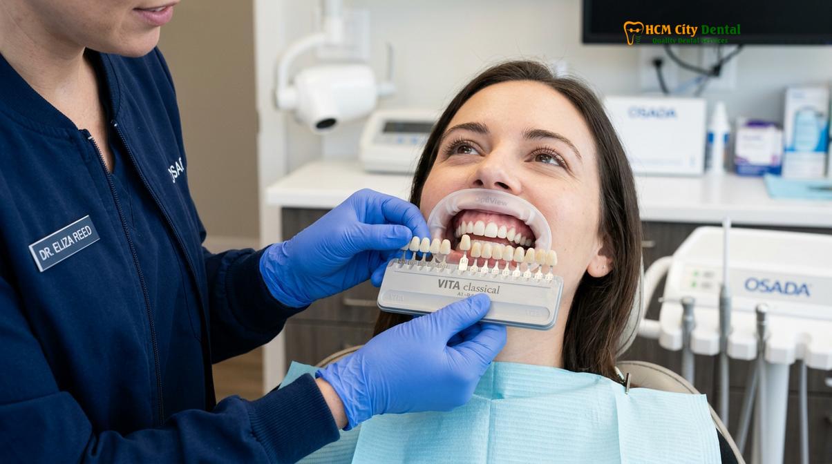
While the classic system remains prevalent, advanced cosmetic practices often utilize the 3D-Master system, which prioritizes value over hue. Human eyes are significantly more sensitive to changes in brightness than to subtle shifts in color. If a crown’s value is too high (too bright), it will immediately appear artificial and “pop” out of the smile line, regardless of how perfectly the hue was matched. Therefore, clinicians meticulously evaluate the value first, often squinting to reduce color distraction and focus solely on the black-and-white brightness scale of the teeth.
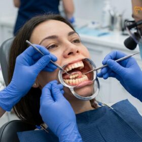
The timing of shade selection is a critical clinical parameter. Teeth naturally dehydrate when a patient’s mouth remains open during dental procedures. As enamel loses moisture, it temporarily becomes more opaque and significantly lighter in value. To prevent a mismatch, Dr. Nguyen Van Cuong advocates for capturing the shade at the very beginning of the appointment, before any isolation techniques or tooth preparation begins. Furthermore, the ambient lighting in the operatory must be carefully controlled. Standard fluorescent clinic lights can distort color perception, a phenomenon known as metamerism. To counteract this, shade matching is ideally performed under color-corrected 5500K lighting, which mimics natural midday sunlight, ensuring the crown will look natural in various everyday environments.
“The true artistry in prosthodontics lies not just in selecting a color from a tab, but in understanding how that color will behave under different lighting conditions and how it will interact with the patient’s unique soft tissue architecture.”
Digital Shade Selection with Intraoral Cameras
Modern intraoral scanners capture precise digital color maps of the dentition, eliminating subjective visual errors and providing the laboratory with objective light-reflection data.
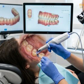
The transition from analog to digital dentistry has revolutionized the predictability of aesthetic restorations. Historically, shade matching relied heavily on the subjective visual acuity of the dentist, which could be influenced by eye fatigue, ambient lighting, and even the color of the patient’s clothing. Today, state-of-the-art digital scanning technology, such as the iTero + Medit i700 Intraoral Scanners, has largely replaced messy traditional silicone impressions while simultaneously capturing objective color data [2].
These advanced scanners utilize confocal microscopy and structured light to create highly accurate 3D models of the prepared tooth and surrounding dentition. Beyond capturing the physical geometry required for CAD/CAM milling, these devices feature integrated spectrophotometric capabilities. As the wand passes over the teeth, it measures the exact wavelengths of light reflected from the enamel surface, generating a comprehensive digital color map. This map details the varying shades across the cervical (gumline), middle, and incisal (biting edge) thirds of the tooth.
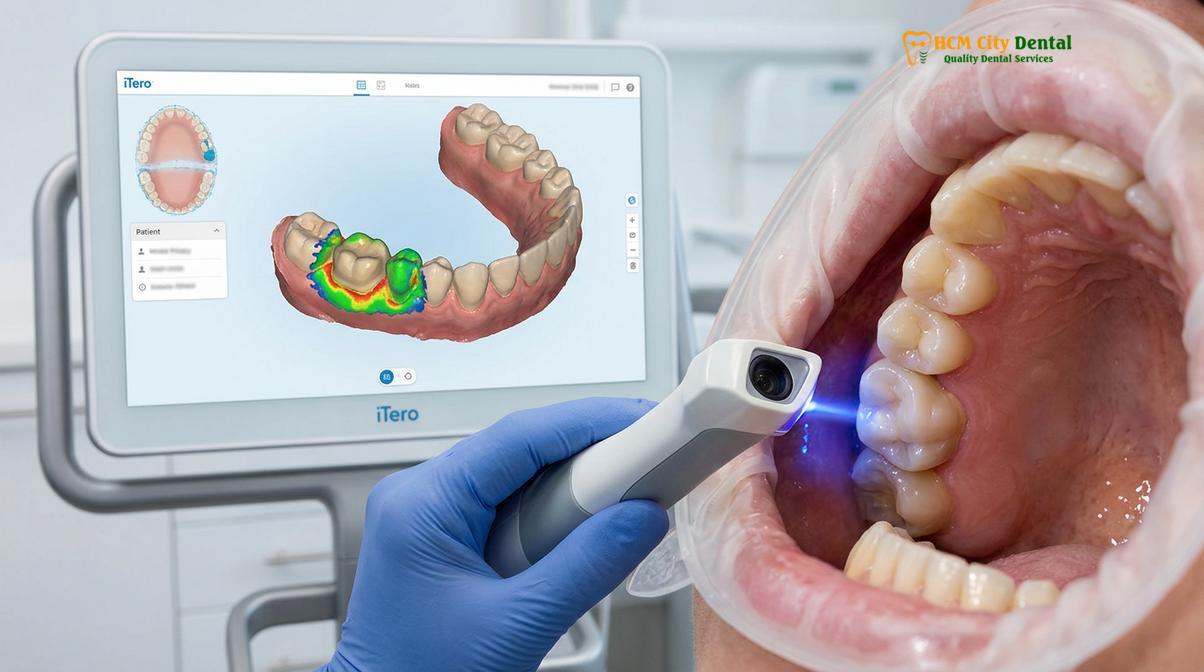
The integration of digital shade selection is particularly beneficial for complex cases involving multiple restorations or when treating patients with highly characterized natural teeth. The digital file is instantly transmitted to the dental laboratory via a secure cloud network. This direct communication eliminates the risk of information loss that can occur with handwritten lab prescriptions. When combined with high-resolution macro photography—where the selected shade tab is photographed edge-to-edge with the natural tooth—the ceramist receives a wealth of objective data. This comprehensive digital workflow ensures maximum comfort for the patient while providing the 3D precision necessary for flawless crown design and shade integration.
Factors Affecting Crown Color (Lighting, Gum Translucency, Adjacent Teeth)
A crown’s final appearance is heavily influenced by ambient lighting, the underlying tooth structure, and the optical properties of neighboring teeth, making holistic aesthetic evaluation essential.
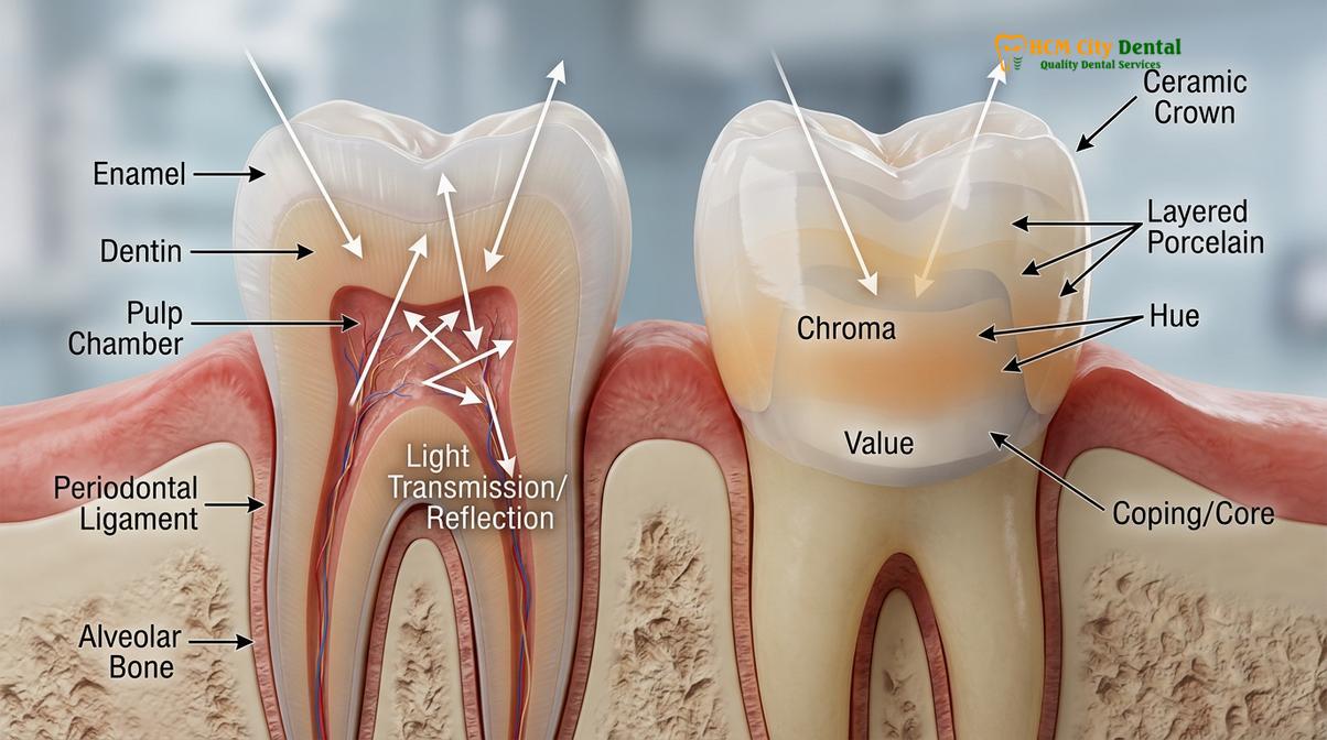
Achieving a perfect match extends far beyond the surface of the crown itself. The final perceived color of a dental restoration is a complex interplay between the ceramic material, the underlying tooth structure, the surrounding gingival tissues, and the adjacent dentition. One of the most challenging aspects of aesthetic dentistry is managing translucency matching crown properties. Natural enamel is not a solid, opaque substance; it is highly translucent, allowing light to penetrate, scatter within the dentin, and reflect back out. If a crown is fabricated from a densely opaque material, it will look lifeless and flat, completely lacking the depth and vitality of a natural tooth.
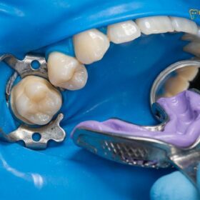
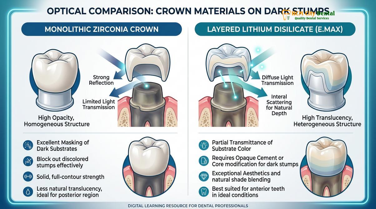
The condition of the underlying tooth preparation is a critical factor. When a tooth has undergone a root canal treatment, it often darkens over time due to the breakdown of blood byproducts or the presence of certain endodontic sealers. Similarly, teeth rebuilt with large metal amalgam cores or dark composite resins present a significant aesthetic hurdle. If a highly translucent material like lithium disilicate is placed over a dark stump, the underlying discoloration will show through, resulting in a grayish, mismatched crown [3]. In these scenarios, the clinician must utilize a more opaque substructure, such as a zirconia core, to block out the darkness before layering translucent porcelain on top.
Clinical Warning: Teeth Whitening and Dental Crowns
Dental ceramics, including zirconia and porcelain, do not respond to bleaching agents. If you are planning to whiten your teeth, you must complete the whitening process and wait at least 10 to 14 days for the new shade to stabilize before your dentist selects the color for your new crown. Failing to do so will result in a crown that appears noticeably darker than your newly whitened natural teeth.
Furthermore, the health and biotype of the gingival tissue play a vital role in the overall aesthetic frame. The concept of biologic width must be strictly respected during tooth preparation. If the crown margin invades this delicate attachment zone, it will trigger chronic inflammation, leading to red, swollen, or purplish gums that severely detract from the beauty of the restoration. In cases where the gum line is uneven or a “gummy smile” is present, functional and esthetic crown lengthening may be required prior to crown placement to establish a harmonious gingival zenith. The color of the gums themselves also affects perception; healthy, stippled pink tissue provides a contrasting background that enhances the brightness and vitality of the ceramic.
Why Custom Staining in the Lab Matters
Custom staining allows master ceramists to add lifelike characterizations, such as incisal halos and subtle craze lines, transforming a monochromatic block into a dynamic, natural-looking tooth.
While CAD/CAM milling technology can rapidly produce a crown with perfect physical dimensions, a freshly milled ceramic block is inherently monochromatic. It lacks the subtle nuances, gradients, and characterizations that make a tooth look real. This is where the artistry of custom staining ceramic becomes indispensable. A Direct Lab Partnership, such as the one maintained by leading clinics in Ho Chi Minh City, enables complete quality control and direct collaboration between the prosthodontist and the master ceramist.
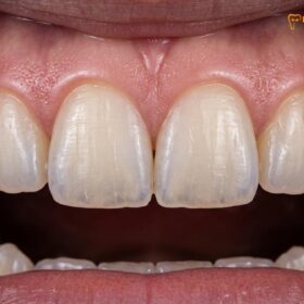
During the custom staining process, the ceramist acts as a micro-painter, using specialized ceramic stains and glazes to replicate the unique features of the patient’s adjacent teeth. Natural teeth are rarely uniform in color. The cervical third near the gumline is typically warmer and more saturated (higher chroma) because the enamel is thinner, allowing the yellowish dentin to show through. The middle third represents the true base shade, while the incisal third is often highly translucent, sometimes displaying a bluish or grayish halo where there is only enamel and no underlying dentin.
Clinical Case Review: Anterior Aesthetic Rehabilitation
A 34-year-old patient visited HCMC Dental Clinic seeking to replace a discolored, decades-old PFM crown on a central incisor. The old crown lacked translucency and exhibited a dark metal margin at the gumline. Utilizing the Medit i700 scanner and high-resolution photography, the clinical team captured the exact surface texture and internal mamelon structure of the adjacent natural incisor. The laboratory fabricated a custom-layered E.max crown. Through meticulous custom staining, the ceramist replicated the subtle white fluorosis spots and the precise incisal translucency of the neighboring tooth, resulting in a virtually seamless integration.
Beyond color gradients, custom staining allows for the replication of specific characterizations. As teeth age, they develop micro-cracks (craze lines), subtle wear facets, and variations in surface texture. A perfectly smooth, high-gloss crown will look artificial next to natural teeth that possess a slightly matte, textured surface. By carefully applying stains and controlling the final glazing temperature, the ceramist can mimic these micro-textures, ensuring the way light reflects off the crown perfectly matches the surrounding dentition. This level of customization is what elevates a standard dental crown to a premium aesthetic restoration.
Cross-Matching Shades Across Multiple Crowns
Restoring multiple teeth requires harmonizing the shade across different materials and abutments, ensuring a uniform smile line, especially when mixing implants with natural tooth restorations.
The complexity of shade matching increases exponentially when a patient requires multiple restorations, particularly when those restorations span different types of underlying structures. A common clinical challenge is color matching porcelain across a combination of natural teeth, root canal-treated teeth, and dental implants within the same smile line. Each of these foundations interacts with light differently, and achieving a cohesive final appearance requires meticulous material selection and advanced laboratory techniques [4].
For example, consider a patient receiving a 4-unit bridge alongside a single implant crown. The natural abutment teeth supporting the bridge may have a different stump shade than the titanium or zirconia abutment supporting the implant. If the same translucent ceramic is used for all restorations without modifying the substructure, the implant crown may appear grayish compared to the bridge. To resolve this, clinicians must carefully manage the opacity of the framework. For implant restorations, the choice between screw-retained vs. cement-retained implant crowns also impacts aesthetics. Screw-retained crowns require an access hole that must be sealed with composite resin, which must be perfectly color-matched to the surrounding ceramic to remain invisible.
The design of the tooth preparation margin is equally critical when cross-matching shades. A heavy chamfer or a modified shoulder preparation is required to provide the ceramist with adequate physical space (bulk) to layer the porcelain. If the preparation is too conservative, the ceramic will be too thin, resulting in an opaque, lifeless appearance as the lab attempts to mask the underlying tooth within a restricted dimension. By ensuring uniform preparation depths across all teeth involved in the treatment plan, the clinician provides the laboratory with the ideal canvas to create a harmonious, multi-unit restoration that exhibits consistent value, chroma, and translucency across the entire arch.
Clinical Workflows, Materials, and Cost Considerations
Selecting the right material balances esthetics, strength, and budget, with advanced clinics offering express turnarounds and transparent pricing for premium certified restorations.
The selection of the restorative material is the foundation of a successful aesthetic outcome. Modern prosthodontics relies on authentic, certified materials to ensure both biocompatibility and long-term optical stability. For anterior (front) teeth, where aesthetic demands are highest, lithium disilicate (E.max by Ivoclar, Liechtenstein) is often the material of choice due to its outstanding lifelike translucency and ability to bond adhesively to the tooth structure. For posterior (back) teeth, or for patients with heavy bite forces (bruxism), Zirconia HT (Cercon HT, Germany) provides ultimate flexural strength while still offering high translucency compared to older, opaque zirconia generations [5].
For international patients and expats, dental tourism has made premium aesthetic dentistry highly accessible. Clinics offering an Express 3-5 Day Turnaround allow patients to undergo complete smile makeovers or receive multiple crowns and bridges within a single week. This rapid workflow is facilitated by in-house digital design and direct laboratory partnerships, ensuring that the temporary crown phase is minimized and the final bite adjustment and cementation are completed efficiently.
Understanding the financial investment is crucial for patients planning restorative care. According to the latest clinic fee schedules, transparent pricing structures often include significant incentives for advanced booking. For example, the pricing parameters (incorporating a 40% pre-arrival discount for WhatsApp bookings) are structured as follows:
| Restoration Type | Standard Walk-in Price | WhatsApp Pre-booking (-40%) |
|---|---|---|
| PFM Crown | ~$140 (3.5M VND) | From $84 (2.1M VND) |
| Zirconia Crown (Cercon) | ~$200 (5.0M VND) | From $120 (3.0M VND) |
| Zirconia HT Crown (Cercon HT) | ~$260 (6.5M VND) | From $156 (3.9M VND) |
| E.max Crown (Ivoclar) | ~$280 (7.0M VND) | From $168 (4.2M VND) |
| 3-Unit Bridge (Zirconia HT) | ~$480 (12.0M VND) | From $288 (7.2M VND) |
| 4-Unit Bridge (Zirconia HT) | ~$640 (16.0M VND) | From $384 (9.6M VND) |
| Inlay / Onlay (Sứ Lab) | ~$180 (4.5M VND) | From $108 (2.7M VND) |
Beyond the initial cost, long-term peace of mind is a vital component of patient care. Reputable institutions provide a global warranty policy, ensuring that the structural integrity of the certified materials is guaranteed. Furthermore, remote follow-up support via WhatsApp (utilizing specific photo and video assessment protocols) allows clinicians to monitor gingival healing and aesthetic integration even after the patient has returned home. For local residents, flexible financials, including 0% interest monthly credit card installment plans, make comprehensive aesthetic rehabilitations highly attainable. In the event of an unforeseen issue, such as an emergency loose crown, prompt recementation or replacement protocols are readily available to protect the underlying tooth structure.
“A successful dental restoration is one that the patient forgets is there. It must function flawlessly within the dynamic occlusion and disappear visually into the natural smile, reflecting light exactly as healthy enamel would.”
When to See a Doctor
While modern dental crowns are designed for long-term durability and aesthetic stability, certain clinical signs warrant an immediate professional evaluation. You should schedule a consultation with a prosthodontist if you notice a distinct dark line appearing at the gum margin of an older PFM crown, which indicates gingival recession exposing the metal substructure. Additionally, if you experience sudden temperature sensitivity, pain upon biting, or if the crown feels loose, the underlying cement seal may have been compromised, requiring emergency loose crown recementation to prevent bacterial microleakage and decay. Furthermore, if the shade of your crown appears drastically mismatched following a recent teeth whitening procedure, a clinical assessment can determine if replacing the restoration with a shade-matched E.max or Zirconia HT crown is the appropriate aesthetic solution.

If you are considering a cosmetic upgrade or require restorative care, exploring your options with a specialist is the first step toward a flawless smile. We invite you to learn more about our comprehensive Dental Crowns & Bridges protocols and discover how advanced digital shade matching can transform your dental experience.
Frequently Asked Questions
How do dentists match crown color to natural teeth?
Dentists match crown color by evaluating hue, chroma, and value using standardized shade guides and digital spectrophotometers before the tooth dehydrates. They also capture high-resolution photographs to communicate subtle translucency and surface texture details to the dental laboratory, ensuring the final ceramic restoration mimics the exact optical properties of the surrounding natural enamel.
Can a dental crown shade be changed after cementation?
No, once a ceramic crown is permanently cemented, its intrinsic shade cannot be altered or whitened. If the color is significantly mismatched after final placement, the only clinical solution is to cut off the existing restoration and fabricate a completely new crown, which is why meticulous shade verification during the try-in phase is critical.
Why does my crown look whiter than my other teeth?
A crown may look whiter if it was matched while your natural teeth were dehydrated, if you recently consumed staining foods that darkened your natural enamel, or if the laboratory used a ceramic block with a higher value (brightness) than your adjacent dentition. Natural teeth also naturally darken with age, while ceramic crowns maintain their original shade indefinitely.
How does the underlying tooth affect the crown’s final color?
The underlying tooth structure heavily influences the final color of highly translucent restorations like E.max. If the natural tooth is darkened from a previous root canal or amalgam filling, the dark stump can show through, requiring the dentist to use an opaque zirconia core to mask the discoloration before layering translucent porcelain on top.
Should I whiten my teeth before or after getting a dental crown?
You must always complete any teeth whitening treatments before getting a dental crown. Because dental ceramics do not respond to bleaching agents, whitening beforehand allows the dentist to match the new crown to your newly brightened, stabilized natural enamel shade, preventing a noticeable color mismatch in the future.
References
- Journal of Prosthetic Dentistry. Visual and instrumental shade matching in clinical dentistry. (2022).
- International Journal of Prosthodontics. Optical properties of monolithic zirconia and lithium disilicate. (2021).
- Journal of Esthetic and Restorative Dentistry. The effect of background color on the final shade of all-ceramic restorations. (2020).
- American Dental Association. Clinical guidelines for shade selection and custom characterization. (2019).
- Journal of Dentistry. Accuracy of intraoral scanners in digital shade determination. (2023).