Periodontal pocket depth is the precise millimeter measurement of the space between your gum tissue and the tooth root. Healthy depths range from 1 to 3 millimeters, while measurements of 4 millimeters or higher indicate bacterial infection, tissue detachment, and progressive gum disease requiring professional clinical intervention.
Clinical Summary:
Periodontal pocket depth serves as the primary clinical indicator of gum health and periodontal disease progression. When bacterial biofilm accumulates beneath the gumline, it triggers an inflammatory response that destroys the junctional epithelium and alveolar bone, causing the gums to detach from the teeth. Dentists utilize a calibrated periodontal probe to measure these spaces in millimeters. Depths of 1 to 3 millimeters represent a healthy gingival sulcus. Measurements of 4 to 5 millimeters indicate early to moderate periodontitis, often requiring non-surgical scaling and root planing. Depths of 6 millimeters or greater signify severe disease with significant bone loss, necessitating advanced surgical interventions, bone grafting, or laser therapy to halt progression, eradicate anaerobic bacteria, and preserve the natural dentition.
Key Takeaways:
- A healthy gingival sulcus measures between 1 and 3 millimeters with no bleeding upon probing.
- Depths of 4 millimeters or more indicate active periodontal disease and structural tissue detachment.
- Clinical attachment loss and bleeding are critical secondary indicators of active bacterial infection.
- Non-surgical treatments like scaling and root planing are highly effective for moderate pocket depths.
- Severe pockets (6mm+) often require surgical intervention to regenerate lost bone and re-establish tissue health.
What is a Periodontal Pocket?
A periodontal pocket is an abnormally deep space between the tooth and surrounding gum tissue caused by bacterial infection. This pathological deepening occurs when inflammatory responses destroy the connective tissue and bone anchoring the tooth.
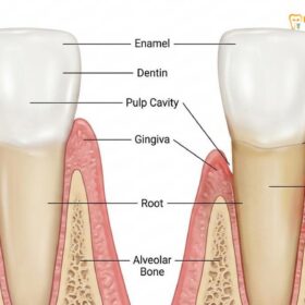
To fully comprehend the clinical significance of a periodontal pocket, it is essential to first understand the anatomy of a healthy periodontium. The periodontium consists of the specialized tissues that both surround and support the teeth, maintaining them in the maxillary and mandibular bones. These tissues include the gingiva (gums), the periodontal ligament, the cementum (the calcified substance covering the root of a tooth), and the alveolar bone. In a healthy state, the gingiva fits tightly around the neck of the tooth, creating a very shallow, V-shaped crevice known as the gingival sulcus [1].
A periodontal pocket develops when this healthy sulcus is compromised by a persistent bacterial infection. The process begins with the formation of dental plaque, a sticky, colorless biofilm composed of various bacterial species, salivary proteins, and food debris. If this plaque is not adequately removed through daily oral hygiene practices, it begins to mineralize into a hard substance known as dental calculus, or tartar. Calculus provides a rough, porous surface that encourages even more bacterial accumulation, particularly below the gumline where oxygen levels are low.
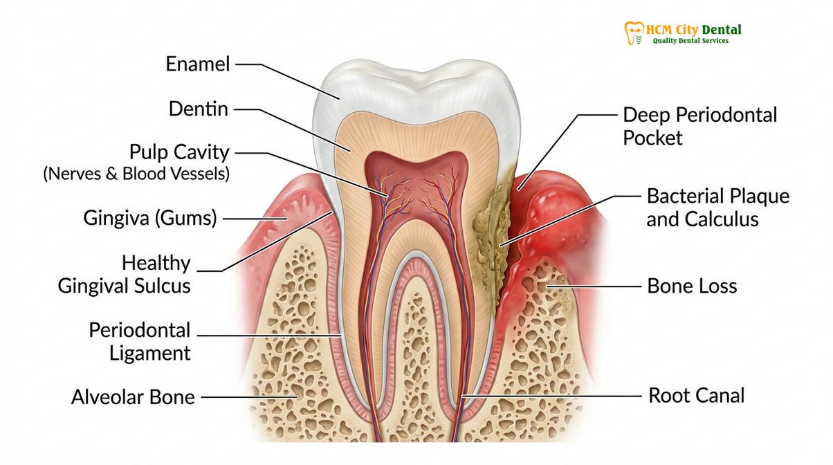
As the bacterial biofilm matures within this subgingival environment, it shifts from predominantly aerobic, Gram-positive bacteria to highly pathogenic, anaerobic, Gram-negative bacteria. These virulent microorganisms release toxins, enzymes, and metabolic byproducts that directly irritate the gingival tissues. However, the most significant tissue destruction is actually caused by the host’s own immune response. In an attempt to fight off the invading bacteria, the body’s immune system releases inflammatory mediators, such as cytokines, prostaglandins, and matrix metalloproteinases (MMPs). While these substances are intended to neutralize the infection, their chronic presence leads to the collateral degradation of the collagen fibers in the periodontal ligament and the resorption of the alveolar bone [2].
As the connective tissue fibers are destroyed, the junctional epithelium—the band of tissue that attaches the gum to the tooth—migrates apically (downward toward the root tip). This apical migration, combined with the swelling of the inflamed gingival tissue, transforms the shallow healthy sulcus into a deep, pathological space. These deep periodontal pockets become highly efficient traps for further plaque and calculus accumulation, creating a vicious cycle of infection and tissue destruction that cannot be resolved through at-home brushing and flossing alone. The deeper the pocket, the more anaerobic the environment becomes, fostering the growth of the most destructive periodontal pathogens.
How Your Dentist Measures Gum Pockets (Periodontal Probing)
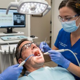
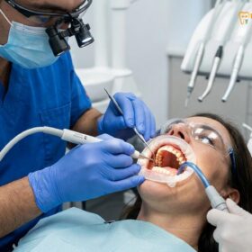
Dentists measure gum pockets using a specialized instrument called a periodontal probe, gently inserting it into the space between the tooth and gum. The probe acts like a tiny ruler, recording the depth in millimeters at six specific points around each tooth.
The process of measuring gum pockets, clinically referred to as periodontal probing, is the gold standard diagnostic procedure for evaluating the health of the periodontium. During a comprehensive periodontal examination, a dentist or dental hygienist utilizes a periodontal probe—a slender, hand-held instrument with a blunt, rounded tip and calibrated millimeter markings along its shaft. Common types of probes include the UNC-15 probe, which has millimeter markings up to 15mm, and the Marquis color-coded probe, which features alternating colored bands to facilitate quick visual reading of the depths.
To obtain accurate measurements, the clinician gently inserts the tip of the probe into the gingival sulcus or pocket, keeping the instrument parallel to the long axis of the tooth. The probe is carefully advanced until slight tissue resistance is felt at the base of the pocket, which corresponds to the coronal-most portion of the junctional epithelium. The clinician applies a standardized, very light pressure—typically between 10 and 20 grams—to avoid penetrating healthy tissue and causing unnecessary discomfort or false readings. The probe is then “walked” around the entire circumference of the tooth using short, bobbing strokes to ensure that the deepest areas of detachment are not missed [3].
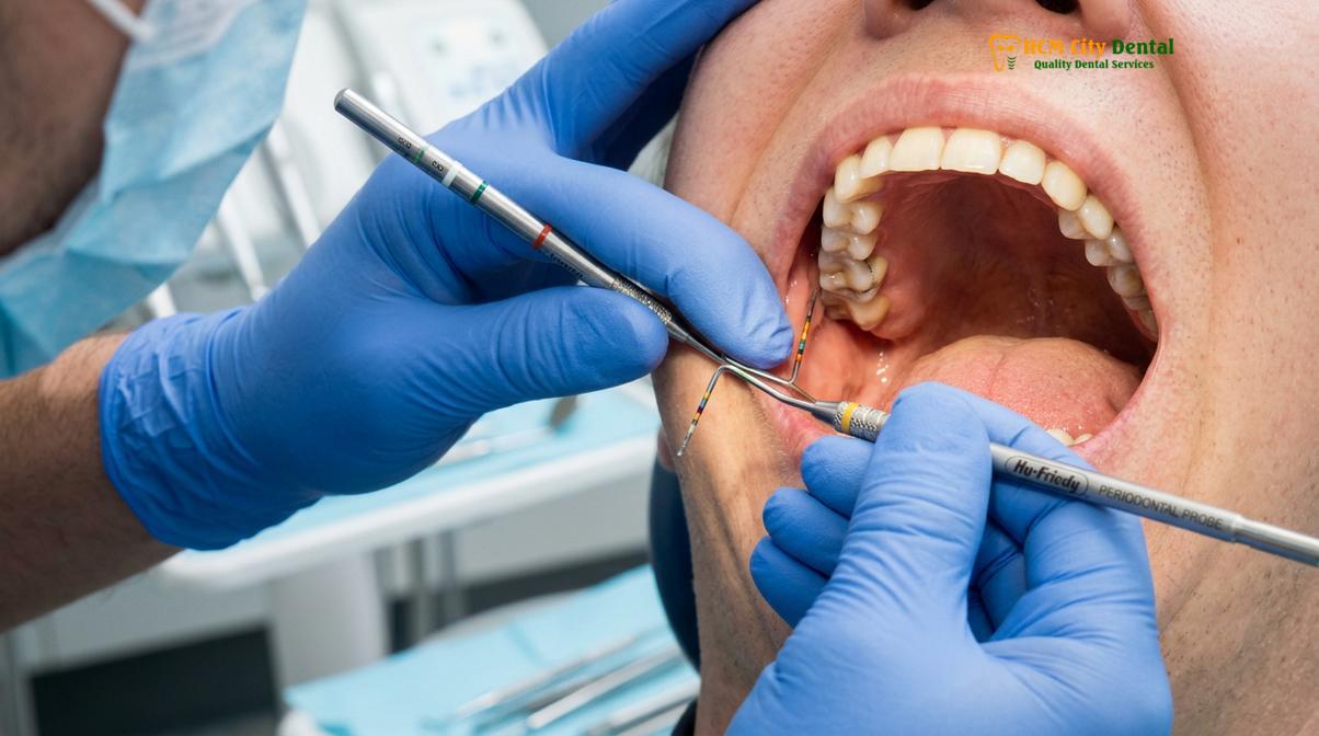
During this meticulous examination, the clinician is not only recording the numerical depth of the pockets but also assessing several other critical clinical parameters. One of the most important secondary indicators is Bleeding on Probing (BOP). If the gingival tissue bleeds when the probe is gently introduced, it is a definitive sign of active inflammation and micro-ulceration of the pocket lining. Healthy gums do not bleed upon probing. The presence of BOP indicates that the bacterial infection is currently active and that the site is at a higher risk for future attachment loss.
Dr. Nguyen Van Cuong, a leading expert in periodontal therapies, emphasizes that probing depth alone does not tell the entire story. “When we evaluate a patient’s periodontal status, we must also calculate the Clinical Attachment Level (CAL),” explains Dr. Cuong. “CAL takes into account both the probing depth and the position of the gingival margin relative to the cementoenamel junction (CEJ). This gives us a much more accurate picture of the true amount of structural support the tooth has lost over time, regardless of whether the gums are swollen or receding.” This comprehensive approach to diagnostics is a cornerstone of the General Dentistry protocols utilized to ensure accurate treatment planning.
The Periodontal Pocket Depth Chart Explained
A periodontal chart is a comprehensive clinical record that maps the exact millimeter depth of gum pockets around every tooth. This visual grid helps dental professionals track disease progression, identify localized infections, and evaluate the success of ongoing treatments.
Once the clinical measurements are obtained, they are meticulously recorded in a specialized document known as a periodontal pocket chart. This chart serves as a detailed topographical map of the patient’s oral health, providing a baseline against which all future examinations will be compared. In modern dental practices, this charting is often done digitally, with software that visually highlights areas of concern using color-coded systems—typically green for healthy depths and red for pathological pockets.
For every single tooth present in the oral cavity, the clinician records six distinct measurements. These six sites ensure that the entire circumference of the tooth is evaluated, as periodontal destruction is rarely uniform. A tooth may have healthy attachment on the cheek side but severe bone loss on the tongue side or between the teeth. The six standard measurement sites are:
- Mesiobuccal: The front corner of the tooth on the cheek/lip side.
- Midbuccal: The direct middle of the tooth on the cheek/lip side.
- Distobuccal: The back corner of the tooth on the cheek/lip side.
- Mesiolingual: The front corner of the tooth on the tongue/palate side.
- Midlingual: The direct middle of the tooth on the tongue/palate side.
- Distolingual: The back corner of the tooth on the tongue/palate side.
In addition to the numerical depths, the periodontal chart also records the presence of bleeding, suppuration (pus), furcation involvement (bone loss between the roots of multi-rooted teeth), tooth mobility, and areas of gingival recession. By synthesizing all this data, the dental team can formulate a highly targeted, site-specific treatment plan.
| Measurement Site | Probing Depth (mm) | Bleeding on Probing (BOP) | Clinical Status |
|---|---|---|---|
| Mesiobuccal | 3 mm | Negative | Healthy |
| Midbuccal | 2 mm | Negative | Healthy |
| Distobuccal | 5 mm | Positive (+) | Moderate Periodontitis |
| Mesiolingual | 4 mm | Positive (+) | Early Periodontitis |
| Midlingual | 3 mm | Negative | Healthy |
| Distolingual | 6 mm | Positive (+) | Severe Periodontitis |
Understanding your gum pocket numbers meaning is crucial for patient compliance and education. When a patient can visually see the areas of their mouth that are failing, they are much more likely to adhere to the recommended home care routines and accept the necessary clinical interventions. The chart transforms an invisible, painless infection into a tangible, measurable medical condition.
What Do Numbers 1 to 3 Mean? (Healthy Gums)
Pocket depths measuring between 1 and 3 millimeters indicate a healthy, intact periodontium where the gum tissue fits snugly against the tooth. At these depths, standard at-home brushing and flossing can effectively remove daily bacterial plaque.
When your dentist calls out numbers ranging from 1 to 3 during a periodontal examination, it is a highly positive clinical finding. These measurements represent a state of periodontal health and stability. Histologically, a probing depth of 1 to 3 millimeters corresponds to a normal, uninflamed gingival sulcus. In this state, the junctional epithelium is firmly attached to the enamel or cementum at the cementoenamel junction, and the underlying network of collagen fibers (the gingival connective tissue attachment) is dense, organized, and intact.
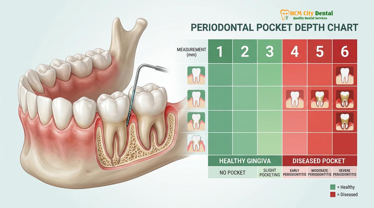
At these shallow depths, the environment within the sulcus is predominantly aerobic, meaning it is exposed to oxygen. This oxygen-rich environment naturally suppresses the growth of the highly destructive, anaerobic bacteria responsible for severe periodontitis. Furthermore, the shallow depth allows the bristles of a standard toothbrush and the mechanical action of dental floss to easily reach the base of the sulcus. This means that with proper daily oral hygiene techniques—such as the modified Bass brushing method and consistent interdental cleaning—a patient can effectively disrupt and remove the bacterial biofilm before it has the opportunity to mature and calcify into tartar.
It is important to note that even within the 1 to 3 millimeter range, the presence of bleeding changes the clinical diagnosis. A 3-millimeter pocket that bleeds upon probing indicates gingivitis—the earliest, reversible stage of gum disease. Gingivitis means that while the bacterial plaque has caused inflammation and micro-ulceration of the soft tissue, it has not yet caused any permanent destruction of the underlying bone or connective tissue attachment. Prompt improvement in daily oral hygiene and a routine professional prophylaxis (cleaning) can easily reverse gingivitis and restore the tissues to a state of complete health.
What Do Numbers 4 to 5 Mean? (Early to Moderate Disease)
Measurements of 4 to 5 millimeters signal early to moderate periodontal disease, where bacterial toxins have begun detaching the gums from the teeth. These depths create anaerobic environments that trap plaque, making professional deep cleaning necessary for resolution.
When probing depths increase to 4 or 5 millimeters, the clinical diagnosis shifts from health or simple gingivitis to early or moderate periodontitis. At this stage, the chronic inflammatory response triggered by the bacterial biofilm has begun to cause irreversible damage to the periodontium. The junctional epithelium has migrated apically down the root surface, and the crestal portion of the alveolar bone has begun to resorb. The tight seal between the gum and the tooth has been broken, creating a true periodontal pocket.
The transition from a 3mm sulcus to a 5mm pocket represents a critical ecological shift within the subgingival environment. As the pocket deepens, oxygen levels plummet, creating an ideal incubator for pathogenic, anaerobic bacteria such as Porphyromonas gingivalis, Treponema denticola, and Tannerella forsythia—collectively known as the “red complex” bacteria. These microorganisms are highly virulent and produce tissue-destroying enzymes that accelerate the breakdown of the periodontal ligament and bone.
“The shift from a healthy 3mm sulcus to a diseased 5mm pocket is not merely a change in depth; it is a fundamental shift in the local microbiome. The environment becomes highly anaerobic, allowing the most aggressive periodontal pathogens to thrive and multiply, driving a chronic inflammatory response that destroys the supporting bone.”
Crucially, a 4 to 5 millimeter pocket is beyond the reach of standard home care tools. Toothbrush bristles generally cannot penetrate more than 1 to 2 millimeters below the gumline, and dental floss cannot effectively clean the concave root surfaces exposed within a deep pocket. Therefore, once a pocket reaches this depth, the bacterial biofilm and calculus trapped at the base can only be removed through professional clinical intervention, typically a procedure known as scaling and root planing. Without this intervention, the disease will inevitably progress.
In some clinical scenarios, a 4 or 5 millimeter reading may represent a “pseudo-pocket.” This occurs when the gingival tissue becomes severely inflamed and swollen (hyperplasia), rising higher on the crown of the tooth without any actual loss of bone or connective tissue attachment. Pseudo-pockets are common in cases of severe gingivitis, during orthodontic treatment, or as a side effect of certain medications (such as calcium channel blockers or anti-seizure drugs). A thorough clinical and radiographic evaluation is necessary to differentiate between a pseudo-pocket and true periodontal attachment loss.
What Do Numbers 6 and Above Mean? (Severe Periodontitis)
Depths of 6 millimeters or greater represent severe, advanced periodontitis characterized by significant destruction of the alveolar bone and periodontal ligament. Without immediate and aggressive clinical intervention, these deep pockets frequently lead to tooth mobility and eventual tooth loss.
Probing depths of 6 millimeters, 7 millimeters, or beyond indicate a severe, advanced stage of periodontal disease. At these depths, the destructive inflammatory process has caused extensive, irreversible damage to the tooth’s supporting structures. A significant volume of the alveolar bone has been resorbed, and the periodontal ligament fibers that anchor the tooth root to the bone have been severely compromised. In many cases, these deep pockets are associated with complex anatomical challenges, such as furcation involvement, where the bone loss extends into the space between the roots of multi-rooted teeth like molars.
The clinical presentation of severe periodontitis often includes noticeable symptoms, although the disease can still remain surprisingly painless for some patients. Common signs associated with 6mm+ pockets include chronic halitosis (bad breath) caused by the volatile sulfur compounds produced by anaerobic bacteria, purulent exudate (pus) expressing from the gums, severe gingival recession exposing the sensitive tooth roots, and pathological tooth migration (teeth shifting or flaring out of their normal alignment). As the bone support diminishes, the teeth may exhibit varying degrees of clinical mobility, feeling loose during chewing or even to the touch.
Treating pockets of this magnitude is highly complex and rarely successful with non-surgical scaling and root planing alone. The sheer depth and the intricate anatomy of the root surfaces make it physically impossible for a clinician to completely remove the calcified deposits and bacterial biofilm using closed, non-surgical techniques. Therefore, measurements of 6mm and above almost universally require advanced surgical interventions to gain visual access to the roots, debride the infection, and attempt to regenerate the lost bone and tissue [4].
Clinical Treatments to Reduce Periodontal Pocket Depth
Treatment strategies aim to eliminate bacterial infection, reduce pocket depths, and facilitate tissue reattachment through both non-surgical and surgical modalities. The specific clinical approach is determined by the severity of the pocket depth and the extent of underlying bone loss.
The primary objective of all periodontal therapy is to halt the progression of the disease, eliminate the bacterial infection, and reduce the pocket depths to a maintainable level (ideally 3mm or less). The chosen treatment modality depends heavily on the initial probing depths, the patient’s systemic health, and the anatomical configuration of the bone loss. At modern facilities like HCMC Dental Clinic, a tiered approach is utilized, starting with the most conservative therapies and progressing to advanced surgical techniques only when necessary.
Non-Surgical Therapy: Scaling and Root Planing (SRP)
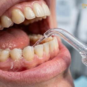
For early to moderate pockets (4 to 5 millimeters), the first line of defense is Scaling and Root Planing, commonly known as a deep cleaning. This procedure is typically performed under local anesthesia to ensure patient comfort. The clinician uses ultrasonic instruments, which utilize high-frequency vibrations and water irrigation, to shatter and flush away large calculus deposits and bacterial biofilm from the crown and root surfaces. Following the ultrasonic debridement, specialized hand instruments called curettes are used to meticulously plane (smooth) the root surfaces. Smoothing the roots removes the microscopic toxins embedded in the cementum and creates a clean, glass-like surface that encourages the gingival tissue to heal and reattach to the tooth, thereby shrinking the pocket depth.
Laser-Assisted Periodontal Therapy
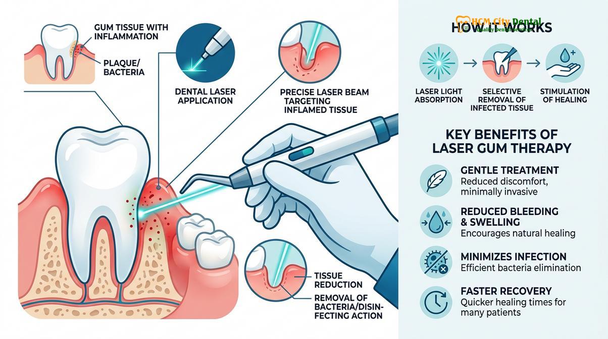
Advancements in dental technology have introduced laser therapy as a powerful adjunct or alternative to traditional treatments. Procedures utilizing specific wavelengths, such as the Nd:YAG or Diode lasers, can selectively target and vaporize the dark-pigmented, diseased tissue lining the pocket while leaving the healthy tissue unharmed. The laser energy also provides profound bactericidal effects, penetrating the dentinal tubules to eradicate bacteria that mechanical instruments cannot reach. Furthermore, the laser stimulates the formation of a stable fibrin clot, which seals the pocket and provides a scaffold for new tissue attachment. This minimally invasive approach often results in less postoperative discomfort and faster healing times [5].
Pocket Reduction Surgery (Flap Surgery)
When pockets exceed 6 millimeters and do not respond adequately to non-surgical therapy, pocket reduction surgery (osseous surgery) is often indicated. During this procedure, a periodontist gently separates the gum tissue from the teeth and folds it back, creating a “flap.” This provides direct visual access to the root surfaces and the underlying bone. The roots are thoroughly debrided of all calculus and granulation tissue. If the bacterial infection has caused the bone to heal in an irregular, jagged pattern, the surgeon will recontour and smooth the bone (osteoectomy and osteoplasty) to eliminate areas where bacteria can hide. The gum tissue is then repositioned and sutured tightly against the teeth, effectively eliminating the deep pocket.

Regenerative Procedures (Bone Grafting)
In cases where the periodontal disease has caused deep, vertical defects in the alveolar bone, regenerative procedures may be employed to rebuild the lost structural support. After the flap is reflected and the defect is meticulously cleaned, the surgeon places a bone graft material (which may be autogenous, allograft, xenograft, or synthetic) into the bony defect. Often, a specialized barrier membrane is placed over the graft. This membrane prevents the fast-growing soft gum tissue from migrating into the defect, allowing the slower-growing bone cells the time and space they need to regenerate. Biologic mediators, such as enamel matrix derivatives (Emdogain), may also be applied to the root surface to stimulate the formation of new cementum, periodontal ligament, and alveolar bone.
Following any of these interventions, the patient must be placed on a strict Periodontal Maintenance program. Because periodontal disease is a chronic condition, patients are typically seen every three to four months for specialized cleanings and re-probing to ensure the pockets remain stable and the infection does not return. For those seeking comprehensive care, exploring specialized gum disease therapies is the first step toward long-term oral health stability.
When to See a Doctor (Important Clinical Considerations)
You should seek immediate clinical evaluation if you experience persistent bleeding while brushing, chronic bad breath, receding gums, or teeth that feel loose or are shifting position.
Periodontal disease is often described as a “silent” disease because it can progress to advanced stages without causing significant pain. Relying on pain as an indicator of gum health is a dangerous strategy that often leads to irreversible bone loss. It is critical to schedule a comprehensive periodontal evaluation if you notice any of the following warning signs:
- Gums that are red, swollen, or tender to the touch.
- Gums that bleed easily during normal brushing or flossing.
- Gums that appear to be pulling away from the teeth, making the teeth look longer (recession).
- Persistent bad breath or a constant bad taste in the mouth that does not resolve with mouthwash.
- Changes in the way your teeth fit together when you bite down.
- Pus visible between your teeth and gums when pressure is applied.
- Teeth that feel loose or mobile.
If you have a family history of severe gum disease, or if you suffer from systemic conditions such as diabetes or cardiovascular disease, you are at a higher risk and should be monitored more closely. While modern periodontal therapies have a high success rate in stabilizing the disease and preserving teeth, there are no absolute guarantees, especially in cases of extreme bone loss. Early diagnosis and intervention are the most critical factors in achieving a successful clinical outcome. We encourage patients to visit HCMC Dental Clinic for a personalized diagnostic assessment to determine the most appropriate course of action.
Frequently Asked Questions
Can deep gum pockets heal and shrink?
Yes, deep gum pockets can heal and shrink, but they require professional clinical intervention to do so. While the lost bone may not always regenerate completely without surgical grafting, professional deep cleaning (scaling and root planing) removes the bacterial biofilm and calculus trapped beneath the gumline. Once the infection is cleared, the gingival tissue undergoes a healing phase, reducing inflammation and allowing the gums to reattach more tightly to the tooth root, thereby decreasing the overall pocket depth.
What is a normal periodontal pocket depth?
A normal, healthy periodontal pocket depth measures between 1 and 3 millimeters. At this depth, the gingival tissue is firmly attached to the underlying tooth structure, and there is no bleeding upon probing. These shallow depths allow the bristles of a standard toothbrush and dental floss to effectively reach and clean the base of the sulcus, preventing the accumulation of anaerobic bacteria that cause progressive gum disease.
Does measuring gum pockets hurt?
Measuring gum pockets is generally a painless procedure for patients with healthy gums, though it may cause mild discomfort if the tissues are inflamed. The dentist or hygienist uses a periodontal probe with a blunt, rounded tip, applying very light pressure (approximately 10 to 20 grams) to walk the instrument around the tooth. If you have active gingivitis or periodontitis, the inflamed, ulcerated tissue inside the pocket may feel sensitive or bleed slightly during the examination.
How do dentists treat 5mm pockets?
Dentists typically treat 5mm pockets using a non-surgical procedure known as scaling and root planing (deep cleaning). This treatment utilizes ultrasonic scalers and specialized hand instruments to meticulously remove bacterial plaque and hard calculus from the root surfaces beneath the gumline. Following the mechanical debridement, the tooth roots are smoothed to discourage further bacterial adherence. In some cases, localized antimicrobial therapies or laser decontamination may be used as adjuncts to promote optimal tissue healing.
How often should periodontal pockets be measured?
Periodontal pockets should be measured at least once a year during a comprehensive dental examination for patients with healthy gums. However, for individuals who have been diagnosed with periodontal disease or have a history of deep pockets, dentists strongly recommend periodontal charting every three to four months. This frequent monitoring, known as periodontal maintenance therapy, is crucial for detecting early signs of disease relapse and ensuring that the pocket depths remain stable over time. If you are overdue for an evaluation, please contact our clinical team to schedule a comprehensive assessment.
References
- Journal of Clinical Periodontology. Pathogenesis of periodontal disease and pocket formation. (2021).
- American Academy of Periodontology. Comprehensive periodontal therapy: a statement on standard of care. (2020).
- International Journal of Dental Hygiene. Efficacy of scaling and root planing in moderate to severe periodontitis. (2019).
- Journal of Periodontal Research. The oral-systemic link: cardiovascular implications of severe attachment loss. (2022).
- British Dental Journal. Laser-assisted non-surgical periodontal therapy: clinical outcomes and tissue healing. (2018).