A coronectomy for wisdom teeth is a specialized surgical procedure where only the crown of the tooth is removed, intentionally leaving the roots intact. This technique is specifically utilized when extracting the entire tooth poses a high risk of damaging the inferior alveolar nerve.
Clinical Summary:
A coronectomy is an advanced, minimally invasive alternative to traditional complete wisdom tooth extraction, indicated primarily for mandibular third molars whose roots are intimately associated with the inferior alveolar nerve (IAN). By surgically sectioning and removing only the clinical crown—and reducing the remaining root structure below the crestal bone—surgeons can eliminate the source of pericoronitis or decay while avoiding the mechanical trauma of root elevation. Clinical evidence demonstrates that retaining vital, uninfected roots significantly reduces the incidence of postoperative paresthesia, dysesthesia, and permanent nerve injury. While the procedure requires meticulous preoperative 3D imaging and precise surgical execution, long-term outcomes show high success rates, with the retained roots typically becoming encased in regenerated alveolar bone.
Key Takeaways:
- A coronectomy removes the tooth crown but leaves the roots to protect the underlying nerve.
- It is strictly indicated for vital teeth without deep root infections or periapical pathology.
- 3D CBCT imaging is mandatory to confirm the root’s proximity to the inferior alveolar nerve.
- Retained roots usually remain healthy and are eventually covered by new bone tissue.
- If a retained root migrates and erupts later, it can be safely extracted with minimal nerve risk.
What is a Coronectomy and How Does It Protect Your Nerves?
A coronectomy protects the inferior alveolar nerve by removing only the problematic crown, leaving the deep roots undisturbed in the jawbone to prevent mechanical nerve trauma.
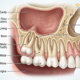
The human mandible (lower jaw) houses a critical anatomical structure known as the inferior alveolar nerve (IAN). This nerve travels through a bony tunnel called the mandibular canal, providing sensory innervation to the lower teeth, the lower lip, and the chin. In many adults, the roots of the lower third molars (wisdom teeth) develop in extremely close proximity to this canal. In some complex cases, the roots may even hook around the nerve, compress the canal, or grow directly through it. When a traditional Tooth Extraction is performed on such a tooth, the mechanical forces required to elevate and deliver the roots can crush, stretch, or sever the nerve.[1]
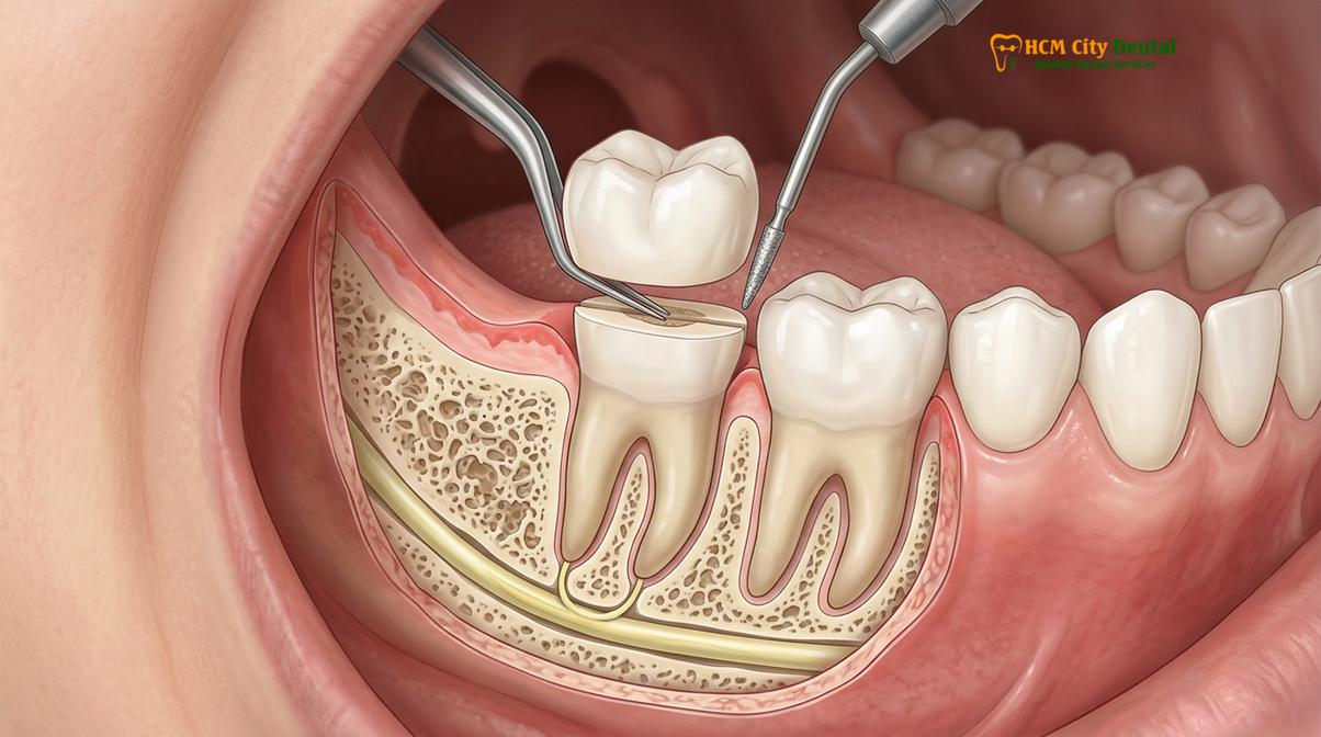
A coronectomy, also referred to as a wisdom tooth partial root removal, was developed as a defensive surgical strategy to mitigate this exact risk. The fundamental principle of the procedure is to eliminate the portion of the tooth that is causing clinical symptoms—the crown—while leaving the hazardous portion—the roots—safely untouched. The crown is typically the source of problems such as recurrent pericoronitis (gum infection), dental caries (decay), or damage to the adjacent second molar. By sectioning the tooth at the cementoenamel junction (where the crown meets the root), the surgeon removes the disease vector without disturbing the delicate neurovascular bundle below.
To understand how alveolar nerve damage coronectomy protocols work, one must look at the mechanics of nerve injury. Neuropraxia (temporary nerve bruising) or neurotmesis (complete nerve severing) during extraction usually occurs when the root apex is forcefully levered against the roof of the mandibular canal. By intentionally leaving the root in situ, the surgeon completely avoids applying any apical pressure. The retained root acts as a biological placeholder. As long as the pulp tissue within the root remains vital and uninfected at the time of surgery, the body’s immune system recognizes the root as native tissue rather than a foreign body, allowing normal bone healing to occur over the surgical site.[2]
This procedure is not a shortcut or a failed extraction; it is a highly calculated, intentional surgical decision. It requires a deep understanding of oral anatomy, precise surgical instrumentation, and strict adherence to biological principles. When performed correctly, it transforms a high-risk surgical scenario into a predictable, safe outcome, preserving the patient’s sensory function and quality of life.
Coronectomy vs. Standard Extraction: The Decision Process
The choice between a coronectomy and a standard extraction depends entirely on the root’s proximity to the nerve, the vitality of the tooth, and the presence of any deep infections.
The decision-making process in oral surgery is heavily reliant on risk-benefit analysis. When evaluating a patient for a coronectomy vs extraction wisdom tooth procedure, the surgeon must carefully weigh the risk of nerve injury against the risk of leaving root tissue behind. Not every impacted wisdom tooth is a candidate for a coronectomy. In fact, standard complete extraction remains the gold standard for the vast majority of wisdom teeth. A coronectomy is reserved exclusively for a specific subset of high-risk cases.
A standard extraction is mandatory when the wisdom tooth is non-vital (dead), has a deep carious lesion extending into the root pulp, or is associated with a periapical infection (an abscess at the tip of the root). Leaving an infected root behind would inevitably lead to severe postoperative complications, systemic infection, and the failure of the surgical site to heal. Furthermore, teeth that are highly mobile or horizontally impacted in a way that the roots would remain exposed to the oral cavity after crown removal are not suitable for a coronectomy. In these scenarios, the entire tooth must be removed, and the surgeon must employ advanced, delicate techniques to navigate the nerve proximity.[3]
Conversely, a coronectomy is the treatment of choice when the tooth is vital, free of deep infection, and radiographically confirmed to be in dangerous proximity to the inferior alveolar nerve. It is particularly beneficial for vertically or mesioangularly impacted teeth where the crown can be cleanly sectioned without mobilizing the roots. The decision also factors in the patient’s age and medical history. For instance, older patients often have denser, less elastic bone, which increases the force required for extraction and subsequently increases the risk of nerve trauma, making a coronectomy a highly attractive option. Patients seeking a Tooth Extraction in their 30s or 40s frequently present with these exact anatomical challenges.
| Clinical Factor | Standard Complete Extraction | Intentional Coronectomy |
|---|---|---|
| Nerve Proximity | Low to moderate risk; clear separation visible on imaging. | High risk; roots touching, compressing, or wrapping the nerve. |
| Tooth Vitality | Can be performed on vital or non-vital (infected) teeth. | Strictly limited to vital teeth without deep pulpal infection. |
| Root Pathology | Required if periapical abscess or cysts are present. | Contraindicated if any periapical pathology exists. |
| Tooth Mobility | Standard procedure for mobile teeth. | Contraindicated; if roots become mobile, they must be extracted. |
| Primary Goal | Complete removal of all dental tissue to prevent future issues. | Elimination of the crown while preserving sensory nerve function. |
At specialized centers like HCMC Dental Clinic, the clinical team conducts a rigorous preoperative assessment to determine the most appropriate surgical pathway. This involves not only clinical examination but also advanced radiographic analysis. The goal is always to provide the safest, most effective treatment tailored to the individual’s unique anatomical presentation.
How 3D CBCT Imaging Identifies High-Risk Nerve Proximity
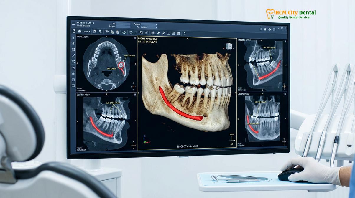
Cone Beam Computed Tomography (CBCT) provides a critical three-dimensional view, allowing surgeons to precisely map the relationship between the wisdom tooth roots and the nerve canal.
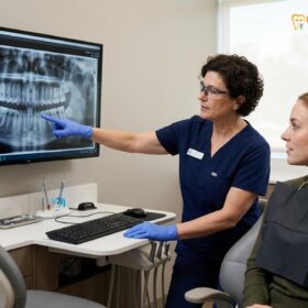
The foundation of a successful coronectomy lies in accurate preoperative diagnostics. Historically, oral surgeons relied on two-dimensional panoramic X-rays (orthopantomograms) to assess wisdom teeth. While panoramic X-rays are excellent for a general overview of the jaws, they suffer from a significant limitation: superimposition. In a 2D image, a wisdom tooth root may appear to overlap the mandibular canal, but it is impossible to determine whether the root is positioned in front of the nerve (buccally), behind the nerve (lingually), or directly intersecting it. This ambiguity can lead to unexpected surgical complications.
To eliminate this guesswork, modern clinical protocols mandate the use of Cone Beam Computed Tomography (CBCT) for any lower wisdom tooth that exhibits high-risk markers on a standard X-ray. These markers, often referred to as the Rood and Shehab criteria, include darkening of the root where it crosses the canal, interruption of the white cortical lines of the canal, diversion or narrowing of the canal, and narrowing of the root itself. When any of these signs are present, a CBCT scan is required to visualize the anatomy in three dimensions (axial, coronal, and sagittal planes).[4]
“The advent of CBCT imaging has revolutionized third molar surgery. It transitions our approach from reactive complication management to proactive risk mitigation, allowing us to definitively identify which patients will benefit most from a coronectomy.”
A CBCT scan allows the surgeon to measure the exact distance, in fractions of a millimeter, between the root apex and the neurovascular bundle. It reveals the exact morphology of the roots—whether they are fused, divergent, or hooked. Furthermore, it shows the thickness of the surrounding cortical bone, particularly the lingual plate, which is crucial for protecting the lingual nerve during surgery. If the CBCT confirms that the roots are intimately entangled with the inferior alveolar nerve, the surgeon will confidently recommend a coronectomy.
Dr. Nguyen Van Cuong emphasizes that high-resolution imaging is non-negotiable when planning complex extractions. By utilizing advanced CBCT technology, the surgical team can virtually plan the exact angle and depth of the tooth sectioning before the patient even sits in the surgical chair. This level of preparation is what separates a standard dental visit from a premier Tooth Extraction experience, ensuring maximum safety and predictability.
The Step-by-Step Coronectomy Procedure Explained
The procedure involves elevating a surgical flap, precisely sectioning the tooth crown, reducing the remaining roots below the bone level, and achieving primary tissue closure.
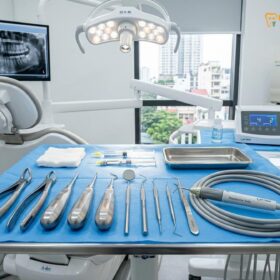
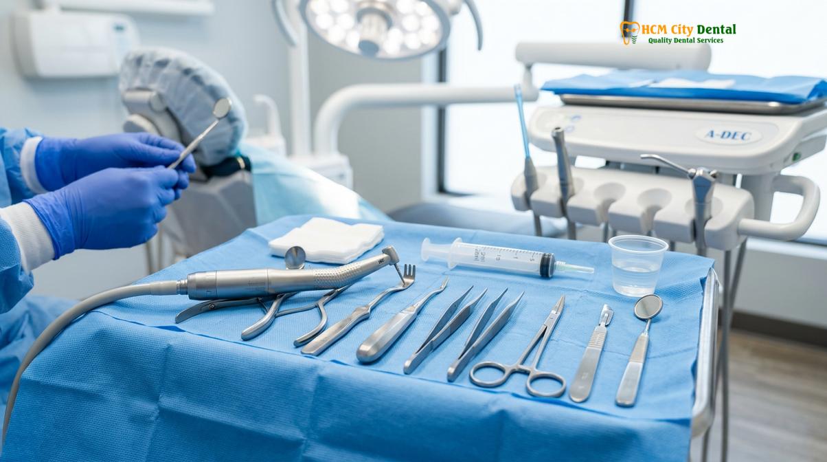
The surgical execution of a coronectomy requires meticulous technique and specialized instrumentation. The procedure is typically performed under local anesthesia, often supplemented with conscious sedation to ensure patient comfort. The workflow is highly structured to minimize tissue trauma and prevent the inadvertent mobilization of the roots.
Step 1: Flap Design and Elevation
The surgeon begins by creating a full-thickness mucoperiosteal flap. An incision is made along the gum line, and the tissue is gently reflected to expose the jawbone and the impacted wisdom tooth. Careful attention is paid to the lingual aspect (the side facing the tongue) to avoid disturbing the lingual nerve, which often runs very close to the surface in this area.
Step 2: Bone Removal (Guttering)
If the tooth is deeply impacted, a small amount of surrounding bone must be removed to gain access to the crown. This is usually done using a surgical handpiece with a round bur and copious sterile saline irrigation to prevent bone necrosis from heat generation. Alternatively, advanced clinics may use a Piezotome—an ultrasonic bone-cutting device that selectively cuts hard tissue while preserving soft tissue, offering a Tooth Extraction experience with reduced postoperative swelling.
Step 3: Tooth Sectioning
This is the most critical phase of the operation. The surgeon uses a cross-cut fissure bur to section the tooth horizontally, typically at a 45-degree angle, just below the cementoenamel junction. The cut is made approximately three-quarters of the way through the tooth. The surgeon intentionally avoids cutting completely through the tooth to prevent the bur from penetrating the lingual cortical plate and damaging the lingual nerve. A surgical elevator is then inserted into the groove, and a gentle rotational force is applied to fracture off the crown.
Step 4: Root Reduction
Once the crown is removed, the remaining root surface must be evaluated. For a coronectomy to be successful, the cut surface of the roots must be reduced to at least 2 to 3 millimeters below the crest of the surrounding alveolar bone. This space is essential because it allows a blood clot to form and new bone to regenerate over the roots, permanently burying them. The surgeon uses a specialized bur to smooth the root surface, ensuring there are no sharp enamel spicules left behind, as enamel does not integrate with bone and can cause delayed healing.
Clinical Case Study: High-Risk Impaction Management
A 28-year-old patient presented to HCMC Dental Clinic in Ho Chi Minh City with recurrent pericoronitis around a partially erupted lower right wisdom tooth. CBCT imaging revealed the mesial root was hooked directly around the inferior alveolar nerve canal. Dr. Nguyen Van Cuong performed an intentional coronectomy using ultrasonic piezosurgery. The crown was successfully sectioned without mobilizing the roots. The patient experienced zero postoperative numbness and returned to normal function within days, demonstrating the efficacy of the procedure in high-risk anatomical scenarios.
Step 5: Irrigation and Closure
The surgical site is thoroughly irrigated with sterile saline or an antimicrobial solution to remove any bone dust or tooth debris. Proper Tooth Extraction irrigation protocols are vital to prevent postoperative infection. Finally, the gum tissue is repositioned and sutured closed. Primary closure (completely sealing the wound) is preferred to protect the retained roots from the oral environment while the bone heals.
Long-Term Retention: What Happens to the Retained Roots?
Retained roots typically remain vital and become encased in newly formed bone, though a small percentage may slowly migrate away from the nerve over several years.
One of the most common concerns patients have regarding a coronectomy is the fate of the roots left behind in the jaw. From a biological perspective, as long as the tooth was vital and the surgical technique was sterile, the retained roots do not act as foreign bodies. The dental pulp within the root canal maintains its blood supply from the apical foramen (the opening at the tip of the root). The cut surface of the root undergoes a healing process where cementum (the calcified tissue covering the root) and bone gradually grow over the exposed dentin.
Over the first six to twelve months following the surgery, osteogenesis (bone formation) occurs in the space created above the reduced roots. Radiographically, this appears as a solid layer of trabecular bone bridging the gap, permanently encasing the roots within the mandible. In the vast majority of cases, these roots remain completely asymptomatic for the rest of the patient’s life.[5]
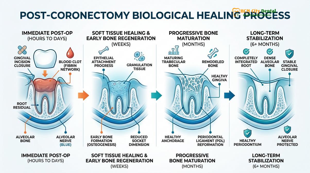
“The biological integration of retained vital roots is a well-documented phenomenon. The body’s innate healing capacity recognizes the healthy root structure, sealing it beneath a layer of newly regenerated alveolar bone, effectively neutralizing any future pathological risk.”
However, clinical studies have documented a phenomenon known as root migration. In approximately 15% to 30% of coronectomy cases, the retained roots may slowly migrate away from the inferior alveolar nerve, usually moving in a coronal (upward) or mesial (forward) direction. This migration typically occurs within the first two years post-surgery and then stabilizes. Root migration is not inherently a complication; in fact, it is often viewed as a positive outcome. If the roots migrate far enough to erupt through the bone and gum tissue, they may require a secondary extraction. Crucially, because the roots have moved significantly away from the nerve canal, this secondary extraction is a simple, low-risk procedure with virtually no chance of nerve damage.
Patients are advised to undergo routine radiographic monitoring (usually a panoramic X-ray at 6 months, 12 months, and 24 months post-op) to track the position of the roots and confirm healthy bone fill. This long-term follow-up is a standard component of comprehensive oral surgical care.
Recovery, Healing, and Potential Coronectomy Complications
Recovery closely mirrors standard extraction, requiring careful pain management and soft diet adherence, while monitoring for specific risks like root mobilization or delayed infection.
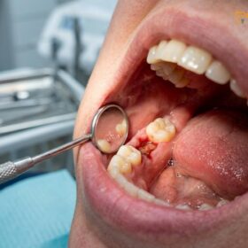
The immediate postoperative recovery from a coronectomy is very similar to that of a standard surgical extraction. Patients can expect mild to moderate swelling, localized discomfort, and limited jaw opening (trismus) for the first 3 to 5 days. Management involves cold compresses, prescribed analgesics (such as ibuprofen or acetaminophen), and a strict adherence to a soft food diet. Knowing the right Tooth Extraction dietary guidelines is essential to prevent food debris from traumatizing the surgical site.
While coronectomy recovery and infection risk profiles are generally favorable, the procedure is not without potential complications. The most immediate intraoperative complication is root mobilization. During the sectioning and removal of the crown, the roots may inadvertently become loose. If the roots are mobilized, the blood supply to the pulp is severed, and the roots will eventually necrose (die) if left in place. Therefore, if mobilization occurs, the surgeon must abandon the coronectomy protocol and proceed with a complete extraction, accepting the associated nerve risks.
Important Clinical Warning: Late-Stage Infection
Although rare, a retained root can develop an infection months or even years after a coronectomy. This usually occurs if the initial surgical closure was incomplete, allowing oral bacteria to penetrate the bone, or if the tooth pulp was unknowingly compromised prior to surgery. Symptoms include localized swelling, a bad taste in the mouth, and dull aching pain. If a late-stage infection is diagnosed, the retained roots must be surgically extracted.
Another potential complication is dry socket (alveolar osteitis), which occurs when the blood clot over the surgical site dislodges or dissolves prematurely. This exposes the underlying bone and nerve endings, causing severe, radiating pain. Management involves irrigating the socket and placing a medicated dressing to soothe the tissue while secondary healing occurs. Patients with underlying health conditions must also be managed carefully; for example, protocols for Tooth Extraction in patients with diabetes or those on blood thinners require specific preoperative adjustments to ensure proper hemostasis and immune response.
When to See a Doctor (Clinical Considerations)
Immediate clinical evaluation is required if you experience prolonged numbness, severe escalating pain, or signs of systemic infection following your surgical procedure.
While a coronectomy is designed to minimize risks, patients must remain vigilant during the healing phase. You should contact your oral surgeon immediately if you experience any of the following symptoms:
- Prolonged Altered Sensation: While the goal is to prevent nerve damage, temporary swelling can sometimes compress the nerve. If you experience numbness, tingling, or a burning sensation in your lower lip, chin, or tongue that lasts beyond the first 24 hours, clinical evaluation is necessary.
- Escalating Pain: Discomfort should peak around day 2 or 3 and then steadily improve. Severe, throbbing pain that worsens after day 4 may indicate a dry socket or an emerging infection.
- Signs of Infection: The presence of pus, a foul odor or taste, persistent fever, or swelling that increases significantly after the third day requires immediate antibiotic therapy and possible surgical intervention.
A coronectomy is a highly technique-sensitive procedure that should only be performed by experienced dental surgeons. A thorough clinical examination, including 3D imaging, is mandatory to determine if you are a suitable candidate. Always rely on personalized diagnostic advice rather than general educational information when making surgical decisions.
Frequently Asked Questions
What is the difference between a coronectomy and a extraction?
A standard extraction removes the entire wisdom tooth, including the roots, whereas a coronectomy removes only the crown of the tooth, intentionally leaving the roots embedded in the jawbone. This specialized approach is chosen specifically to prevent nerve damage when roots are entangled with the inferior alveolar nerve. By leaving the roots undisturbed, the surgeon avoids the mechanical trauma that causes long-term numbness.
Is the root left behind in a coronectomy safe?
Yes, the retained root is generally safe as long as the tooth was vital and free from deep infection prior to the surgery. Clinical studies show that the healthy root tissue typically remains alive, and the jawbone eventually heals and regenerates over the cut surface, permanently encasing it. The body’s immune system recognizes the vital root as natural tissue, preventing rejection.
Can a coronectomy root get infected later?
While rare, a retained root can develop an infection later if bacteria penetrate the surgical site before the bone fully heals, or if the root was already compromised. If a late-stage infection occurs, the root must be surgically extracted. However, the risk of nerve damage during this secondary extraction is usually much lower, as the root often migrates away from the nerve over time.
Does a coronectomy prevent permanent nerve damage?
A coronectomy significantly minimizes the risk of permanent nerve damage, but it does not completely eliminate all surgical risks. By avoiding the physical extraction of roots that are wrapped around or compressing the nerve canal, the likelihood of long-term numbness or altered sensation is drastically reduced compared to a standard complete extraction in high-risk cases.
How much does a coronectomy cost in Saigon?
The cost of a coronectomy in Saigon varies depending on the complexity of the impaction, the need for 3D CBCT imaging, and the specific surgical technology utilized. Because it requires advanced surgical precision, specialized equipment like Piezotomes, and meticulous follow-up, it is typically priced slightly higher than a standard surgical extraction. According to the latest clinic fee schedules, a personalized consultation is required for an exact estimate.
References
- Journal of Oral and Maxillofacial Surgery. Intentional Coronectomy: A Clinical Review. (2021).
- International Journal of Oral and Maxillofacial Surgery. Inferior Alveolar Nerve Protection Strategies. (2020).
- British Dental Journal. Long-term Follow-up of Retained Wisdom Tooth Roots. (2019).
- Journal of the American Dental Association. CBCT Diagnostics in Third Molar Surgery. (2022).
- Clinical Oral Investigations. Root Migration Patterns Post-Coronectomy. (2018).