Pericoronitis is a painful inflammation of the gum tissue surrounding a partially erupted wisdom tooth. It occurs when bacteria and food debris become trapped beneath the overlying gum flap, leading to localized infection, swelling, and severe discomfort that requires prompt clinical intervention.
Clinical Summary:
Pericoronitis primarily affects the mandibular third molars when an operculum (gum flap) traps anaerobic bacteria. Acute episodes present with severe pain, trismus, and purulent discharge. Management ranges from localized irrigation and antimicrobial therapy to definitive surgical interventions like operculectomy or complete tooth extraction, depending on the tooth’s eruption trajectory and impaction status.
Key Takeaways:
- Pericoronitis is the leading cause of acute pain in partially erupted lower wisdom teeth.
- The condition is driven by anaerobic bacteria trapped in the inaccessible pocket beneath the operculum.
- Mild cases may temporarily resolve with professional irrigation, but recurrent infections often require surgical intervention.
- Trismus (restricted jaw opening) and facial swelling are critical signs of a spreading infection.
- Definitive treatment usually involves extracting the offending third molar to permanently eliminate the anatomical trap.
What is Pericoronitis?
Pericoronitis is an acute or chronic infection of the soft tissue covering a partially erupted tooth, most commonly occurring around the lower third molars when a flap of gum tissue traps bacteria.
In the field of oral and maxillofacial pathology, pericoronitis represents one of the most frequent dental emergencies among young adults. The term literally translates to inflammation (“itis”) around (“peri”) the crown (“corona”) of a tooth. This condition manifests almost exclusively during the eruption phase of the third molars, commonly known as wisdom teeth, when they fail to fully breach the gingival surface.
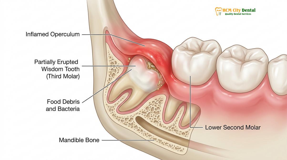
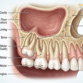
When a wisdom tooth only partially emerges through the gums, a persistent flap of soft tissue remains draped over the biting surface of the tooth. This anatomical structure is clinically referred to as an operculum. Because the operculum is not firmly attached to the tooth enamel, it creates a deep, dark, and moist pseudopocket. This microenvironment is highly conducive to the stagnation of oral fluids, food particles, and plaque. Consequently, patients often experience a severely swollen gum flap wisdom tooth, which becomes highly sensitive to touch and masticatory forces[1].
The pathophysiology of pericoronitis is exacerbated by the opposing maxillary (upper) wisdom tooth. If the upper wisdom tooth is fully erupted, it may repeatedly bite down onto the swollen operculum of the lower tooth during normal chewing or swallowing. This continuous mechanical trauma causes further ulceration, swelling, and compromised blood flow to the tissue, accelerating the infectious process and making spontaneous healing nearly impossible without clinical intervention.
Symptoms of an Infected Gum Flap
Symptoms range from mild localized tenderness and bad breath to severe radiating pain, facial swelling, and restricted jaw movement, depending on the depth and severity of the bacterial infection.
The clinical presentation of pericoronitis can be broadly categorized into acute and chronic phases. Chronic pericoronitis often presents as a low-grade, dull ache accompanied by a persistent bad taste in the mouth. Patients may notice mild halitosis (bad breath) caused by the putrefaction of trapped food debris and the metabolic byproducts of anaerobic bacteria. In this chronic state, the infected gum flap wisdom tooth may appear slightly erythematous (red) but does not typically cause debilitating pain.
However, when the bacterial load exceeds the local immune response, the condition rapidly transitions into acute pericoronitis. Acute symptoms are significantly more aggressive and distressing. Patients typically report a sudden onset of intense, throbbing pain that radiates from the back of the jaw to the ear, temporal region, or down the neck. The operculum becomes highly engorged, visibly red, and exquisitely tender to palpation.
“Patients often mistake the early stages of pericoronitis for a normal teething process, but the presence of purulent discharge and restricted jaw movement indicates an active, potentially spreading bacterial infection that requires immediate clinical evaluation.”
As the acute infection progresses, purulent exudate (pus) may be expressed from beneath the gum flap when gentle pressure is applied. One of the most concerning symptoms is trismus, a condition where the patient experiences severe difficulty opening their mouth. Trismus occurs when the inflammatory exudate infiltrates the medial pterygoid muscle, one of the primary muscles of mastication located adjacent to the lower wisdom teeth[2]. Additionally, patients may experience dysphagia (difficulty swallowing) and swelling of the submandibular lymph nodes as the body attempts to contain the localized infection.
Why Bacteria Collects Under the Operculum
The operculum creates a deep, inaccessible pseudopocket where mechanical brushing and flossing cannot reach, allowing plaque, food debris, and destructive anaerobic bacteria to proliferate rapidly.
To understand why pericoronitis is so prevalent, one must examine the unique anatomy of the retromolar region—the area immediately behind the last standing molar. When a wisdom tooth erupts, it must navigate through dense cortical bone and thick, fibrous gingival tissue. Due to evolutionary changes in human jaw size, there is frequently insufficient arch length to accommodate these final molars, leading to various degrees of impaction.
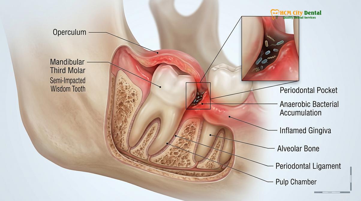
When a tooth is vertically or mesioangularly impacted (tilted forward), a portion of the crown may break through the mucosa while the distal (back) portion remains submerged under the retromolar pad. The resulting operculum acts like a one-way valve. During mastication, food debris is forced under the flap, but the tight adaptation of the tissue prevents the debris from being naturally washed away by saliva or removed by a toothbrush. This creates the perfect storm for operculum tooth pain.
The microbiological profile of the pocket beneath the operculum is highly complex. It is predominantly colonized by obligate anaerobic bacteria—microorganisms that thrive in oxygen-deprived environments. Pathogens such as Prevotella intermedia, Fusobacterium nucleatum, and various Actinomyces species multiply rapidly within this stagnant pocket. As they metabolize trapped food, they release endotoxins and volatile sulfur compounds, triggering a robust inflammatory cascade from the host’s immune system[3].
| Type of Impaction | Anatomical Description | Risk of Pericoronitis | Clinical Rationale |
|---|---|---|---|
| Vertical Impaction | Tooth is upright but partially covered by gum tissue. | High | Creates a classic operculum that easily traps food and is subject to trauma from upper teeth. |
| Mesioangular Impaction | Tooth is tilted forward toward the adjacent second molar. | Very High | Forms a deep, V-shaped periodontal pocket between the two molars, highly prone to severe infection. |
| Horizontal Impaction | Tooth is lying completely flat, parallel to the jawbone. | Moderate | Often fully encased in bone, but if partially erupted, the exposed crown surface acts as a bacterial trap. |
| Distoangular Impaction | Tooth is tilted backward, away from the adjacent molar. | High | The distal crown remains deeply submerged under the thick retromolar pad, making hygiene impossible. |
Immediate Home Care and Oral Rinses
Temporary relief can be achieved through vigorous warm saline rinses and targeted mechanical irrigation to flush out trapped debris and reduce the bacterial load beneath the gum flap.
When acute pericoronitis strikes, immediate professional dental care is the gold standard. However, if a patient cannot reach a clinic immediately, specific home care protocols can help mitigate the severity of the symptoms and prevent the infection from escalating rapidly. The primary goal of home management is mechanical debridement—physically washing away the trapped bacteria and food particles.
The most effective at-home intervention is vigorous rinsing with warm hypertonic saline. Dissolving a half-teaspoon of salt in eight ounces of warm water creates a solution that exerts osmotic pressure on the swollen tissues, helping to draw out excess interstitial fluid and reduce edema. The warmth of the water also promotes local vasodilation, increasing blood flow and delivering immune cells to the site of infection.
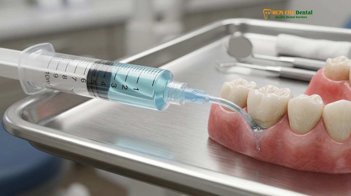
For more targeted cleaning, a curved monojet irrigation syringe (often provided by dentists after oral surgery) can be utilized. By gently placing the plastic tip just at the edge of the operculum and flushing the pocket with warm salt water or an over-the-counter antimicrobial rinse, patients can dislodge stubborn debris. If available, a 0.12% chlorhexidine gluconate mouthwash is highly effective at reducing the anaerobic bacterial count, though it should be used as a short-term adjunct to prevent tooth staining[4].
Over-the-Counter Treatments in Vietnam
Pharmacies offer various non-steroidal anti-inflammatory drugs (NSAIDs) and antiseptic mouthwashes that can temporarily manage the pain and inflammation associated with an infected wisdom tooth.
For patients seeking pericoronitis treatment saigon, local pharmacies provide several over-the-counter (OTC) options to manage the acute discomfort while awaiting a professional dental consultation. Pharmacological management focuses on interrupting the inflammatory pathway and providing systemic analgesia.
Non-steroidal anti-inflammatory drugs (NSAIDs), such as Ibuprofen, are typically the first-line choice for dental pain. Ibuprofen works by inhibiting the cyclooxygenase (COX) enzymes, thereby reducing the production of prostaglandins—the chemical messengers responsible for pain and swelling at the infection site. For patients who cannot tolerate NSAIDs due to gastrointestinal issues or allergies, Paracetamol (Acetaminophen) serves as an effective alternative for pain relief, though it lacks the potent anti-inflammatory properties of Ibuprofen.
It is crucial to understand that while OTC medications can significantly reduce pain and make chewing or swallowing more tolerable, they are strictly palliative. They mask the symptoms without addressing the root cause. The anatomical defect—the operculum—remains intact, meaning the bacterial trap is still present. Relying solely on painkillers for an extended period allows the underlying infection to fester and potentially spread to adjacent anatomical spaces.
Professional Treatments: Operculectomy vs Extraction
Definitive clinical management involves either surgically excising the overlying gum tissue (operculectomy) or completely removing the impacted wisdom tooth to permanently eliminate the infection source.
When a patient presents to a dental clinic with pericoronitis, the immediate objective is to control the acute infection. The dentist will perform a thorough subgingival irrigation using professional-grade antiseptics, often followed by the application of a topical soothing agent or a localized antibiotic gel. If the patient exhibits systemic signs of infection, such as fever, severe trismus, or facial cellulitis, a course of oral antibiotics (typically Amoxicillin or Clindamycin) will be prescribed.
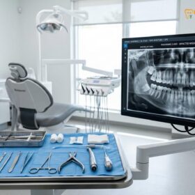
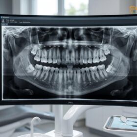
Once the acute phase is managed, a definitive long-term treatment plan must be established to prevent recurrence. The clinician will rely on digital panoramic or 3D CBCT imaging to assess the angulation, root morphology, and available arch space for the offending wisdom tooth. Based on these diagnostics, the treatment diverges into two primary surgical pathways: operculectomy or extraction.
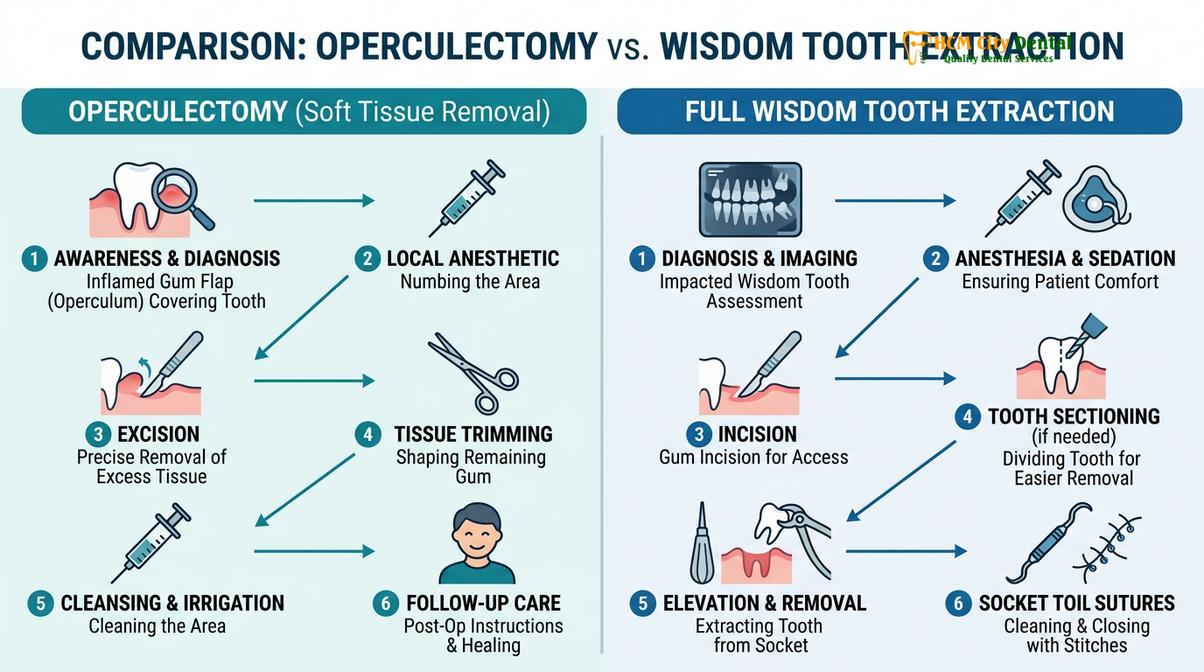
An operculectomy is a minor surgical procedure where the dentist excises the redundant gum flap covering the tooth. This is typically performed using a soft tissue diode laser, electrocautery, or a traditional scalpel. By removing the operculum, the deep pseudopocket is eliminated, transforming the area into a self-cleansing surface. However, an operculectomy is only indicated if the wisdom tooth is erupting in a perfectly vertical trajectory, has sufficient space to fully emerge, and is opposed by a functional upper molar. If these strict criteria are not met, the gum tissue often regrows over the tooth, leading to a relapse of pericoronitis.
In the vast majority of cases, wisdom tooth extraction is the most predictable and definitive treatment. If the tooth is impacted, poorly angled, or non-functional, removing it entirely is the standard of care. Modern extraction techniques utilize advanced instrumentation to gently section the tooth and remove it with minimal trauma to the surrounding bone. For a detailed understanding of this surgical workflow, patients can review comprehensive guides on wisdom tooth extraction to prepare for their procedure[5].
When to See a Doctor
While mild irritation around an erupting tooth can sometimes be managed with diligent hygiene, true pericoronitis is a progressive bacterial infection that demands professional oversight. Delaying treatment can lead to severe, life-threatening complications, particularly if the infection breaches the localized tissue barriers and enters the deep fascial spaces of the head and neck.
“If you experience difficulty swallowing, notice swelling extending down your neck, or cannot open your mouth more than a few centimeters, seek emergency dental care immediately. These are critical signs that the infection is spreading toward the airway,” advises Dr. Nguyen Van Cuong.
You should schedule an immediate clinical evaluation if you experience any of the following red flag symptoms:
- A persistent fever or chills accompanying your jaw pain.
- Visible asymmetry or swelling of the cheek, jawline, or neck.
- Severe trismus, making it difficult to open your mouth wide enough to eat or speak.
- Difficulty breathing or swallowing (dysphagia), which may indicate the onset of Ludwig’s Angina, a rapid and dangerous submandibular space infection.
- Pus or foul-tasting discharge continuously leaking from the gum tissue.
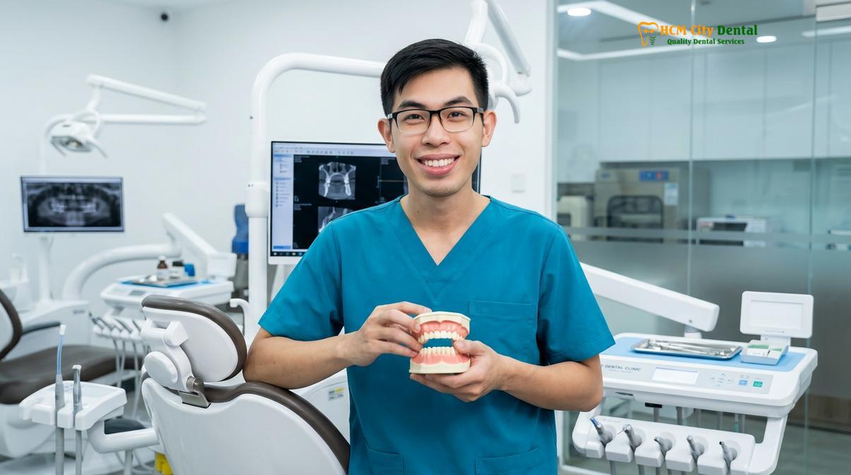
At HCMC Dental Clinic, our oral surgery team utilizes advanced 3D imaging to accurately diagnose the extent of the infection and the precise position of the impacted tooth. Early intervention not only alleviates excruciating pain but also prevents the need for more invasive hospital-based treatments. If you are experiencing symptoms of an infected wisdom tooth, do not rely solely on home remedies; secure a professional diagnostic consultation to protect your overall health.
Frequently Asked Questions
Does pericoronitis go away on its own?
Mild pericoronitis may temporarily subside with rigorous oral hygiene, but it rarely resolves permanently on its own. As long as the anatomical gum flap (operculum) remains over the partially erupted tooth, bacteria will continue to accumulate. This structural defect means that even if the pain disappears for a few weeks or months, the infection is highly likely to return, often with increased severity, until the tooth fully erupts or is surgically addressed.
How do you treat a swollen gum flap over a wisdom tooth?
Initial treatment involves flushing the area with warm saline or chlorhexidine to remove trapped debris, followed by anti-inflammatory medications. A dental professional will perform a deep subgingival irrigation to debride the pocket. Definitive treatment requires a dentist to either surgically remove the excess gum tissue (operculectomy) or extract the underlying wisdom tooth to prevent future bacterial trapping and recurrent swelling.
Can I pop a swollen gum flap over my wisdom tooth?
No, you should never attempt to pop, puncture, or cut a swollen gum flap yourself. Doing so can introduce new bacteria deep into the soft tissue, cause severe uncontrolled bleeding, and push the existing infection further into the fascial spaces of your neck and jaw. The swelling is not a simple pimple; it is highly vascularized gingival tissue engorged with inflammatory fluid that requires sterile, professional management.
What antibiotics are prescribed for pericoronitis?
Dentists typically prescribe broad-spectrum antibiotics like Amoxicillin, often combined with Metronidazole, or Clindamycin for penicillin-allergic patients. However, antibiotics are only indicated if there is systemic involvement, such as fever, swollen lymph nodes, or facial swelling. It is critical to understand that antibiotics only suppress the bacterial load temporarily; they do not cure the underlying anatomical defect (the operculum) causing the infection.
Is operculectomy better than extracting the wisdom tooth?
Operculectomy is only better if the wisdom tooth is erupting perfectly straight, has adequate room in the jaw, and serves a functional chewing purpose. In these rare cases, removing the gum flap allows the tooth to function normally. However, in most cases involving impacted or poorly angled third molars, the gum tissue will simply regrow after an operculectomy. Therefore, complete extraction is generally the superior, more predictable long-term solution.
References
- Journal of Oral and Maxillofacial Surgery. Pathogenesis and microbiology of pericoronitis. (2021).
- International Journal of Dentistry. Clinical management of acute pericoronitis and trismus. (2020).
- British Dental Journal. Anatomical risk factors for third molar impaction and infection. (2019).
- Journal of Clinical Periodontology. Efficacy of chlorhexidine irrigation in acute gingival infections. (2022).
- American Association of Oral and Maxillofacial Surgeons. Guidelines for the management of impacted third molars. (2023).