Complications after tooth extraction range from normal postoperative swelling to severe conditions like dry socket, nerve injury, or systemic infection. While mild discomfort is expected, persistent pain, excessive bleeding, or purulent discharge requires immediate clinical evaluation to prevent localized bone loss and ensure proper socket healing.
Clinical Summary:
Tooth extraction is a routine oral surgery, but the disruption of alveolar bone and gingival tissue initiates a complex biological healing cascade. While the majority of extractions heal uneventfully, complications can arise due to anatomical complexities, systemic health factors, or postoperative care failures. The most frequent complication is alveolar osteitis (dry socket), characterized by the premature loss of the protective blood clot. More severe complications include secondary hemorrhage, localized osteomyelitis, oroantral communication, and nerve paresthesia. Effective clinical management relies on accurate differential diagnosis—distinguishing normal inflammatory edema from pathological cellulitis—and implementing targeted interventions such as antimicrobial debridement, medicated dressings, and appropriate pharmacological support. Patient compliance with postoperative protocols remains the most critical factor in preventing adverse outcomes.
Key Takeaways:
- Normal healing involves a stable blood clot, followed by granulation tissue and eventual bone remodeling over several months.
- Alveolar osteitis (dry socket) occurs when the blood clot dislodges, exposing sensitive bone and nerve endings to the oral environment.
- Severe, spreading swelling or a fever after 72 hours are red flags for systemic infection requiring urgent antibiotic therapy.
- Nerve injuries, though rare, can cause temporary or permanent numbness in the lip, chin, or tongue, especially after lower wisdom tooth removal.
- Strict adherence to soft diets, avoiding suction (like straws), and gentle oral hygiene are essential to protect the extraction site.
Healing Milestones After a Tooth Extraction
Normal socket healing progresses through immediate blood clot formation, granulation tissue development within a week, and complete bone regeneration over several months.
To accurately identify when a recovery trajectory has deviated into pathology, one must first understand the precise biological milestones of normal socket healing. The extraction of a tooth leaves a void in the alveolar bone, initiating a highly orchestrated sequence of hemostasis, inflammation, proliferation, and remodeling. This cascade is heavily dependent on the patient’s immune function, local vascularity, and the surgical technique employed during the procedure[1].
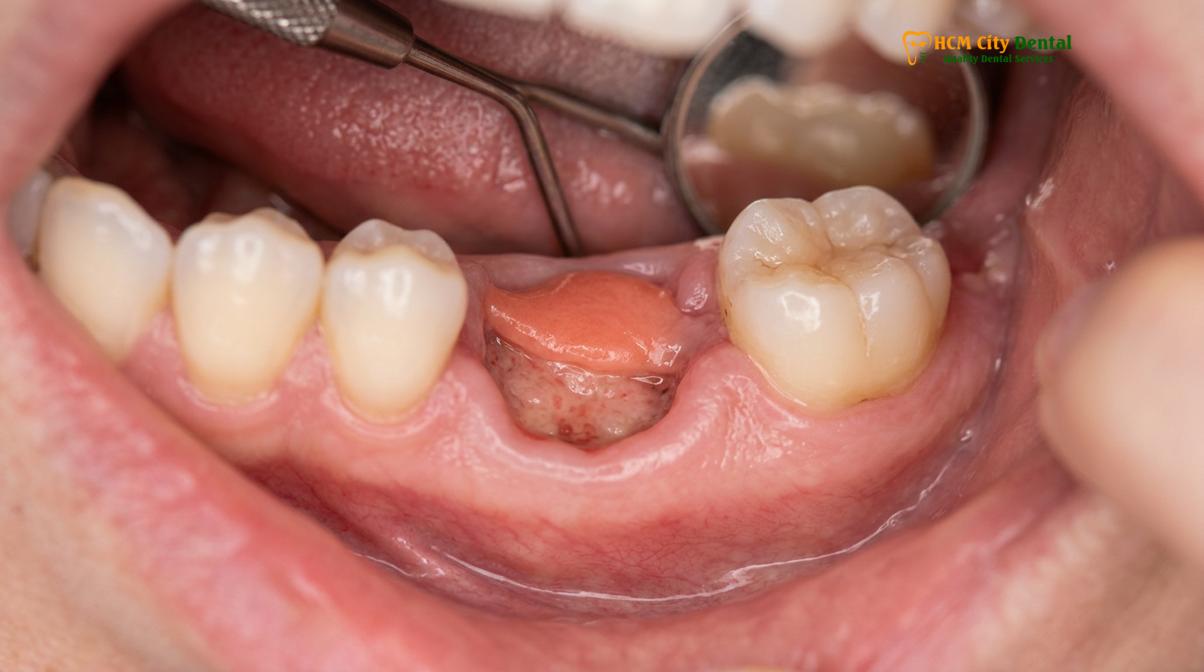
Immediately following the luxation and delivery of the tooth, the severed periodontal ligament vessels and alveolar bone marrow spaces bleed into the socket. Within the first 15 to 30 minutes, primary hemostasis occurs. Platelets aggregate and a fibrin meshwork forms, creating a robust blood clot. This clot is the foundational matrix for all subsequent healing; it acts as a biological scaffold for cellular migration and a physical barrier against oral bacteria and food debris. Protecting this clot is the primary objective of all immediate postoperative instructions.
During the first 24 to 72 hours, the inflammatory phase dominates. The body sends neutrophils and macrophages to the surgical site to phagocytize necrotic tissue fragments and bacteria. This cellular activity, combined with localized vasodilation, is responsible for the clinical presentation of normal swelling after extraction. Patients will typically experience peak edema and discomfort around day two or three. This is a physiological necessity, not a complication, provided the swelling remains localized and is not accompanied by purulent exudate or high fever.
By the end of the first week, the proliferative phase is well underway. Fibroblasts migrate into the fibrin clot, synthesizing collagen to form granulation tissue. Concurrently, angiogenesis occurs, establishing a new capillary network to supply oxygen and nutrients to the healing tissues. Clinically, the dark red blood clot is replaced by a whitish-yellow or pinkish tissue matrix. Patients often mistake this normal granulation tissue for an infection after pulling tooth, but it is a sign of healthy epithelialization. Over the next two to three weeks, the gingival mucosa will migrate across the surface, completely sealing the socket from the oral cavity.
The final phase, bone remodeling, is a prolonged process that is invisible to the naked eye. Osteoblasts begin laying down woven bone at the base and lateral walls of the socket. Over the course of three to six months, this immature bone is remodeled into mature, load-bearing trabecular bone. While the patient feels completely healed within a few weeks, the architectural restoration of the jawbone takes significantly longer[2].
| Timeframe | Biological Process | Clinical Presentation |
|---|---|---|
| 0 – 24 Hours | Hemostasis and Clot Formation | Bleeding stops, dark red clot fills the socket, mild oozing. |
| 1 – 3 Days | Inflammatory Phase | Peak swelling, localized throbbing, tissue warmth. |
| 4 – 7 Days | Proliferative Phase (Granulation) | Swelling subsides, whitish/pink tissue replaces the dark clot. |
| 2 – 4 Weeks | Epithelialization | Gum tissue fully covers the socket, no pain during eating. |
| 3 – 6 Months | Bone Remodeling | Socket fills with mature alveolar bone (visible only on X-ray). |
Identifying Common Extraction Complications
Clinical complications include alveolar osteitis (dry socket), excessive bleeding, nerve paresthesia, and sinus communication, each requiring specific diagnostic criteria for proper management.
Despite meticulous surgical technique, complications can and do occur. The oral cavity is a harsh environment, constantly subjected to mechanical forces, thermal changes, and a diverse microbiome. Recognizing extraction complications signs early allows for conservative management and prevents the escalation of localized issues into systemic health threats.
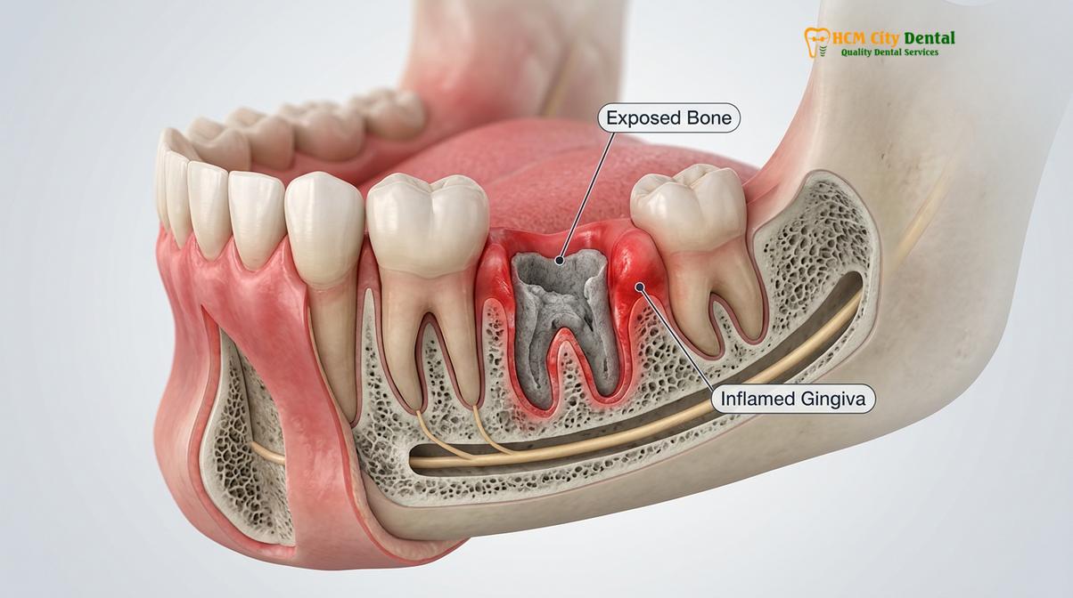
The most notorious complication is Alveolar Osteitis, universally known as dry socket. This condition occurs in approximately 2% to 5% of routine extractions but can spike to over 20% in the surgical removal of impacted mandibular third molars. Pathophysiologically, dry socket is not an infection; it is a failure of the healing cascade caused by premature fibrinolysis. The blood clot either fails to form adequately or is lysed and dislodged before granulation tissue can replace it. This leaves the alveolar bone and highly sensitive nerve endings completely exposed to the oral environment. Patients typically report a sudden, severe, radiating pain that begins three to five days post-extraction, often accompanied by a foul taste and halitosis. The pain is notoriously refractory to standard over-the-counter analgesics.
“Alveolar osteitis remains the most common postoperative complication following dental extractions. Its etiology is multifactorial, involving surgical trauma, bacterial pyrogens, and localized fibrinolytic activity. Prompt palliative intervention with medicated intra-alveolar dressings is the gold standard for managing the acute pain associated with this condition.”
— Journal of Oral and Maxillofacial Surgery
Postoperative hemorrhage is another significant concern. While slight blood-tinged saliva is normal for the first 24 hours, active, uncontrolled bleeding is a complication. Primary hemorrhage occurs immediately during or after the surgery, often due to the severing of a small arteriole or failure to achieve initial hemostasis. Secondary hemorrhage occurs days later, usually as a result of an underlying infection eroding a blood vessel or the mechanical dislodgment of the clot. Patients on anticoagulant therapies (such as Warfarin or novel oral anticoagulants) require specific perioperative management protocols to mitigate this risk[3].
Nerve injury is a rare but profoundly distressing complication, primarily associated with the extraction of mandibular molars. The inferior alveolar nerve (IAN) and the lingual nerve run in close proximity to the roots of the lower teeth. Surgical trauma, compression from edema, or direct severing of the nerve fibers can result in paresthesia (altered sensation), dysesthesia (painful sensation), or anesthesia (complete numbness) of the lower lip, chin, or tongue. Most nerve injuries are neuropraxias—temporary conduction blocks caused by bruising or swelling—that resolve spontaneously within weeks to months. However, severe axonotmesis or neurotmesis may result in permanent sensory deficits.
For maxillary (upper) molars, the primary anatomical risk is oroantral communication (OAC). The roots of the upper molars often protrude into the maxillary sinus cavity, separated only by a paper-thin layer of bone and the Schneiderian membrane. When the tooth is extracted, this delicate barrier can tear, creating a direct fistula between the oral cavity and the sinus. If left untreated, oral bacteria and fluids can enter the sinus, leading to acute or chronic sinusitis. Small communications may close spontaneously with the formation of a blood clot, but larger defects require surgical closure using buccal advancement flaps or buccal fat pad grafts.
Red Flags: Fever, Severe Swelling, and Bad Breath
Systemic symptoms such as elevated body temperature, spreading facial cellulitis, and putrid halitosis indicate a severe bacterial infection requiring urgent pharmacological and surgical intervention.
Distinguishing between the normal inflammatory response and a pathological infection is a critical clinical skill. While localized edema and mild discomfort are expected, the presence of systemic symptoms or rapidly progressive local signs constitutes a dental emergency. Odontogenic infections can spread rapidly through the fascial spaces of the head and neck, leading to life-threatening conditions if not aggressively managed.
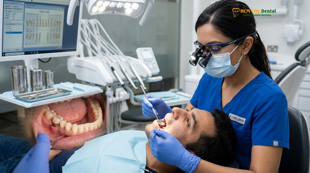
A primary red flag is the nature and progression of facial swelling. Normal surgical edema peaks at 48 to 72 hours and then begins to subside. If the swelling continues to increase after the third day, becomes indurated (hard to the touch), erythematous (red and inflamed), or spreads beyond the immediate surgical site, it is indicative of cellulitis. Cellulitis is a diffuse bacterial infection of the connective tissue. If an infection in the lower jaw spreads bilaterally into the submandibular, sublingual, and submental spaces, it can develop into Ludwig’s angina—a rapidly progressing cellulitis that can elevate the floor of the mouth, displace the tongue posteriorly, and compromise the airway[4].
Fever is a definitive systemic marker of infection. A transient, low-grade temperature (below 38°C or 100.4°F) in the first 24 hours can be a normal metabolic response to surgical trauma. However, a persistent or high-grade fever, accompanied by chills, malaise, and lethargy, strongly suggests that bacteria from the extraction site have overwhelmed local immune defenses and entered the systemic circulation. This requires immediate evaluation for broad-spectrum antibiotic therapy.
Clinical Warning: Difficulty swallowing (dysphagia), difficulty breathing (dyspnea), or an inability to open the mouth more than a few millimeters (severe trismus) are absolute medical emergencies. These symptoms suggest that an odontogenic infection has spread into deep fascial spaces, potentially threatening the airway, and requires immediate emergency room evaluation.
The presence of purulent exudate (pus) is an unequivocal sign of bacterial infection. Patients may notice a thick, yellow, green, or white discharge oozing from the socket, often accompanied by a profoundly foul odor and a bitter, metallic taste in the mouth. This putrid halitosis is typically caused by anaerobic bacteria, such as *Prevotella* and *Fusobacterium* species, which thrive in the oxygen-depleted environment of a necrotic socket.
In rare instances, particularly in patients with compromised immune systems or those taking specific medications, an infection can progress into the bone marrow, causing osteomyelitis. Furthermore, patients with a history of intravenous bisphosphonate therapy or high-dose radiation to the head and neck are at risk for Medication-Related Osteonecrosis of the Jaw (MRONJ) or Osteoradionecrosis (ORN). In these conditions, the bone fails to heal, becomes necrotic, and remains exposed through the gingiva, leading to chronic pain and recurrent infections. Any exposed bone that persists for more than eight weeks post-extraction warrants a thorough investigation for these severe pathologies.
How Dentists Treat Socket Infections and Complications
Treatment protocols involve socket debridement, antimicrobial irrigation, placement of medicated dressings, and targeted antibiotic therapy based on the severity of the complication.
When a patient presents with post extraction problems, the clinical workflow begins with a comprehensive diagnostic assessment. This includes a detailed review of the patient’s symptoms, a visual and tactile examination of the surgical site, and often, radiographic imaging (such as a periapical X-ray or CBCT scan) to rule out retained root tips, bony sequestra, or damage to adjacent anatomical structures.
For the management of alveolar osteitis (dry socket), the primary goal is palliative pain relief, as the condition is self-limiting but excruciating. The dentist will first isolate the area and gently irrigate the socket with warm sterile saline or a chlorhexidine gluconate solution. It is crucial that this irrigation is performed without excessive pressure to avoid driving debris deeper into the marrow spaces. Once the socket is cleansed of necrotic food debris and bacterial biofilm, a medicated dressing is placed. Traditionally, this involves a resorbable sponge or gauze impregnated with a paste containing eugenol (a clove oil derivative with potent analgesic and obtundent properties), an anesthetic like benzocaine, and sometimes an antimicrobial agent. This dressing provides almost immediate pain relief by shielding the exposed nerve endings. The patient is usually monitored, and the dressing may be replaced every few days until the patient’s natural granulation tissue begins to cover the bone.
If the diagnosis is an acute localized infection, the treatment shifts from palliative to curative. The cornerstone of treating any purulent odontogenic infection is establishing drainage. The dentist may need to administer local anesthesia—which can be challenging in infected tissues due to the acidic environment altering the efficacy of the anesthetic—and perform an incision and drainage (I&D) procedure. The socket is thoroughly debrided (curettage) to remove infected granulation tissue and induce fresh bleeding, effectively restarting the healing cascade. Following debridement, the site is copiously irrigated.
Pharmacological intervention is tailored to the clinical presentation. For localized infections without systemic involvement, local debridement and antimicrobial rinses may suffice. However, if there is evidence of spreading cellulitis, lymphadenopathy, or fever, systemic antibiotics are prescribed. Empirical therapy typically begins with broad-spectrum penicillins, such as Amoxicillin, often combined with Clavulanate to combat beta-lactamase-producing bacteria. For penicillin-allergic patients, Clindamycin or Azithromycin are common alternatives. It is imperative that patients complete the entire course of antibiotics to prevent the development of resistant bacterial strains[5].
Clinical Case Review: Managing Delayed Healing
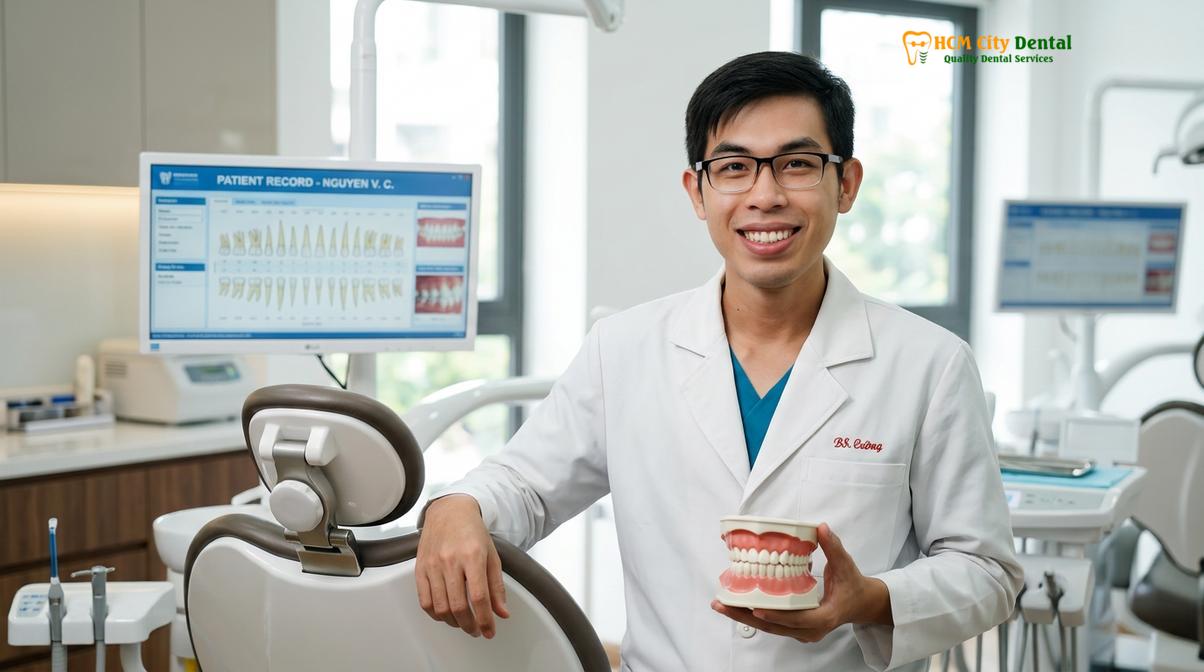
A 45-year-old patient presented to HCMC Dental Clinic in Ho Chi Minh City complaining of severe, radiating jaw pain five days after a complex molar extraction. Clinical examination by Dr. Nguyen Van Cuong revealed a complete absence of the blood clot and exposed alveolar bone, confirming a diagnosis of severe alveolar osteitis. Dr. Cuong performed gentle saline irrigation to remove accumulated debris and placed a specialized eugenol-based intra-alveolar dressing. The patient reported a 90% reduction in pain within twenty minutes. Dr. Cuong scheduled a follow-up appointment 48 hours later to replace the dressing and monitor the onset of secondary epithelialization, ensuring the socket healed without further infectious complications.
In modern dental practice, advanced biological adjuncts are increasingly used to manage and prevent complications. Platelet-Rich Fibrin (PRF), derived from the patient’s own blood spun in a centrifuge, can be placed into the socket immediately after extraction or during the treatment of a complication. PRF releases a high concentration of growth factors over several days, significantly accelerating angiogenesis, soft tissue healing, and bone regeneration, while also reducing the incidence of postoperative pain and infection.
For patients requiring comprehensive management of complex cases, exploring specialized emergency dental care protocols ensures that all surgical and pharmacological interventions are handled with the highest clinical standards.
At-Home Care to Prevent Complications
Preventive care relies on maintaining the blood clot through soft diets, avoiding negative oral pressure, and practicing gentle antimicrobial hygiene during the initial healing phase.
The success of a tooth extraction is heavily dependent on the patient’s adherence to postoperative instructions. The primary objective of at-home care is the preservation and protection of the fragile blood clot during the critical first 72 hours. Any mechanical or chemical disruption can halt the healing cascade and precipitate complications.
Pain and swelling management is the first line of at-home care. Non-Steroidal Anti-Inflammatory Drugs (NSAIDs), such as Ibuprofen, are the pharmacological agents of choice. They work by inhibiting the cyclooxygenase (COX) enzymes, thereby reducing the synthesis of prostaglandins—the lipid compounds responsible for mediating inflammation and pain. For optimal efficacy, NSAIDs should be taken on a strict schedule (e.g., every 6 to 8 hours) rather than waiting for the pain to become severe. If NSAIDs are contraindicated due to gastrointestinal or renal issues, Acetaminophen is a safe alternative, though it lacks the anti-inflammatory properties. For detailed pharmacological protocols, patients can refer to a comprehensive Dental Pain Medication Guide.
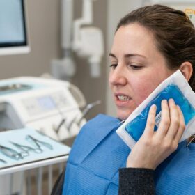
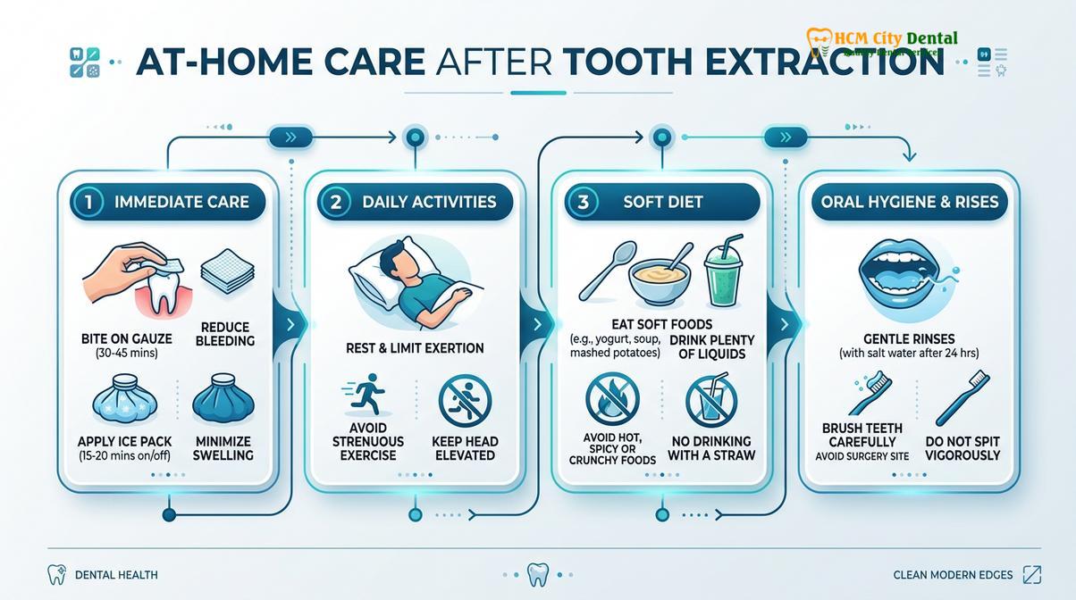
To manage edema, cold therapy is highly effective. Applying an ice pack to the outside of the face over the surgical site for 15-minute intervals during the first 24 hours causes local vasoconstriction, reducing blood flow and limiting the extravasation of fluid into the tissues. After 48 hours, switching to warm, moist compresses can promote vasodilation, helping the lymphatic system clear the accumulated inflammatory exudate.
“Patient compliance with postoperative hygiene and dietary restrictions is the most significant variable in preventing alveolar osteitis and secondary infections. The mechanical preservation of the primary blood clot must be the focal point of all patient education following exodontia.”
— International Journal of Oral Science
Dietary modifications are essential. Patients must adhere strictly to a soft diet—such as yogurt, applesauce, scrambled eggs, and mashed potatoes—for the first few days. Hard, crunchy, or sharp foods (like chips or nuts) can physically traumatize the healing tissue or become lodged in the socket. Furthermore, patients must avoid any action that creates negative pressure in the oral cavity. This means absolutely no drinking through straws, no forceful spitting, and no vigorous rinsing. The suction created by these actions can easily dislodge the blood clot.
Smoking is perhaps the most significant modifiable risk factor for extraction complications. The negative pressure from inhaling smoke can dislodge the clot, while the carbon monoxide and nicotine in cigarette smoke cause severe peripheral vasoconstriction. This drastically reduces the blood supply to the healing bone, starving the tissue of oxygen and immune cells. Dr. Nguyen Van Cuong strongly advises patients to cease smoking for a minimum of 72 hours post-extraction to mitigate the high risk of dry socket and delayed healing.
Oral hygiene must be maintained to reduce the bacterial load, but it must be modified. Patients should continue to brush and floss their remaining teeth normally but must avoid the surgical site. After 24 hours, gentle rinsing with warm salt water (half a teaspoon of salt in a cup of warm water) or a prescribed Chlorhexidine mouthwash can help keep the socket clean without disrupting the clot.
When to Contact Your Dentist for a Follow-Up
While minor discomfort and swelling are part of the normal healing journey, certain symptoms require immediate professional evaluation. You should contact your dental clinic if you experience any of the following:
- Uncontrolled Bleeding: If heavy bleeding continues despite applying firm, continuous pressure with a gauze pad for 45 to 60 minutes.
- Severe, Worsening Pain: Pain that dramatically increases after the third day and is not relieved by prescribed pain medications, indicating a potential dry socket.
- Signs of Spreading Infection: A fever exceeding 38°C (100.4°F), swelling that spreads to the neck or eye, or difficulty swallowing and breathing. (See our Swollen Face Tooth Infection Emergency Guide for more details).
- Prolonged Numbness: Any loss of sensation, tingling, or numbness in the lips, chin, or tongue that persists for more than 24 hours after the local anesthesia should have worn off.
- Foul Discharge: The presence of yellow or green pus oozing from the extraction site, accompanied by a persistent bad taste. Recognizing Tooth Infection Spreading Signs early is crucial for effective treatment.
If you are experiencing any of these symptoms, or if you have concerns about your recovery following a Tooth Extraction, do not attempt to manage it solely at home. Prompt clinical intervention is the safest way to ensure your oral health is protected and your recovery gets back on track.
Frequently Asked Questions
How do I know if my tooth extraction socket is infected?
An infected extraction socket typically presents with throbbing pain that worsens after a few days, accompanied by yellow or green purulent discharge, severe facial swelling, and a foul odor or taste. Unlike normal healing, an infection often causes systemic symptoms like a low-grade fever and swollen lymph nodes in the neck. If you experience these signs, immediate clinical evaluation is required for antibiotic therapy and socket debridement. A dentist will need to clean the site and prescribe appropriate medications to halt the bacterial spread.
Is severe swelling normal 3 days after tooth extraction?
Moderate swelling peaking at 48 to 72 hours is a normal physiological response to surgical trauma. However, severe swelling that rapidly expands, causes difficulty swallowing, or is hot to the touch is not normal. If the swelling continues to increase significantly past the third day or spreads toward your eye or neck, it may indicate a spreading fascial space infection requiring urgent dental intervention. Cold compresses are effective initially, but pathological swelling requires clinical diagnosis.
What does dry socket or a failed clot look like?
A dry socket appears as an empty, dark hole where the tooth was extracted, often with visible whitish or grayish bone exposed at the base, rather than a dark red blood clot. Clinically known as alveolar osteitis, it is characterized by the absence of normal granulation tissue and is accompanied by intense, radiating pain that typically begins three to four days post-extraction. The pain often radiates to the ear or temple and is not easily managed with standard painkillers.
Can a tooth extraction cause jaw stiffness (trismus)?
Yes, jaw stiffness, clinically referred to as trismus, is a common complication following the extraction of lower molars, particularly impacted wisdom teeth. It occurs due to inflammation and muscle spasms in the muscles of mastication (chewing muscles) caused by surgical trauma or prolonged mouth opening during the procedure. Trismus usually resolves gradually within one to two weeks with warm compresses and gentle stretching exercises, though severe cases may require muscle relaxants.
When is it safe to stop worrying about complications after extraction?
For most routine extractions, the critical window for acute complications like dry socket or primary infection passes after the first seven to ten days. Once the socket is covered by a layer of new pink epithelial tissue and you can eat comfortably without pain, the risk drops significantly. However, complete internal bone regeneration takes several months, during which you should maintain excellent oral hygiene. If you have had a complex surgical extraction, your dentist will advise you on your specific healing timeline.
References
- Journal of Oral and Maxillofacial Surgery. Pathophysiology and management of alveolar osteitis. (2021).
- American Dental Association (ADA). Clinical guidelines for post-extraction complications. (2022).
- International Journal of Oral Science. Inflammatory responses and bone healing post-extraction. (2020).
- British Journal of Oral and Maxillofacial Surgery. Nerve injuries in dental extraction: prevention and management. (2019).
- Journal of Endodontics. Odontogenic infections and fascial space spread. (2023).