For decades, the rehabilitation of the severely atrophic posterior maxilla has presented one of the most formidable challenges in oral and maxillofacial surgery. When patients lose their upper molars, the combination of rapid alveolar bone resorption and the downward pneumatization of the maxillary sinus often leaves a paper-thin ridge of bone that is entirely inadequate for traditional dental implants. Historically, the only solution was highly invasive bilateral sinus lift surgery—a procedure requiring extensive bone grafting, prolonged healing periods of up to nine to twelve months, and significant patient morbidity. Today, advanced surgical protocols have revolutionized this paradigm. By utilizing the dense cortical bone pillars located deep in the posterior facial skeleton, specialists can bypass the sinus cavity entirely. The clinical protocol for pterygoid implants tuberosity anchorage no sinus lift all on 6 restorations represents the pinnacle of modern graftless implantology, allowing patients to achieve immediate, fully functional, and biomechanically superior full-arch teeth in a single surgical intervention.
At HCMC Dental Clinic, located in the medical epicenter of District 1, Ho Chi Minh City, our surgical and prosthodontic teams specialize in these complex graftless rehabilitations. Under the clinical direction of Dr. Nguyen Van Cuong, DDS (Clinical Director — MOH License 0003265/BL-CCHN), international patients, dental travelers, and expatriates have access to world-class implantology that adheres strictly to global standards. By integrating 3D Cone Beam Computed Tomography (CBCT), Dynamic Optical Navigation, and advanced biomaterials, we provide a predictable, safe, and highly cost-effective alternative to traditional bone grafting. This comprehensive clinical review explores the anatomical foundations, biomechanical advantages, and surgical execution of pterygoid dental implants in Vietnam.
Clinical Summary:
Pterygoid implants are specialized, elongated dental implants (15mm to 20mm) designed to anchor into the dense cortical bone of the pyramidal process of the palatine bone and the pterygoid process of the sphenoid bone. By engaging this highly mineralized posterior pillar, surgeons can achieve exceptional primary stability (>45 Ncm) in the severely resorbed upper jaw without the need for sinus lifts or bone grafting. This graftless approach facilitates immediate loading protocols, eliminates detrimental distal prosthetic cantilevers, and provides robust posterior support for full-arch monolithic zirconia restorations. At HCMC Dental Clinic in District 1, Ho Chi Minh City, Dr. Nguyen Van Cuong, DDS, utilizes 3D CBCT mapping and dynamic navigation to safely execute these complex procedures, offering international patients a highly predictable, minimally invasive, and cost-effective pathway to complete maxillary rehabilitation.
Key Takeaways:
- Graftless Posterior Anchorage: Pterygoid implants completely bypass the pneumatized maxillary sinus, eliminating the need for invasive lateral window sinus lifts and prolonged bone graft maturation periods.
- Bicortical Stability: By anchoring into the dense cortical bone of the sphenoid and palatine bones, pterygoid implants routinely achieve insertion torques exceeding 45 Ncm, enabling immediate loading of provisional prostheses.
- Biomechanical Superiority: Placing implants in the far posterior region eliminates distal cantilevers, optimizing the anterior-posterior (A-P) spread and protecting the final monolithic zirconia bridge from occlusal fracture.
- Advanced Surgical Safety: Precise 3D CBCT vascular mapping and Dynamic Optical Navigation are mandatory to safely navigate the complex anatomy of the pterygopalatine fossa and avoid the greater palatine artery.
- Transparent Global Pricing: HCMC Dental Clinic offers Official 2026 Dental Price List & -40% Online Booking Fee Schedule, providing premium graftless full-arch rehabilitations at a fraction of the cost found in Australia, the US, or the UK.
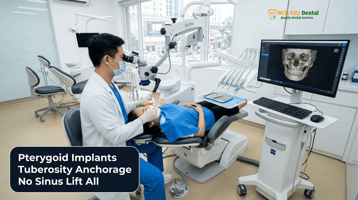
Why the Posterior Upper Jaw (Maxillary Tuberosity) Has the Softest D4 Bone and Highest Traditional Implant Failure Rate
To understand the profound clinical value of pterygoid implants, one must first examine the unique and challenging biology of the posterior maxilla. The human upper jaw is structurally designed to absorb and dissipate the heavy compressive forces of mastication. However, the bone density in the posterior maxilla—specifically the maxillary tuberosity region—is notoriously poor. According to the widely accepted Lekholm and Zarb classification of bone quality, the posterior maxilla is predominantly composed of Type III and Type IV bone. Type IV bone, often described clinically as resembling “balsa wood” or “styrofoam,” consists of a very thin layer of cortical bone surrounding a sparse, highly porous core of low-density trabecular (cancellous) bone.
When a patient loses their maxillary molars, two distinct physiological processes immediately begin to degrade the remaining bone volume. First, the lack of mechanical stimulation (periodontal ligament tension) triggers rapid osteoclastic activity, leading to severe horizontal and vertical alveolar ridge resorption. Second, the maxillary sinus cavity, which sits directly above the molar roots, begins to expand downward in a process known as sinus pneumatization. As air pressure within the sinus cavity exerts continuous force on the sinus floor, the bone resorbs from the inside out. Within a few years of tooth loss, the residual bone height between the oral cavity and the sinus floor can diminish to less than 2 to 3 millimeters[1].
Attempting to place standard cylindrical or tapered dental implants into this compromised environment is fraught with high failure rates. Traditional implants rely on primary stability—the mechanical friction between the implant threads and the surrounding bone—to remain immobile during the critical osseointegration phase. In D4 bone, achieving adequate insertion torque (typically >35 Ncm) is exceedingly difficult. Furthermore, if the residual bone height is less than 4mm, a standard implant will perforate the Schneiderian membrane of the maxillary sinus, potentially leading to acute sinusitis, graft infection, and catastrophic implant failure. For decades, the standard of care for this anatomical deficit was the lateral window sinus lift—a procedure where the sinus membrane is surgically elevated and a xenograft or allograft bone substitute is packed into the space. While effective, sinus lifts require 6 to 9 months of healing before implants can be placed, subjecting the patient to prolonged edentulism, multiple surgeries, and significant postoperative swelling and morbidity.
What Is a Pterygoid Implant? Engaging the Dense Cortical Pillar of the Maxillary Tuberosity, Palatine Bone, and Pterygoid Process
The pterygoid implant was first conceptualized in the late 1980s and formally introduced to the surgical community by Dr. Jean-François Tulasne in 1989. The fundamental philosophy behind this technique is to abandon the soft, resorbed bone of the alveolar ridge and instead seek anchorage in the dense, highly mineralized basal bone of the facial skeleton that does not resorb following tooth loss. The target for this anchorage is the pterygomaxillary region, a complex anatomical junction where three distinct bony structures converge: the posterior aspect of the maxillary tuberosity, the pyramidal process of the palatine bone, and the pterygoid process of the sphenoid bone.
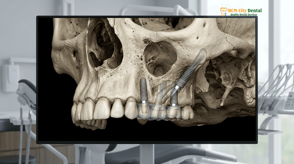
A pterygoid implant is specifically engineered for this unique anatomical pathway. Unlike standard implants which range from 8mm to 12mm in length, pterygoid implants are significantly longer, typically measuring between 15mm and 20mm. The surgical trajectory begins at the maxillary tuberosity (usually in the region of the second or third molar). The osteotomy is directed posteriorly, superiorly, and slightly medially, typically at a 45-degree angle to the Frankfort horizontal plane. The implant passes through the soft cancellous bone of the tuberosity, traverses the pyramidal process of the palatine bone, and finally engages the dense cortical plates of the pterygoid process of the sphenoid bone. The apex of the implant often protrudes 1mm to 2mm into the pterygoid fossa to ensure maximum bicortical engagement[2].
This bicortical anchorage is the secret to the pterygoid implant’s success. The pyramidal and pterygoid processes are composed of dense, Type I and Type II cortical bone. The average thickness of the bone at this juncture is between 6mm and 6.7mm, providing a robust, unyielding pillar of support. By engaging this dense cortical bone, the surgeon can routinely achieve insertion torques well in excess of 45 Ncm, even in patients with the most severe maxillary atrophy. This high primary stability is the absolute prerequisite for immediate loading protocols, allowing the prosthodontic team to attach a fixed provisional bridge on the same day as surgery.
Biomechanical Triumph: Eliminating Distal Prosthetic Cantilevers and Delivering Full 14-Tooth First-and-Second Molar Chewing
The placement of pterygoid implants is not merely a surgical workaround for missing bone; it is a profound biomechanical upgrade for the final prosthetic restoration. In traditional 3D-Guided All-on-4 & All-on-6 Full-Arch Dental Implants in Vietnam protocols, the most posterior implants are often tilted distally to avoid the anterior wall of the maxillary sinus. However, even with this angulation, the implants typically emerge in the second premolar or first molar region. To provide the patient with a full complement of chewing teeth (up to the second molars), the prosthetic bridge must extend backward beyond the last implant. This unsupported extension is known as a distal cantilever.
In implant prosthodontics, distal cantilevers are biomechanically hazardous. They act as Class I levers. When a patient bites down on the cantilevered molar, the occlusal force is multiplied, creating immense compressive stress on the most posterior implant and severe tensile stress on the most anterior implants. Over time, this cantilever effect can lead to a cascade of complications, including screw loosening, fracture of the prosthetic framework, marginal bone loss (saucerization) around the implant neck, and ultimately, loss of osseointegration. To mitigate this risk, traditional All-on-4 prostheses are often shortened to only 10 or 12 teeth, depriving the patient of full molar masticatory efficiency.
Pterygoid implants completely neutralize this biomechanical threat. By placing the terminal support in the far posterior region of the maxilla (the second or third molar position), the anterior-posterior (A-P) spread of the implants is dramatically increased. The A-P spread is the distance from the center of the most anterior implant to the distal aspect of the most posterior implant; a larger A-P spread provides a more stable foundation for the prosthesis. With pterygoid implants, the distal cantilever is entirely eliminated. The prosthesis is fully supported from the central incisors all the way back to the second molars.
“The elimination of the distal cantilever through pterygoid anchorage is a game-changer in full-arch prosthodontics. It transforms the biomechanical environment from a high-stress lever system into a fully supported, rigid platform. This allows us to confidently deliver a full 14-tooth monolithic zirconia restoration that can withstand the heaviest bruxism forces without the fear of catastrophic fracture.”
— Dr. Nguyen Van Cuong, DDS (Clinical Director, HCMC Dental Clinic)
This robust support is particularly critical when utilizing advanced restorative materials. At HCMC Dental Clinic, our final restorations adhere to Misch’s FP1, FP2, and FP3 classifications for fixed prostheses. For patients with severe bone loss requiring an FP3 prosthesis (which replaces both missing teeth and missing gingival tissues), we utilize 1,100–1,200 MPa monolithic 3Y-TZP zirconia. This ultra-high-strength ceramic is virtually indestructible when properly supported, but it is brittle under tensile stress caused by cantilevers. By utilizing pterygoid implants, we ensure the zirconia is kept under pure compression. Furthermore, we meticulously design the emergence profile using the Su & Gonzalez-Martin Critical/Subcritical contour principles via custom PEEK healing abutments and PMMA provisionals, ensuring optimal soft tissue health and aesthetics. For patients with heavy bruxism, a hard Michigan occlusal splint is provided to protect the restoration during sleep[3].
Pterygoid Implants vs. Bilateral Sinus Lifts vs. Zygomatic Implants: Morbidity, Immediate Loading (>45 Ncm), and Indications
When a patient presents with a severely atrophic posterior maxilla, the surgical team must evaluate three primary treatment pathways: traditional sinus augmentation, extra-maxillary zygomatic implants, or pterygomaxillary implants. Each approach has distinct indications, advantages, and morbidity profiles.
1. Bilateral Sinus Lifts with Bone Grafting:
The traditional approach involves elevating the Schneiderian membrane and placing a bone graft (often utilizing a chairside Smart Dentin Grinder to create a 300–1,200 µm Autogenous Demineralized Dentin Matrix that releases native BMP-2 for accelerated healing). While highly predictable, this approach is time-consuming. The graft must mature for 6 to 9 months before implants can be placed, and another 3 to 4 months must pass before the implants can be loaded. This means the patient must wear a removable denture for up to a year. Furthermore, sinus lifts carry risks of membrane perforation, graft infection, and postoperative sinusitis. For patients seeking Same-Day Dental Implants vs Traditional 2-Stage Implants in Vietnam, sinus lifts are often a deterrent.
2. Zygomatic Implants:
Zygomatic implants are ultra-long fixtures (35mm to 55mm) that anchor into the cheekbone. They are an excellent graftless solution for extreme maxillary atrophy. However, zygomatic implant surgery is highly invasive. It requires extensive flap reflection, often necessitates general anesthesia, and carries higher risks of postoperative complications, including severe facial swelling, hematoma, sinusitis, and in rare cases, altered sensation in the infraorbital nerve. Zygomatic implants are generally reserved for patients who have lost virtually all bone in both the anterior and posterior maxilla.
3. Pterygoid Implants:
Pterygoid implants offer a “middle path” that provides the immediate loading benefits of zygomatic implants without the extreme surgical morbidity. Because the surgical site is confined to the intraoral retromolar region, there is no need for extra-maxillary dissection. The procedure can routinely be performed under local anesthesia with oral sedation. The dense cortical anchorage guarantees the >45 Ncm torque required for immediate loading, allowing the patient to receive fixed teeth on the day of surgery. Pterygoid implants are specifically indicated when the anterior maxilla has sufficient bone for standard implants, but the posterior maxilla is deficient.
In some borderline cases where the residual bone height under the sinus is between 4mm and 6mm, an alternative minimally invasive approach is the use of 6mm–8mm Straumann Roxolid (Ti-Zr) splinted short implants. These ultra-strong, short implants can sometimes negate the need for both sinus lifts and pterygoid implants, provided the occlusal forces are carefully managed[4].
Clinical Case Study: Graftless Maxillary Rehabilitation in District 1
Patient Profile: A 62-year-old expatriate from Australia presented to HCMC Dental Clinic with a failing upper removable denture. He had been told by a local clinic in Sydney that he required bilateral sinus lifts, bone grafting, and a 14-month treatment timeline at a cost exceeding $35,000 AUD.
Clinical Assessment: 3D CBCT analysis revealed severe posterior maxillary atrophy (2mm residual bone height below the sinuses) but adequate bone in the anterior premaxilla.
Treatment Protocol: Dr. Nguyen Van Cuong designed a graftless All-on-6 protocol utilizing four standard anterior implants and two posterior Pterygoid implants (18mm length). The surgery was performed under local anesthesia using Dynamic Optical Navigation.
Outcome: All six implants achieved >50 Ncm insertion torque. A fixed PMMA provisional bridge was delivered on the same day. Four months later, a final 14-tooth monolithic 3Y-TZP Zirconia bridge was seated. The patient avoided sinus surgery, eliminated distal cantilevers, and completed the entire treatment for under $11,000 USD, utilizing our Dental Implants in Vietnam: Complete Cost Breakdown & How to Avoid Hidden Fees transparency policy.
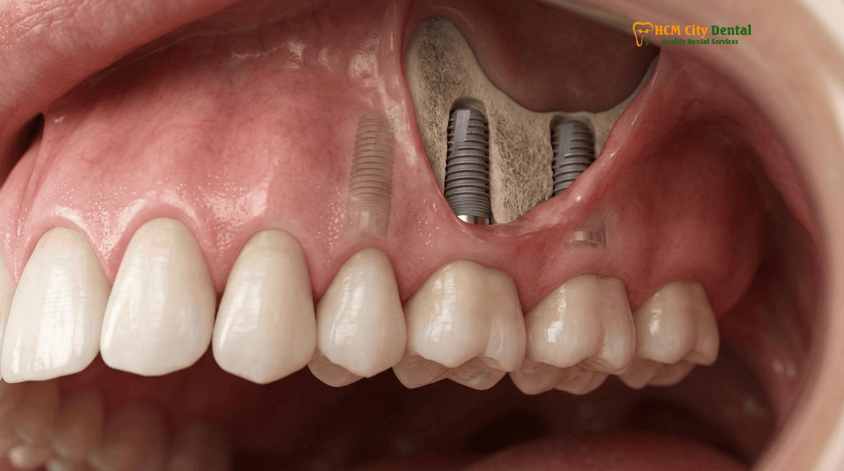
3D CBCT Vascular & Anatomical Safety Mapping: Avoiding the Greater Palatine Artery and Internal Maxillary Plexus
While the biomechanical benefits of pterygoid implants are undeniable, the surgical execution is highly technique-sensitive. The pterygomaxillary region is a complex anatomical crossroad housing several critical neurovascular structures. The primary concern during osteotomy preparation is the pterygopalatine fossa, which lies immediately superior and medial to the pterygoid process. This fossa contains the maxillary division of the trigeminal nerve (V2), the pterygopalatine ganglion, and the terminal branches of the internal maxillary artery, including the descending palatine artery.
If a surgeon deviates medially or penetrates too deeply into the pterygopalatine fossa, there is a risk of severe arterial hemorrhage from the internal maxillary plexus or the greater palatine artery. To mitigate these risks, blind or freehand placement of pterygoid implants is strongly discouraged. At HCMC Dental Clinic, the utilization of high-resolution 3D CBCT imaging is mandatory for every pterygoid case. The CBCT scan allows the surgical team to precisely map the trajectory of the descending palatine canal and measure the exact distance from the tuberosity to the pterygoid plate.
To translate this digital plan into surgical reality, we employ advanced surgical guidance systems. While static 3D-printed sleeve guides are excellent for anterior implants (as detailed in our guide to 3D-Printed Surgical Guide & Flapless Computer-Guided Dental Implants in Vietnam: Precision & Cost), the posterior location of the tuberosity often makes it difficult to accommodate bulky surgical stents, especially in patients with limited mouth opening (trismus). Therefore, Dynamic Optical Navigation is often the preferred modality. This technology uses overhead optical cameras to track the surgical drill in real-time, overlaying its exact position onto the patient’s 3D CBCT scan on a monitor. This allows Dr. Cuong to navigate the drill with sub-millimeter accuracy, ensuring the implant engages the dense cortical bone while remaining safely away from the vascular plexus.
Furthermore, long-term maintenance of these posterior implants is critical. Due to their distal location, oral hygiene can be challenging for the patient. We strictly adhere to Lang’s CIST Protocol (Cumulative Interceptive Supportive Therapy) for the management of peri-implant mucositis and peri-implantitis. If inflammation is detected, our protocol includes erythritol air-polishing (Swiss EMS AirFlow GBT), PEEK ultrasonic tips, and diode/Er:YAG laser decontamination. In severe cases of peri-implantitis, titanium brush implantoplasty combined with L-PRF Sticky Bone Guided Bone Regeneration (GBR) is utilized to arrest bone loss and regenerate the supporting tissues[5].
Clinical Precaution: When to Contact Your Specialist
Pterygoid implant surgery is highly advanced and should only be performed by experienced implantologists. Patients with severe trismus (limited mouth opening less than 35mm) may not be candidates, as the surgical instruments cannot physically reach the posterior maxilla. If you experience excessive, pulsatile bleeding, severe prolonged numbness in the palate, or difficulty swallowing following posterior maxillary surgery, contact Dr. Nguyen Van Cuong immediately via our 24/7 WhatsApp Hotline at +84 853 020 003 for urgent clinical evaluation.
2026 Pterygoid Implant & Graftless Full-Arch Maxillary Rehab Fee Schedule at HCMC Dental Clinic (District 1)
One of the most compelling reasons international patients travel to Vietnam for complex implantology is the profound cost efficiency without any compromise in clinical quality, biomaterial safety, or technological precision. In Western nations, a graftless full-arch rehabilitation utilizing pterygoid implants and a monolithic zirconia bridge can easily exceed $30,000 to $45,000 USD per arch. At HCMC Dental Clinic, our transparent pricing model ensures that patients receive premium European and Korean implant systems at a fraction of the global cost.
Below is our official 2026 fee schedule. By utilizing our online booking system, international patients can secure a -40% reduction compared to standard walk-in rates. We guarantee that there are no hidden fees; the prices listed for full-arch packages include the surgical placement, the temporary provisional bridge, and the final monolithic Zirconia restoration.
| Clinical Procedure / Implant System | 2026 Online Booking Fee (USD) | Standard Walk-In Fee (USD) | VND Equivalent (Online) |
|---|---|---|---|
| Single Korean Implant (Dentis / Neo Biotech / Dentium) Includes fixture + standard abutment | $462 – $654 | $769 – $1,090 | 12,000,000 – 17,000,000 VND |
| Single European / Swiss Implant (Straumann BLT-SLA / BLA-Active) Premium titanium-zirconium alloy | $962 – $1,346 | $1,603 – $2,244 | 25,000,000 – 35,000,000 VND |
| All-on-4 Full-Arch Implants (Economy) Includes temporary & final Zirconia bridge | From $6,346 / arch | N/A | 165,000,000 VND |
| All-on-6 Full-Arch Implants (Economy) Ideal for Pterygoid posterior anchorage | From $8,654 / arch | N/A | 225,000,000 VND |
| All-on-6 Full-Arch Implants (Straumann SLA/Active) Premium Swiss graftless rehabilitation | $12,308 – $13,462 / arch | N/A | 320,000,000 – 350,000,000 VND |
| 3D CBCT Scan (Diagnostic Imaging) Mandatory for Pterygoid surgical planning | $18 | $18 | 480,000 VND |
| Surgical Wisdom Tooth Extraction (Piezotome) Required if impacted in the tuberosity | $58 – $115 / tooth | N/A | 1,500,000 – 3,000,000 VND |
“Our mission at HCMC Dental Clinic is to democratize access to elite-level implantology. By leveraging digital workflows, in-house CAD/CAM milling, and direct partnerships with global implant manufacturers like Straumann and Dentium, we can offer life-changing graftless procedures like pterygoid implants at a price point that makes dental tourism to Vietnam a highly logical and safe medical decision.”
— Dr. Nguyen Van Cuong, DDS
For patients who require preliminary procedures before implant placement, such as the removal of impacted third molars in the tuberosity region, we utilize advanced ultrasonic bone surgery. You can learn more about this atraumatic technique in our guide to Piezosurgery Ultrasonic Bone Surgery for Sinus Lifts & Impacted Wisdom Teeth in Vietnam: Safety & Cost. If you have been told you do not have enough bone for implants, or if you wish to avoid the prolonged healing times of sinus grafting, contact our international coordination team today to schedule a comprehensive 3D CBCT evaluation and discover if pterygoid implants are the right solution for your smile.
Frequently Asked Questions
How can a Pterygoid implant achieve immediate loading stability (>45 Ncm) when the back of the upper jaw has soft bone?
While the maxillary tuberosity itself consists of soft Type IV cancellous bone, a Pterygoid implant is specifically designed to bypass this porous region. It is inserted at a 45-degree angle to engage the dense, highly mineralized cortical bone of the pyramidal process of the palatine bone and the pterygoid process of the sphenoid bone. This bicortical anchorage provides exceptional primary stability, routinely exceeding 45 Ncm, which is the critical threshold required for immediate loading protocols in full-arch restorations.
What is the clinical difference between a Pterygoid implant and a Zygomatic (cheekbone) implant?
Both are graftless solutions for severe maxillary atrophy, but they target different anatomical structures. A Pterygoid implant (typically 15mm to 20mm in length) is placed in the posterior maxilla, anchoring into the sphenoid and palatine bones to eliminate distal cantilevers. A Zygomatic implant is much longer (35mm to 55mm) and is anchored into the zygomatic arch (cheekbone). Zygomatic implants are generally reserved for the most extreme cases of anterior and posterior bone loss, whereas Pterygoid implants specifically resolve posterior bone deficiencies without the higher surgical morbidity associated with extra-maxillary zygomatic placement.
Does placing a Pterygoid implant behind the maxillary sinus cause sinus infections or congestion?
No. When planned correctly using 3D CBCT imaging and Dynamic Optical Navigation, a Pterygoid implant completely bypasses the maxillary sinus cavity. Because the implant trajectory runs posterior to the posterior wall of the sinus, it does not perforate the Schneiderian membrane. Consequently, it does not interfere with normal sinus drainage or ciliary function, effectively eliminating the risk of implant-related sinusitis that can sometimes occur with traditional sinus lift bone grafting procedures.
Why does adding two posterior Pterygoid implants protect a full-arch Monolithic Zirconia bridge from fracturing?
In a standard All-on-4 configuration, the most posterior implants usually emerge around the premolar region. To provide molar chewing function, the prosthetic bridge must extend backward without underlying bone support—a design known as a distal cantilever. Long cantilevers act as levers, multiplying occlusal forces and increasing the risk of zirconia fracture or implant failure. By placing Pterygoid implants in the far posterior region, the distal cantilever is entirely eliminated. This creates a biomechanically stable, fully supported anterior-posterior (A-P) spread, allowing the 1,100–1,200 MPa monolithic 3Y-TZP zirconia bridge to safely absorb heavy bite forces.
How much does graftless full-arch upper jaw rehabilitation with Pterygoid implants cost at HCMC Dental Clinic in District 1?
At HCMC Dental Clinic in District 1, Ho Chi Minh City, our 2026 online booking fee schedule offers a complete All-on-6 Full-Arch Implant package (which can incorporate Pterygoid implants for posterior anchorage) starting from $8,654 USD per arch (225,000,000 VND). This comprehensive package includes the surgical placement of the implants, the temporary provisional bridge, and the final monolithic Zirconia bridge. Premium options utilizing Straumann SLA/Active implants range from $12,308 to $13,462 USD per arch, representing a savings of approximately 40% compared to walk-in rates or standard fees in Australia and the United States.
References
- Journal of Prosthetic Dentistry. Biomechanical evaluation of pterygoid implants in graftless maxillary rehabilitation: A finite element analysis. (2023).
- International Journal of Oral & Maxillofacial Implants. Survival rates and marginal bone loss of pterygomaxillary implants: A 10-year retrospective clinical study. (2022).
- Clinical Implant Dentistry and Related Research. Immediate loading of pterygoid implants in the severely atrophic maxilla: Clinical outcomes and prosthetic complications. (2024).
- Journal of Cranio-Maxillofacial Surgery. Anatomical considerations and 3D CBCT mapping of the pterygopalatine fossa for safe pterygoid implant placement. (2021).
- Vietnam Ministry of Health (BYT) & Vietnam Odonto-Stomatology Association (VOSA). National Clinical Guidelines on Specialist Dental Care, Biomaterial Traceability & EN 13060 Class B Infection Control. (2024).