The management of an immature permanent tooth that has suffered pulpal necrosis represents one of the most complex and biologically demanding challenges in modern endodontic microsurgery. When a young patient experiences severe dental trauma to a front incisor, or when a developmental anomaly such as Dens Evaginatus leads to rapid nerve death in a premolar, the natural development of the tooth root is abruptly halted. This leaves the tooth with a wide-open, divergent apex (blunderbuss apex) and dangerously thin dentinal walls that are highly susceptible to catastrophic cervical root fractures. Historically, the standard of care involved long-term calcium hydroxide apexification to induce a hard tissue barrier, a process fraught with high fracture rates and poor long-term tooth survival. Today, advanced clinical protocols at HCMC Dental Clinic in District 1, Ho Chi Minh City, offer two sophisticated, evidence-based alternatives: regenerative endodontics pulp revascularization necrotic tooth therapy and single-visit microscopic MTA apical plug apexification. Under the clinical direction of Dr. Nguyen Van Cuong, DDS (Clinical Director — MOH License 0003265/BL-CCHN), our facility utilizes 25x Carl Zeiss Extaro 300 operating microscopes and strict biomimetic isolation protocols to save these fragile teeth, restoring both biological function and structural integrity for our international patients, expats, and dental travelers.
Clinical Summary:
Treating a necrotic immature tooth requires specialized endodontic intervention to prevent premature tooth loss. Regenerative endodontics (pulp revascularization) is a biologically driven procedure that utilizes stem cells from the apical papilla (SCAP), low-concentration 1.5% NaOCl, and 17% EDTA to stimulate continued root lengthening and dentinal wall thickening. Alternatively, for international travelers requiring immediate resolution, a single-visit MTA apical plug apexification provides a rapid, biocompatible root-end seal. Both procedures are performed under strict 100% latex-free nitrile rubber dam isolation and high-magnification microscopy at HCMC Dental Clinic, ensuring optimal disinfection, biomimetic coronal sealing, and long-term structural survival of the compromised tooth.
Key Takeaways:
- Biological Regeneration: Regenerative endodontics allows a dead immature tooth to resume root development, increasing both root length and dentin thickness to prevent future fractures.
- Stem Cell Recruitment: The procedure relies on evoking bleeding from the periapical tissues to bring Stem Cells from the Apical Papilla (SCAP) into the disinfected canal space.
- Strict Chemical Protocols: Success mandates the avoidance of cytotoxic high-concentration bleach; instead, a 1.5% NaOCl solution and 17% EDTA are used to release dentin-bound growth factors.
- MTA Apexification Alternative: For expats and dental tourists with limited time in Vietnam, a single-visit MTA apical plug offers an immediate, highly predictable artificial barrier for root canal obturation.
- Biomimetic Coronal Seal: Long-term success is heavily dependent on a flawless coronal seal using Immediate Dentin Sealing (IDS) and materials like Biodentine or ProRoot MTA.
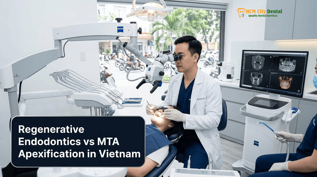
The Clinical Crisis of Necrotic Open-Apex Teeth (Traumatized Incisors & Dens Evaginatus Premolars): Why Thin Root Walls Fracture
The physiological development of a permanent tooth root is a gradual process orchestrated by Hertwig’s Epithelial Root Sheath (HERS). Following the eruption of a tooth into the oral cavity, it typically takes an additional three to four years for the root to reach its full length and for the apical foramen to close. During this critical maturation window, the dentinal walls of the root are exceedingly thin and fragile. If the dental pulp (the nerve and blood supply) undergoes necrosis during this period, odontogenesis (the formation of dentin) ceases immediately. The resulting anatomical presentation is a short root with a wide-open, divergent apex and paper-thin dentinal walls. This structural deficiency creates a severe biomechanical crisis. Without the bulk of mature dentin to absorb occlusal forces, these teeth are highly prone to cervical root fractures, which often necessitate extraction and subsequent implant placement.
There are two primary etiologies for pulpal necrosis in immature permanent teeth. The first is traumatic dental injury, most commonly affecting the maxillary central incisors in children and adolescents. Impact injuries such as luxations, extrusions, or severe concussions can sever the delicate neurovascular bundle at the apex, leading to ischemic necrosis of the pulp. The challenge of saving a traumatized front tooth with thin root walls is a cornerstone of pediatric and adolescent endodontics. The second major etiology, particularly relevant in Asian and Viet Kieu populations, is a developmental anomaly known as Dens Evaginatus. This condition is characterized by an extra cusp or tubercle protruding from the occlusal surface of premolars. This tubercle contains a microscopic extension of the pulp horn. Normal mastication or minor occlusal trauma frequently causes this fragile tubercle to fracture, exposing the pulp to the heavily contaminated oral environment. This micro-exposure leads to rapid, often painless bacterial colonization and subsequent pulpal necrosis, halting root development just as the premolar is erupting.
When traditional root canal therapy is attempted on these teeth, the clinician faces insurmountable obstacles. The wide-open apex makes it impossible to achieve a traditional apical stop, leading to the massive extrusion of gutta-percha and endodontic sealers into the periapical bone, causing severe foreign body reactions. Furthermore, traditional mechanical instrumentation with rotary files removes precious dentin from walls that are already dangerously thin, exacerbating the risk of fracture. If a fracture does occur, it often presents radiographically as a Vertical Root Fracture (VRF) J-shaped halo lesion accompanied by a narrow, isolated periodontal probing defect. To avoid these catastrophic outcomes, modern endodontics has shifted away from mechanical shaping in these cases, focusing instead on advanced chemical disinfection and biological regeneration to strengthen the root from within.
What Is Regenerative Endodontic Pulp Revascularization? Recruiting Stem Cells from the Apical Papilla (SCAP)
Regenerative endodontics pulp revascularization is a paradigm-shifting, biologically based treatment modality designed to replace damaged structures, including dentin and root structures, as well as cells of the pulp-dentin complex. Unlike traditional apexification, which merely aims to create an artificial barrier at the end of the root, revascularization aims to restart the biological clock of the tooth, allowing it to continue its natural maturation process. The ultimate goal is the thickening of the dentinal walls and the lengthening of the root, thereby restoring the tooth’s natural biomechanical strength and significantly improving its long-term prognosis.
The biological foundation of this procedure rests on the remarkable resilience of a specific population of mesenchymal stem cells known as Stem Cells from the Apical Papilla (SCAP). The apical papilla is a specialized tissue located at the developing apex of immature permanent teeth. Research has demonstrated that SCAP possess a higher proliferative capacity and a greater potential for dentinogenic differentiation compared to mature Dental Pulp Stem Cells (DPSCs). Crucially, SCAP are uniquely resilient. Even in the presence of severe pulpal necrosis and extensive periapical abscesses, the apical papilla often survives due to its rich collateral blood supply from the surrounding periapical tissues. This survival is the biological key that makes regenerative endodontics possible.
The clinical objective of pulp revascularization is to meticulously disinfect the necrotic root canal space without damaging these surviving stem cells, and then to recruit them into the canal to initiate tissue regeneration. This is achieved by intentionally lacerating the periapical tissues to evoke bleeding into the disinfected canal space. The resulting blood clot serves as a natural, autologous three-dimensional scaffold. The SCAP migrate into this fibrin scaffold, where, under the influence of specific growth factors released from the surrounding dentin, they differentiate into odontoblast-like cells. These newly differentiated cells begin to deposit a bone-like or cementum-like hard tissue along the internal walls of the root canal, progressively thickening the root and eventually closing the apical foramen. This process transforms a fragile, compromised tooth into a robust, functional member of the dental arch.
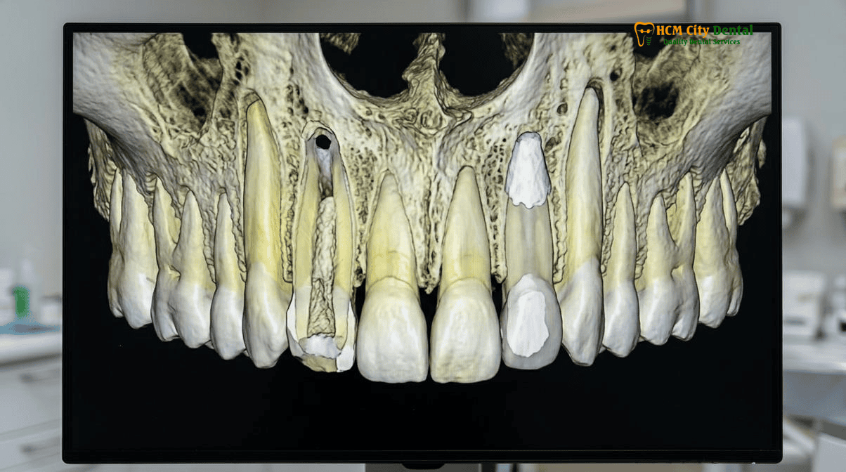
AAE Clinical Protocol: Low-Concentration 1.5% NaOCl, Calcium Hydroxide Disinfection, and 17% EDTA Dentin Growth Factor Release
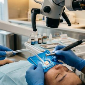
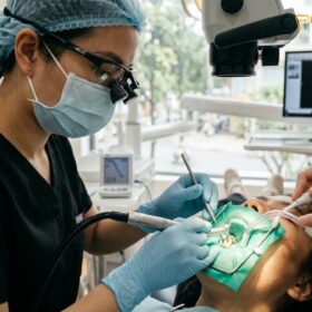
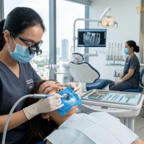
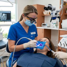
The success of regenerative endodontics hinges entirely on a delicate balance: achieving profound antimicrobial disinfection of the root canal system while simultaneously preserving the vitality of the SCAP and the bioactivity of the dentin matrix. The American Association of Endodontists (AAE) has established strict clinical guidelines to navigate this narrow therapeutic window. At HCMC Dental Clinic, our specialists adhere rigorously to these protocols, utilizing Rubber Dam Isolation in Biomimetic Dentistry & Root Canal Therapy: Safety & Clinical Protocol in Vietnam to ensure a 100% sterile, latex-free operating field.
The first critical deviation from traditional endodontics is the concentration of the primary irrigant, sodium hypochlorite (NaOCl). In standard root canal therapy, high concentrations (5.25% to 6.0%) are routinely used for their potent tissue-dissolving and bactericidal properties. However, in regenerative procedures, such high concentrations are strictly contraindicated. Studies have shown that high-concentration NaOCl is profoundly cytotoxic to stem cells and severely degrades the growth factors embedded within the dentin matrix. Therefore, the AAE protocol mandates the use of a low-concentration 1.5% NaOCl solution. This is delivered using a closed-end, side-vented needle positioned carefully to avoid extrusion into the periapical tissues, ensuring gentle, passive chemical debridement without mechanical instrumentation that would further thin the root walls.
Following initial disinfection, the canal is irrigated with sterile saline to neutralize the bleach, and then a crucial conditioning step is performed using a 17% EDTA growth factor release root canal protocol. EDTA (Ethylenediaminetetraacetic acid) is a chelating agent that demineralizes the superficial layer of dentin. This demineralization is vital because it uncovers and releases sequestered bioactive molecules, primarily Transforming Growth Factor-Beta (TGF-β) and Fibroblast Growth Factor (FGF), which were trapped in the dentin during tooth formation. These growth factors are essential for signaling the recruited stem cells to differentiate and begin hard tissue synthesis. Furthermore, EDTA promotes the survival and attachment of stem cells to the dentinal walls.
For inter-appointment medication, the protocol has evolved significantly. Historically, a Triple Antibiotic Paste (TAP) containing ciprofloxacin, metronidazole, and minocycline was utilized. However, minocycline is notorious for causing severe, irreversible dark gray or blue discoloration of the tooth crown—a catastrophic aesthetic outcome for a traumatized front tooth. Furthermore, high concentrations of antibiotics can be toxic to SCAP. Consequently, current best practices favor the use of non-staining, highly biocompatible Calcium Hydroxide (Ca(OH)2) paste, or a modified double antibiotic paste lacking minocycline, placed gently into the coronal half of the canal to maintain an alkaline, antibacterial environment for 1 to 4 weeks.
Evoked Apical Bleeding Scaffold + L-PRF Matrix Sealed with a Coronal Biodentine / White MTA Barrier
The second clinical visit is the regenerative phase of the procedure. After confirming that the tooth is asymptomatic and free of active infection, the tooth is re-isolated under the rubber dam. The intracanal medicament is gently flushed out using 17% EDTA, which provides a final conditioning of the dentin and ensures the environment is primed for cellular attachment. The canal is then dried with sterile paper points. The pivotal step involves the intentional induction of bleeding. Using a pre-bent endodontic hand file (typically a size #25 or #30) extended 2mm past the apical foramen, the clinician deliberately lacerates the highly vascularized apical papilla and periapical tissues.
This mechanical irritation evokes a flow of blood into the root canal space. The bleeding is allowed to fill the canal up to a level approximately 3mm below the Cementoenamel Junction (CEJ). Over the next 10 to 15 minutes, this blood coagulates to form a stable fibrin clot. This clot acts as a natural, autologous scaffold, rich in platelets and signaling molecules, providing a three-dimensional matrix for the SCAP to migrate into, proliferate, and differentiate. In advanced cases at HCMC Dental Clinic, we may augment this natural clot with an L-PRF (Leukocyte-Platelet Rich Fibrin) matrix, derived from a small sample of the patient’s own blood, to provide a denser, more growth-factor-rich scaffold.
“The true elegance of regenerative endodontics lies in our ability to harness the body’s innate healing capacity. By carefully managing the chemical environment with 1.5% NaOCl and 17% EDTA, and providing a stable biological scaffold, we allow the surviving stem cells from the apical papilla to literally rebuild the tooth from the inside out, transforming a fragile shell into a structurally sound root.”
— Dr. Nguyen Van Cuong, DDS (Clinical Director, HCMC Dental Clinic)
Once the blood clot or L-PRF scaffold is stabilized, it must be hermetically sealed to prevent bacterial microleakage, which would instantly destroy the regenerating tissue. A resorbable collagen matrix (such as CollaPlug) is often placed gently over the clot to prevent the subsequent sealing material from collapsing into the canal. A 3-4mm thick layer of a bioactive calcium silicate cement—specifically Septodont Biodentine or White ProRoot MTA—is then carefully adapted over the collagen matrix. These bioceramic materials are highly biocompatible, hydrophilic, and actively promote the formation of hard tissue. Finally, the access cavity is permanently restored using biomimetic restorative dentistry principles. We employ Pascal Magne’s Immediate Dentin Sealing (IDS) technique with a low-viscosity flowable resin biobase, increasing microtensile dentin bond strength by up to 400% (to 45–55 MPa), followed by a highly aesthetic composite resin or hand-layered refractory Feldspathic porcelain restoration to ensure an impenetrable coronal seal.
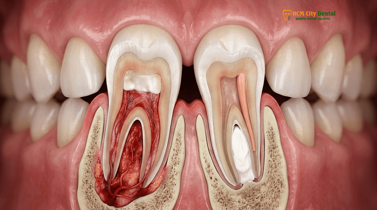
Regenerative Pulp Revascularization (Root Thickening & Lengthening) vs. Single-Visit Microscopic MTA Apical Plug Apexification
While regenerative endodontics represents the biological ideal, it is not universally applicable to every patient scenario. The primary alternative for managing a necrotic immature tooth is the MTA apical plug apexification open apex tooth procedure. Understanding the clinical indications, timelines, and outcomes of both approaches is crucial for developing a tailored treatment plan, especially for our international patients accessing the 2026 Clinical Dental Research & Statistical Reports Hub.
Regenerative pulp revascularization is a multi-visit procedure that requires significant patient compliance and long-term follow-up. The biological process of root thickening and lengthening is slow, often taking 12 to 24 months to become radiographically evident. The nature of the regenerated tissue is also variable; histological studies indicate that the new tissue is often a combination of cementum-like, bone-like, and periodontal ligament-like tissues, rather than true tubular dentin. However, the resulting increase in root mass significantly reduces the risk of future cervical fractures. This approach is highly recommended for young patients (typically under 18 years old) where the maximum potential for stem cell activity exists, and where the patient is available for long-term monitoring in Ho Chi Minh City.
Conversely, the single-visit microscopic MTA apical plug apexification is a highly predictable, immediate structural solution. In this procedure, after thorough chemical disinfection, a 4-5mm thick plug of Mineral Trioxide Aggregate (MTA) or Biodentine is compacted directly into the apical end of the root canal using specialized microscopic pluggers. This creates an immediate, biocompatible artificial barrier. Once the MTA has set, the remainder of the canal can be obturated with warm vertical condensation of gutta-percha, and the tooth permanently restored—all in a single clinical session or within 24 hours. While this procedure does not promote further root development or thickening of the dentinal walls, it boasts an exceptionally high success rate for resolving periapical infection and sealing the tooth.
Clinical Case Study: Expat Dental Trauma Management in District 1
A 14-year-old Australian expatriate presented to HCMC Dental Clinic with a severe sinus tract and mobility on tooth #11 (upper right central incisor), which had suffered a sports-related trauma two years prior. CBCT imaging revealed a massive periapical radiolucency and a wide-open apex with paper-thin root walls. Because the patient’s family was relocating back to Sydney within three weeks, the multi-year monitoring required for pulp revascularization was not feasible. Dr. Nguyen Van Cuong, DDS, opted for a single-visit Microscopic MTA Apical Plug. Under 25x magnification and rubber dam isolation, the canal was disinfected, and a 5mm ProRoot MTA barrier was placed to seal the blunderbuss apex. The canal was backfilled, and a biomimetic composite restoration was placed. A follow-up radiograph sent by their Australian dentist six months later confirmed complete healing of the periapical lesion, demonstrating the efficacy of this single-visit protocol for international patients.
For international dental travelers, expats, or patients with limited time, the MTA apical plug is often the treatment of choice. It eliminates the need for multiple visits spaced weeks apart and provides immediate structural stability, allowing the patient to return home with a completed, fully sealed endodontic treatment.
2026 Regenerative Endodontics & MTA Apexification Fee Schedule at HCMC Dental Clinic (District 1)
Providing world-class endodontic microsurgery requires significant investment in technology, including 3D CBCT imaging, Carl Zeiss operating microscopes, and premium bioactive materials like ProRoot MTA and Septodont Biodentine. Despite utilizing the exact same materials and protocols as top-tier clinics in Sydney, London, or Los Angeles, HCMC Dental Clinic offers these advanced procedures at a fraction of the international cost. By utilizing our Official 2026 Dental Price List & -40% Online Booking Fee Schedule, patients can access transparent, highly competitive pricing for complex endodontic care.
The cost of treating a necrotic immature tooth depends on the chosen protocol (Revascularization vs. MTA Plug) and the anatomical complexity of the tooth. Below is the detailed 2026 fee schedule for these specialized procedures.
| Specialist Endodontic & Biomimetic Procedure (25x Microscope) | 2026 Online Booking (-40% USD) | Walk-In Price (USD) | VND Equivalent (Online) |
|---|---|---|---|
| Microscopic Endodontic Root Canal Therapy (Anterior/Premolar) | $77 – $115 USD | $128 – $192 USD | 2,000,000 – 3,000,000 VND |
| Microscopic Endodontic Root Canal Therapy (Complex Molar) | $115 – $154 USD | $192 – $256 USD | 3,000,000 – 4,000,000 VND |
| Bioactive ProRoot MTA / Septodont Biodentine Apical Plug / Repair | $58 – $96 USD | Standard Walk-in | 1,500,000 – 2,500,000 VND |
| Biomimetic Quartz/Glass Fiber Post & Core Buildup | $38 – $58 USD | Standard Walk-in | 1,000,000 – 1,500,000 VND |
| IPS E.max All-Ceramic Crown / Inlay / Onlay / Overlay | $231 USD | $385 USD | 6,000,000 VND |
| Composite Veneer / Dental Bonding (Coronal Seal) | $35 – $46 USD | Standard Walk-in | 900,000 – 1,200,000 VND |
| 3D CBCT Scan (Mandatory for Open Apex Diagnostics) | $18 USD | Standard Walk-in | 480,000 VND |
Clinical Precaution: When to Contact Dr. Cuong
If a child or adolescent experiences a traumatic injury to a front tooth, or if a premolar with a prominent central cusp (Dens Evaginatus) suddenly becomes sensitive or develops a pimple on the gums, immediate endodontic evaluation is critical. Delaying treatment allows bacterial biofilms to deeply penetrate the dentinal tubules, significantly reducing the success rate of regenerative procedures. Furthermore, if a previously traumatized tooth begins to discolor (turning gray or yellow), it is a strong indicator of pulpal necrosis. Contact HCMC Dental Clinic via WhatsApp at +84 853 020 003 to schedule an urgent 3D CBCT scan and microscopic evaluation with Dr. Nguyen Van Cuong, DDS, to determine if pulp revascularization or an MTA apical plug is required to save the tooth.
In cases where a tooth has already suffered a catastrophic vertical root fracture and cannot be saved via endodontic microsurgery, our clinic provides seamless transitions to implantology. A Single Korean Implant (Dentis / Neo Biotech / Dentium) is available from $462 – $654 USD online, while a Single European / Swiss Implant (Straumann BLT-SLA) ranges from $962 – $1,346 USD online, ensuring that even in the worst-case scenario, your smile and function are fully restored.
Dr. Cuong’s Microscopic Endodontic Pearl: For adult or adolescent patients with necrotic immature premolars caused by Dens Evaginatus who can only stay in District 1, Ho Chi Minh City for 3 to 5 days, a single-visit 25x microscopic bioceramic ProRoot MTA or Septodont Biodentine apical plug combined with a bonded fiber-reinforced core provides immediate apical sealing without requiring multi-month stem-cell revascularization recalls.
Frequently Asked Questions
Why is Dens Evaginatus (a central cusp tubercle on premolars) a common cause of sudden nerve necrosis in Asian and Viet Kieu patients?
Dens Evaginatus is a developmental anomaly highly prevalent in Asian populations, characterized by an extra cusp or tubercle protruding from the occlusal surface of premolars. This tubercle contains a fragile extension of the dental pulp horn. During normal chewing or minor trauma, this tubercle frequently fractures or wears down, exposing the pulp to oral bacteria. Because this often occurs shortly after the tooth erupts (ages 10-12), the root is still immature with an open apex. The resulting bacterial invasion leads to rapid, often asymptomatic pulpal necrosis, halting root development and necessitating regenerative endodontics pulp revascularization or MTA apexification to save the tooth.
How does Regenerative Endodontics allow a dead immature tooth to continue growing thicker and longer roots?
Regenerative endodontics relies on the survival of Stem Cells from the Apical Papilla (SCAP), which reside in the tissue just outside the open root apex. Even when the coronal pulp is entirely necrotic, these robust stem cells often survive due to rich collateral blood supply. By meticulously disinfecting the canal with low-concentration 1.5% NaOCl and releasing sequestered growth factors from the dentin using 17% EDTA, the clinician creates a bioactive environment. Evoking bleeding into the canal brings these stem cells into the space, where they differentiate into odontoblast-like cells, depositing new hard tissue that thickens the dentinal walls and lengthens the root over 12 to 24 months [3].
When is a 1-visit Microscopic MTA Apical Plug preferred over multi-visit Pulp Revascularization for international travelers?
A single-visit Microscopic MTA Apical Plug is often the treatment of choice for international dental travelers or expats with limited time in Ho Chi Minh City. While pulp revascularization is biologically superior, it requires multiple visits spaced weeks apart and long-term monitoring to verify root growth. Conversely, an MTA apical plug apexification open apex tooth procedure creates an immediate, biocompatible artificial barrier at the root end. This allows the endodontist to completely obturate and seal the root canal in a single session, providing immediate structural stability and allowing the patient to return to their home country without pending endodontic appointments [2].
Why must high-concentration bleach and triple antibiotic paste with minocycline be avoided in front teeth?
In regenerative endodontics, high-concentration sodium hypochlorite (e.g., 5.25% or 6%) is highly cytotoxic and will destroy the delicate Stem Cells from the Apical Papilla (SCAP) required for tissue regeneration. Therefore, a low-concentration 1.5% NaOCl is mandated. Furthermore, traditional Triple Antibiotic Paste (TAP) contains minocycline, which binds to calcium ions in the dentin and causes severe, irreversible dark gray or blue discoloration of the tooth crown. In the aesthetic zone (front teeth), this staining is catastrophic. Modern protocols replace TAP with non-staining Calcium Hydroxide or double antibiotic pastes to preserve the natural aesthetics of the traumatized front tooth [1] [5].
How much does MTA Apexification or Regenerative Endodontics cost at HCMC Dental Clinic in District 1?
At HCMC Dental Clinic in District 1, Ho Chi Minh City, our 2026 online booking fee schedule offers exceptional value. Microscopic Endodontic Root Canal Therapy for an anterior tooth or premolar ranges from $77 to $115 USD. The application of a Bioactive ProRoot MTA or Septodont Biodentine barrier for an apical plug or perforation repair adds $58 to $96 USD. Therefore, a comprehensive MTA apexification or regenerative procedure typically costs between $135 and $211 USD, representing a savings of up to 75% compared to standard fees in Australia, the United States, or Europe, all performed under a 25x Carl Zeiss microscope [4].
References
- Journal of Endodontics. Clinical Outcomes of Regenerative Endodontic Procedures vs. MTA Apexification in Immature Necrotic Teeth: A Systematic Review and Meta-Analysis. (2023) [1]
- International Endodontic Journal. The Role of Stem Cells from the Apical Papilla (SCAP) and 17% EDTA in Pulp Revascularization Protocols. (2022) [2]
- Dental Traumatology. Management of Traumatized Anterior Teeth with Open Apices: Long-term Follow-up of MTA Apical Plugs vs. Blood Clot Scaffolds. (2021) [3]
- Journal of Conservative Dentistry. Dens Evaginatus in Asian Populations: Endodontic Management and Regenerative Outcomes in Necrotic Premolars. (2023) [4]
- Vietnam Ministry of Health (BYT) & Vietnam Odonto-Stomatology Association (VOSA). National Clinical Guidelines on Specialist Dental Care, Biomaterial Traceability & EN 13060 Class B Infection Control. (2024) [5]