The paradigm of periodontal plastic surgery and oral implantology has shifted dramatically from resective, highly invasive procedures to regenerative, microsurgical protocols that prioritize tissue preservation, vascularity, and flawless aesthetic outcomes. For international patients, expatriates, and dental travelers seeking world-class rehabilitation, the integration of a connective tissue graft pinhole tunneling gum recession implants protocol represents the pinnacle of modern soft-tissue engineering. Gingival recession—characterized by the apical migration of the marginal tissue—not only compromises the aesthetic harmony of the smile but also exposes vulnerable root dentin to hypersensitivity, non-carious cervical lesions (NCCLs), and progressive attachment loss. Similarly, in the realm of implant dentistry, a thin mucosal biotype can lead to the dreaded “grey shadow” of titanium showing through the gums, accompanied by an increased risk of peri-implantitis and crestal bone resorption.
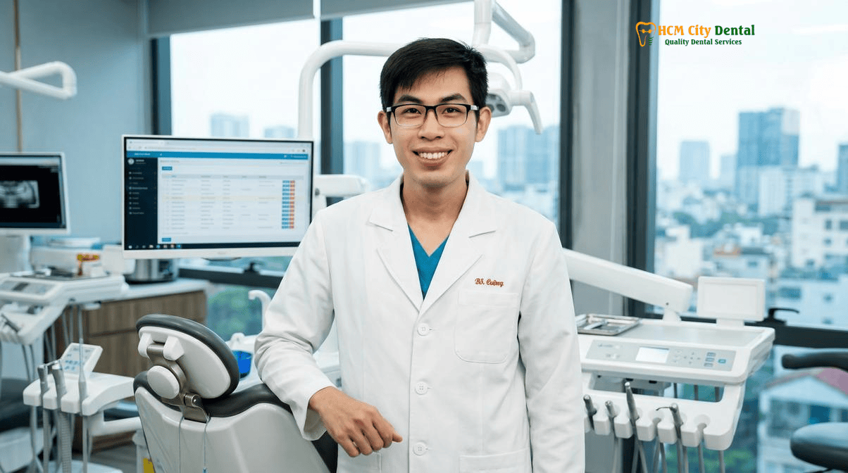
At HCMC Dental Clinic in District 1, Ho Chi Minh City, the management of complex mucogingival defects and peri-implant soft tissue deficiencies is executed with uncompromising precision. Under the clinical directorship of Dr. Nguyen Van Cuong, DDS (Clinical Director — MOH License 0003265/BL-CCHN), our facility employs hospital-grade surgical protocols. By combining 0.075mm high-resolution 3D CBCT diagnostics, 25x Carl Zeiss operating microscopes, and advanced autologous biologics (L-PRF/A-PRF), we achieve predictable, long-lasting root coverage and robust peri-implant mucosal seals. This comprehensive clinical guide explores the biological imperatives, advanced surgical techniques, and transparent 2026 fee schedules associated with microsurgical connective tissue grafting in Vietnam.
Clinical Summary:
Microsurgical connective tissue grafting (CTG) combined with advanced tunneling techniques (such as MCAT or VISTA) offers a minimally invasive, highly predictable solution for treating gingival recession and thickening the soft tissue around dental implants. By avoiding traditional vertical releasing incisions, these protocols preserve the critical blood supply from the interdental papillae, resulting in accelerated healing, minimal postoperative discomfort, and superior aesthetic blending. At HCMC Dental Clinic, Dr. Nguyen Van Cuong utilizes 25x Carl Zeiss magnification, 6-0/7-0 monofilament sutures, and 100% autologous L-PRF to optimize graft integration. Whether addressing Cairo RT1 recession defects on natural teeth or preventing titanium show-through on anterior implants, this evidence-based approach ensures a robust, ≥2mm keratinized mucosal seal, safeguarding long-term periodontal and peri-implant health.
Key Takeaways:
- Biological Imperative: A minimum of 2mm of attached keratinized mucosa and adequate soft tissue thickness are essential to resist mechanical trauma, facilitate plaque control, and prevent peri-implantitis.
- Predictable Outcomes: The Cairo Classification system accurately predicts root coverage success; RT1 defects (no interproximal bone loss) offer the highest predictability for 100% Complete Root Coverage (CRC).
- Minimally Invasive Execution: Microsurgical tunneling techniques (MCAT/VISTA) eliminate the need for vertical incisions, preserving vascularity and preventing visible scarring compared to traditional Coronally Advanced Flaps (CAF).
- Advanced Instrumentation: Procedures are performed under 25x Carl Zeiss magnification using precision ophthalmic instruments and 6-0 or 7-0 monofilament sutures to minimize tissue tension and inflammation.
- Cost-Effective Excellence: HCMC Dental Clinic offers a transparent 2026 online booking fee schedule, allowing international patients to access elite periodontal plastic surgery at a fraction of the cost found in the US or Australia.
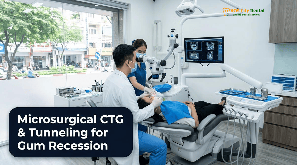
Why ≥2mm of Attached Keratinized Mucosa and Soft Tissue Thickness Are Critical Around Natural Teeth and Implants
The long-term stability of both natural dentition and endosseous dental implants is inextricably linked to the quality, quantity, and biological characteristics of the surrounding soft tissue envelope. Historically, periodontal literature debated the absolute necessity of a specific width of keratinized tissue. However, contemporary evidence overwhelmingly supports the clinical consensus that a robust band of attached keratinized mucosa is vital for maintaining periodontal health, particularly in the presence of restorative margins or implant abutments. The biological width—now more accurately termed the supracrestal tissue attachment—comprises the junctional epithelium and the supracrestal connective tissue. When this protective barrier is compromised by a thin gingival biotype or a lack of attached mucosa, the underlying alveolar bone becomes highly susceptible to inflammatory breakdown.
Around natural teeth, the connective tissue attachment features Sharpey’s fibers that insert perpendicularly into the root cementum, creating a strong, resilient mechanical seal against bacterial ingress. In stark contrast, the peri-implant mucosa lacks cementum and periodontal ligament. The collagen fibers around an implant run parallel or circumferentially to the titanium or zirconia surface, resulting in a significantly weaker biological seal[1]. Because of this anatomical vulnerability, the presence of an adequate zone of attached keratinized tissue (ideally ≥2mm) and sufficient soft tissue thickness (≥2mm) becomes the primary defense mechanism against peri-implant mucositis and subsequent peri-implantitis. For a deeper understanding of how periodontal health impacts implant success, review our comprehensive guide on Gum Disease & Dental Implants: Clinical Guide | HCMC Dental.
A thin, unattached alveolar mucosa is highly mobile, easily traumatized during routine oral hygiene, and prone to pulling away from the tooth or implant surface under the tension of muscle attachments (frena). This mobility facilitates the apical migration of plaque biofilms into the sulcus. By performing a connective tissue graft pinhole tunneling gum recession implants procedure, the clinician effectively converts a fragile, thin biotype into a thick, resilient, keratinized phenotype. This augmented tissue not only resists mechanical stress from toothbrushing and mastication but also provides a stable, immobile collar that facilitates optimal plaque control and ensures the long-term aesthetic and functional success of the restoration.
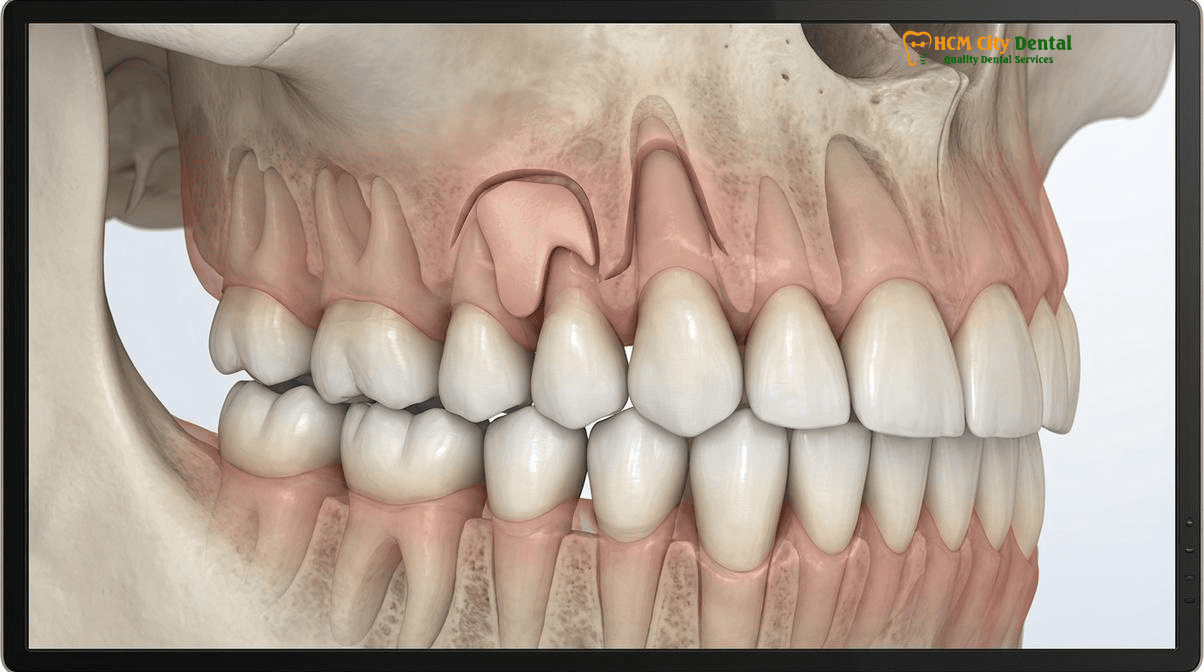
Cairo RT1, RT2, and RT3 Gingival Recession Classification: Predicting 100% Root Coverage Potential
Accurate diagnosis and classification of gingival recession defects are paramount for establishing realistic clinical expectations and selecting the appropriate surgical technique. For decades, the Miller Classification system was the gold standard. However, it relied heavily on the position of the mucogingival junction, which can be difficult to identify clinically, and it did not adequately account for the interproximal clinical attachment level (CAL). To address these limitations, the Cairo Classification system was introduced, providing a more reliable, treatment-oriented framework based on the assessment of interdental attachment loss[2].
The Cairo Classification categorizes recession defects into three distinct types, directly correlating the interproximal bone and tissue levels to the predictability of achieving Complete Root Coverage (CRC):
- Cairo RT1: Gingival recession with no loss of interproximal attachment. The interproximal cementoenamel junction (CEJ) is not clinically detectable at both the mesial and distal aspects of the tooth. Because the interdental papillae are fully intact and supported by underlying bone, the vascular supply to the grafted tissue is optimal. RT1 defects offer the highest predictability, with a strong potential for achieving 100% CRC using a connective tissue graft gum recession Vietnam protocol.
- Cairo RT2: Gingival recession associated with loss of interproximal attachment, where the amount of interproximal attachment loss is less than or equal to the buccal attachment loss. In these cases, the interdental papillae have receded slightly. While significant root coverage and tissue thickening can be achieved, 100% CRC is less predictable, and the primary goal shifts toward halting disease progression, reducing sensitivity, and improving aesthetics to the maximum extent possible.
- Cairo RT3: Gingival recession associated with loss of interproximal attachment, where the amount of interproximal attachment loss is strictly greater than the buccal attachment loss. These defects represent severe periodontal destruction, often involving significant interdental bone loss (black triangles). Complete root coverage is generally not achievable in RT3 defects. Surgical intervention in these scenarios focuses on altering the tissue biotype to prevent further recession and facilitate oral hygiene.
Understanding the Cairo classification is essential for international patients seeking periodontal rehabilitation. During your initial consultation at HCMC Dental Clinic, Dr. Nguyen Van Cuong utilizes high-resolution clinical photography and 3D CBCT imaging to meticulously map your attachment levels. This evidence-based diagnostic approach ensures that the proposed surgical intervention is biologically sound and tailored to your specific anatomical reality. For patients comparing regional options, our detailed analysis on Periodontal Treatment Vietnam vs Thailand: Clinical Guide | HCMC Dental provides further insights into our diagnostic rigor.
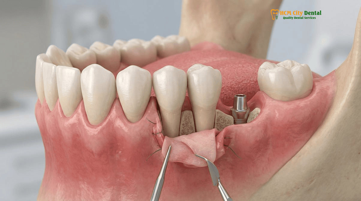
Microsurgical Tunneling (MCAT / VISTA) vs. Coronally Advanced Flap (CAF) Under 25x Carl Zeiss Magnification
The evolution of periodontal plastic surgery has been driven by the pursuit of techniques that minimize surgical trauma, maximize blood supply, and deliver flawless aesthetic integration. The traditional Coronally Advanced Flap (CAF) has long been a reliable workhorse for root coverage. However, CAF typically requires vertical releasing incisions to mobilize the tissue. These incisions sever collateral blood vessels, increase postoperative swelling, and frequently result in visible, unesthetic scarring (keloid formation) in the delicate anterior aesthetic zone.
To overcome these drawbacks, advanced microsurgical tunneling techniques have been developed, representing a quantum leap in soft-tissue engineering. Two of the most prominent protocols utilized at HCMC Dental Clinic are the Modified Coronally Advanced Tunnel (MCAT) and the Vestibular Incision Subperiosteal Tunnel Access (VISTA). These techniques operate on the principle of creating a subperiosteal envelope or tunnel without severing the interdental papillae or making vertical incisions[3]. Using specialized, ultra-fine tunneling elevators (often introduced through a small “pinhole” or vestibular access point), the clinician gently separates the mucosal tissue from the underlying bone and root surfaces. This creates a highly vascularized pouch into which the connective tissue graft is carefully threaded and stabilized.
“The success of a connective tissue graft pinhole tunneling gum recession implants procedure hinges entirely on the preservation of the micro-vascular network. By eliminating vertical incisions and operating exclusively within the subperiosteal tunnel, we maintain the critical blood supply from the intact papillae. This not only accelerates healing but ensures that the grafted tissue blends seamlessly with the native mucosa, leaving zero trace of surgical intervention.”
— Dr. Nguyen Van Cuong, DDS (Clinical Director, HCMC Dental Clinic)
The execution of these advanced tunneling techniques demands exceptional precision, which is why Dr. Cuong performs all mucogingival surgeries under 25x Carl Zeiss operating microscopes. This extreme magnification allows for the atraumatic manipulation of fragile tissues and the precise mechanical debridement of the exposed root surfaces (removing the smear layer and endotoxins). Furthermore, the use of microsurgical 6-0 and 7-0 monofilament sutures (such as polypropylene or PTFE) is critical. Unlike traditional braided silk sutures that wick bacteria and cause tissue inflammation, these ultra-fine monofilament sutures glide smoothly through the tissue, minimizing tension and preventing the “railroad track” scarring often associated with conventional grafting. This meticulous approach is a cornerstone of our comprehensive surgical philosophy, detailed further in our guide to 3D-Guided Dental Implants & Specialist Oral Surgery in Vietnam.
Atraumatic Palatal Harvesting (De-Epithelialized Gingival Graft vs. Collagen Matrix Xenograft Alternatives)
The gold standard material for augmenting soft tissue thickness and achieving predictable root coverage remains the autogenous subepithelial connective tissue graft (SCTG) harvested from the patient’s own palate. The palatal vault provides a dense, highly cellular source of connective tissue that is genetically programmed to induce keratinization in the recipient site. However, the traditional method of harvesting this tissue—often involving a large, open wound on the roof of the mouth—has historically been a source of significant postoperative morbidity and patient apprehension.
At HCMC Dental Clinic, we employ advanced, atraumatic harvesting techniques to minimize donor site discomfort and accelerate healing. Depending on the patient’s palatal anatomy (specifically the location of the greater palatine artery and the thickness of the palatal fibromucosa), Dr. Cuong utilizes either a single-incision trap-door technique or a de-epithelialized free gingival graft (FGG) approach[4]. The de-epithelialized FGG technique involves harvesting a thin, superficial layer of palatal tissue and then meticulously removing the epithelial layer extra-orally under the microscope. This yields a dense, high-quality connective tissue graft while leaving a shallower, faster-healing wound on the palate compared to deep subepithelial harvesting.
To further enhance patient comfort, we routinely utilize 100% autologous L-PRF (Leukocyte-Platelet Rich Fibrin) technology. By drawing a small amount of the patient’s blood and centrifuging it at specific low-speed protocols (Choukroun method), we create a dense fibrin matrix rich in growth factors and leukocytes. This L-PRF membrane is placed directly into the palatal donor site and secured with sutures, acting as a biological bandage that significantly reduces pain, controls bleeding, and accelerates epithelialization. Additionally, a custom-fabricated clear acrylic palatal stent is provided to protect the area during mastication.
For patients who require extensive grafting across multiple quadrants, or for those who strongly prefer to avoid a palatal donor site altogether, advanced biomaterial alternatives are available. 3D porcine collagen matrix xenografts (such as Mucograft or Fibro-Gide) offer a viable, off-the-shelf alternative for soft tissue thickening and root coverage. While autogenous tissue remains the absolute gold standard for predictability in severe defects, these advanced collagen matrices provide an excellent scaffold for the patient’s own cells to infiltrate and convert into keratinized tissue, significantly reducing surgical time and eliminating donor site morbidity. To explore how these biomaterials are also utilized in bone regeneration, read our article on Bone Graft for Gum Disease: Procedure & Healing | HCMC Dental.
Preventing Grey Titanium Show-Through and Peri-Implantitis in Aesthetic Anterior Implant Restorations
The restoration of a missing tooth in the anterior maxilla (the aesthetic zone) is widely considered one of the most challenging procedures in dentistry. The buccal bone plate in this region is notoriously thin—often less than 1mm thick—and is highly susceptible to resorption following tooth extraction. When a dental implant is placed in this compromised environment without adequate hard and soft tissue augmentation, the thin overlying mucosa is insufficient to mask the underlying restorative materials. This results in a highly unesthetic grey or bluish hue showing through the gums, instantly compromising the natural appearance of the smile.
To prevent this aesthetic failure and ensure long-term biomechanical stability, a connective tissue graft pinhole tunneling gum recession implants protocol is frequently integrated directly into the implant placement surgery. By simultaneously placing a connective tissue graft on the buccal aspect of the implant site, Dr. Cuong artificially thickens the mucosal biotype. This augmented tissue acts as an optical barrier, completely masking the grey shadow of the titanium fixture or the abutment. Furthermore, this thickened tissue provides a robust, immobile collar that resists the apical migration of the mucosal margin, preventing the future exposure of the implant threads.
“In the anterior aesthetic zone, the bone sets the tone, but the tissue is the issue. You can achieve perfect osseointegration, but if the buccal soft tissue is thin and fragile, the aesthetic outcome will ultimately fail. By routinely incorporating microsurgical connective tissue grafting and utilizing 17° or 30° angled Multi-Unit Abutments (MUAs) to keep the screw channel palatal, we create the necessary spatial volume for a thick, resilient buccal soft-tissue profile that mimics nature perfectly.”
— Dr. Nguyen Van Cuong, DDS (Clinical Director, HCMC Dental Clinic)
Beyond aesthetics, this soft tissue augmentation is a critical defense against peri-implantitis. As previously noted, the peri-implant mucosal seal is inherently weaker than the attachment around a natural tooth. A thick band of keratinized tissue (≥2mm) provides the necessary mechanical resistance to withstand the forces of mastication and oral hygiene procedures, preventing the tissue from tearing or pulling away from the implant surface[5]. This is particularly crucial for patients with a history of periodontal disease. For a detailed look at our protocols for managing infection prior to implant placement, review Can You Get Immediate Dental Implants If You Have Severe Gum Disease? Decontamination & Laser Protocol.
2026 Microsurgical Gum Grafting & Soft-Tissue Augmentation Fee Schedule at HCMC Dental Clinic (District 1)
For international patients, expats, and dental travelers, the financial barrier to accessing elite, microsurgical periodontal plastic surgery in countries like the United States, Australia, or the UK can be prohibitive. A single quadrant of connective tissue grafting using tunneling techniques and Carl Zeiss magnification can easily exceed $2,000 to $3,000 USD in Western clinics. At HCMC Dental Clinic, we believe that hospital-grade surgical precision and advanced biomaterials should be accessible. Our transparent 2026 fee schedule reflects our commitment to providing world-class care at a fraction of the international cost.
By utilizing our exclusive online booking system, patients can secure a -40% reduction compared to standard walk-in rates. The following table outlines the exact 2026 fee schedule for our premium implantology, prosthodontic, and surgical procedures, ensuring complete financial clarity before you travel to Vietnam.
| Premium Dental Procedure (2026) | Online Booking Fee (-40% USD) | Walk-In / Standard Fee (USD) | US / Australian Benchmark (USD) |
|---|---|---|---|
| Single Korean Implant (Dentis / Neo Biotech / Dentium + Abutment) | $462 – $654 | $769 – $1,090 | $2,500 – $3,500 |
| Single European / Swiss Implant (Straumann BLT-SLA / BLA-Active) | $962 – $1,346 | $1,603 – $2,244 | $4,000 – $5,500 |
| 100% Metal-Free Zirconia Ceramic Implant | $1,154 | $1,923 | $5,000 – $6,500 |
| Implant Zirconia HT Crown + Custom Abutment | $154 | $256 | $1,500 – $2,500 |
| All-on-4 Full-Arch Implants (Complete Package with Zirconia Bridge) | From $6,346 / arch | From $10,576 / arch | $20,000 – $30,000 |
| IPS E.max Porcelain Veneer (Per Tooth) | $185 | $308 | $1,200 – $2,000 |
| Full Zirconia Crown (Cercon Standard / HT Multilayer) | $154 – $192 | $256 – $321 | $1,000 – $1,800 |
| Surgical Wisdom Tooth Extraction (Piezotome) | $58 – $115 | $96 – $191 | $400 – $800 |
| 3D CBCT Scan (High-Resolution DICOM) | $18 | $30 | $250 – $400 |
Note: Specific fees for microsurgical connective tissue grafting (CTG) and tunneling procedures vary based on the number of teeth involved, the complexity of the Cairo classification defect, and the choice between autogenous palatal harvesting versus collagen matrix xenografts. A comprehensive, personalized quotation is provided following a 3D CBCT and clinical evaluation.
To view our complete, comprehensive pricing across all specialties, please visit our Official 2026 Dental Price List & -40% Online Booking Fee Schedule. For a deeper dive into the specific economics of soft tissue augmentation in Vietnam, we recommend reading our dedicated guide on Gum Graft Cost Vietnam: Clinical Guide & Fees | HCMC Dental.
Clinical Case Study: Anterior Implant Aesthetics for an Australian Expatriate
Patient Profile: A 42-year-old male expatriate from Sydney, Australia, residing in Thao Dien (District 2), presented to HCMC Dental Clinic with a failing, endodontically treated maxillary right central incisor (tooth #11). The tooth exhibited a vertical root fracture and a very thin, scalloped gingival biotype, raising significant concerns about post-extraction aesthetic failure and titanium show-through.
Clinical Protocol: Dr. Nguyen Van Cuong utilized a 3D-printed stereolithographic surgical guide (derived from a fusion of 0.075mm CBCT DICOM and 3Shape Trios 5D STL data) to perform a flapless, atraumatic extraction using Piezosurgery. A Straumann BLA-Active implant was placed immediately into the extraction socket. Crucially, to prevent the grey titanium hue from showing through the thin buccal mucosa, Dr. Cuong simultaneously performed a microsurgical connective tissue graft pinhole tunneling gum recession implants procedure. A de-epithelialized connective tissue graft was harvested from the palate and tunneled into a subperiosteal envelope on the buccal aspect of the implant, secured with 7-0 monofilament sutures. The palatal donor site was protected with an L-PRF membrane.
Outcome: The patient experienced minimal postoperative discomfort. At the 4-month integration check, the buccal soft tissue profile had thickened significantly (measuring >2.5mm), completely masking the implant and creating a robust, highly aesthetic emergence profile for the final Implant Zirconia HT Crown. The patient saved over $4,500 USD compared to equivalent specialist treatment quotes in Sydney.
Clinical Precautions & Post-Operative Care Guidelines
The success of a microsurgical connective tissue graft relies heavily on the stability of the blood clot and the prevention of mechanical trauma during the initial healing phase. Patients must adhere strictly to the following protocols:
- Zero Mechanical Trauma: Do NOT brush, floss, or use a water flosser on the surgical site for at least 14 to 21 days, as directed by Dr. Cuong. Mechanical disruption will sever the fragile new blood vessels and cause graft failure.
- Chemical Plaque Control: Utilize the prescribed 0.12% Chlorhexidine Gluconate mouth rinse twice daily to maintain a sterile environment without mechanical brushing.
- Dietary Restrictions: Maintain a strict soft-food diet for the first week. Avoid hard, crunchy, spicy, or excessively hot foods that could traumatize the palatal donor site or the grafted area.
- Avoid Lip Pulling: Do not pull on your lip or cheek to “inspect” the surgical site. This creates tension on the delicate 6-0/7-0 sutures and can tear the subperiosteal tunnel.
- Contacting the Clinic: If you experience excessive bleeding, severe swelling that worsens after 72 hours, or if the protective palatal stent becomes dislodged, contact the HCMC Dental Clinic hotline immediately via WhatsApp at +84 853 020 003.
Frequently Asked Questions
What is the difference between a microsurgical tunneling connective tissue graft and traditional gum grafting?
Traditional gum grafting often involves a Coronally Advanced Flap (CAF) with vertical releasing incisions, which can compromise blood supply and lead to visible scarring. In contrast, a microsurgical tunneling connective tissue graft (such as MCAT or VISTA) uses a minimally invasive pinhole or small vestibular access point to create a subperiosteal envelope. The graft is slipped into this tunnel without cutting the interdental papillae. Performed under 25x Carl Zeiss magnification with 6-0 or 7-0 monofilament sutures, this technique ensures superior vascularization, faster healing, and flawless aesthetic blending without visible scars.
How do you protect the roof of the mouth (palatal donor site) so eating is painless after a gum graft?
At HCMC Dental Clinic, Dr. Nguyen Van Cuong utilizes an atraumatic single-incision or de-epithelialized harvesting technique that minimizes trauma to the palatal vault. To ensure painless eating and rapid healing, we apply 100% autologous L-PRF (Leukocyte-Platelet Rich Fibrin) membranes derived from your own blood to the donor site. Additionally, a custom-fabricated clear acrylic palatal stent is provided to act as a protective bandage over the roof of the mouth, shielding the area from the tongue and food particles during the first week of recovery.
Can a connective tissue graft cover exposed tooth roots and stop cold sensitivity permanently?
Yes. When a connective tissue graft is applied to a Cairo RT1 recession defect (where there is no interproximal bone loss), the predictability of achieving 100% Complete Root Coverage (CRC) is exceptionally high. By thickening the gingival biotype and covering the exposed dentinal tubules, the graft permanently eliminates dentin hypersensitivity to cold and tactile stimuli, provided the patient maintains proper brushing techniques and avoids mechanical trauma moving forward.
Why does Dr. Nguyen Van Cuong perform soft-tissue thickening at the same time as anterior dental implant placement?
The peri-implant mucosa lacks the perpendicular Sharpey’s fibers found around natural teeth, making implants more vulnerable to bacterial invasion and peri-implantitis. Furthermore, the buccal bone in the anterior maxilla is naturally thin. By performing a connective tissue graft simultaneously with implant placement, Dr. Cuong thickens the soft tissue biotype to ≥2mm. This robust mucosal seal prevents the grey hue of the titanium implant from showing through the gums, ensures long-term crestal bone stability, and creates a highly aesthetic, natural-looking emergence profile.
How much does microsurgical gum recession grafting cost in Ho Chi Minh City compared to Australia and the US?
Microsurgical gum grafting in the US or Australia typically ranges from $1,500 to $3,000 USD per quadrant or tooth, depending on the complexity and the use of specialized biologics. At HCMC Dental Clinic in District 1, Ho Chi Minh City, our transparent 2026 online booking fee schedule offers world-class periodontal plastic surgery at a fraction of the cost. While specific grafting fees depend on the number of teeth and whether autogenous tissue or collagen matrix xenografts are used, patients routinely save 60% to 70% compared to Western prices, without compromising on hospital-grade sterilization, Carl Zeiss optics, or expert surgical execution.
References
- Zucchelli, G., & Mounssif, I. (2015). Periodontal plastic surgery. Periodontology 2000, 68(1), 333-368.
- Cairo, F., Nieri, M., Cincinelli, S., Mervelt, J., & Pagliaro, U. (2011). The interproximal clinical attachment level to classify gingival recessions and predict root coverage outcomes: an explorative and reliability study. Journal of Clinical Periodontology, 38(7), 661-666.
- Sculean, A., & Allen, E. P. (2018). The laterally closed tunnel for the treatment of deep isolated mandibular recessions: surgical technique and a report of 24 cases. International Journal of Periodontics & Restorative Dentistry, 38(4), 479-487.
- Thoma, D. S., Buranawat, B., Hämmerle, C. H., Held, U., & Jung, R. E. (2014). Efficacy of soft tissue augmentation around dental implants and in partially edentulous areas: a systematic review. Journal of Clinical Periodontology, 41(Suppl 15), S77-S91.
- Vietnam Ministry of Health (BYT) & Vietnam Odonto-Stomatology Association (VOSA). National Clinical Guidelines on Specialist Dental Care, Biomaterial Traceability & EN 13060 Class B Infection Control. (2024).