Bottom wisdom teeth removal is a surgical procedure to extract the third molars from the lower jaw. Because these teeth often erupt into dense mandibular bone and sit near critical sensory nerves, their extraction requires precise clinical planning to prevent complications like infection, impaction, or nerve damage.
Clinical Summary:
The extraction of mandibular (bottom) wisdom teeth is generally more complex than maxillary (top) extractions due to the dense nature of the lower jawbone and the high prevalence of spatial impaction. Lower third molars frequently lack adequate arch space, leading to partial eruption and a high risk of pericoronitis—a localized gum infection. Furthermore, the roots of these teeth often develop in close proximity to the inferior alveolar nerve (IAN) and lingual nerve, necessitating advanced diagnostic imaging such as Cone Beam Computed Tomography (CBCT) for safe surgical planning. The surgical workflow typically involves raising a mucoperiosteal flap, selective bone removal (ostectomy), and tooth sectioning (odontotomy) to minimize trauma. While recovery involves managing expected swelling and trismus, adherence to clinical aftercare protocols significantly reduces the risk of complications like alveolar osteitis (dry socket).
Key Takeaways:
- Lower wisdom teeth face higher impaction rates due to limited space in the dense mandibular bone.
- Proximity to the inferior alveolar nerve requires careful 3D radiographic mapping prior to surgery.
- Tooth sectioning (cutting the tooth into pieces) is a standard technique to minimize bone trauma during removal.
- Coronectomy is a viable, conservative alternative when full extraction poses a high risk of nerve injury.
- Post-operative recovery focuses on protecting the blood clot to prevent dry socket and managing localized inflammation.
Anatomical Complexities of Lower Wisdom Teeth
Lower wisdom teeth are uniquely challenging to extract due to the dense mandibular bone, high rates of spatial impaction, and close proximity to critical sensory nerves.
The human mandible, or lower jaw, is structurally distinct from the maxilla (upper jaw). It is composed of highly dense cortical bone designed to withstand the immense biomechanical forces of chewing. While this density provides structural integrity, it also presents a significant clinical challenge when a tooth needs to be removed. Unlike upper wisdom teeth, which often sit in softer, more porous bone, bottom wisdom teeth are firmly encased in this rigid structure[1]. This anatomical reality is the primary reason why lower wisdom teeth extractions are frequently classified as surgical procedures rather than simple extractions.
One of the most defining characteristics of lower third molars is their propensity for impaction. As the last teeth to develop—typically erupting between the ages of 17 and 25—they often find that the available space in the dental arch has already been claimed by the preceding molars. For more information on the developmental timeline, you can review our guide on what age do wisdom teeth grow. When space is insufficient, the tooth becomes trapped, or “impacted,” within the jawbone or soft tissue.
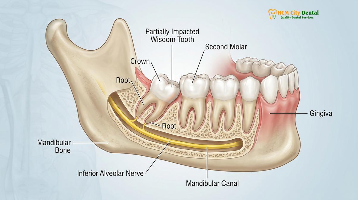
Clinical impactions of bottom wisdom teeth are categorized based on their angulation and depth:
- Mesioangular Impaction: The most common type, where the wisdom tooth is angled forward toward the adjacent second molar. This positioning creates a severe risk of food impaction and subsequent decay in both teeth.
- Vertical Impaction: The tooth is relatively straight but lacks the vertical space to fully clear the gum line.
- Horizontal Impaction: The tooth lies completely flat, parallel to the jawbone, often deeply embedded and pressing directly into the roots of the second molar.
- Distoangular Impaction: The tooth is angled backward, away from the second molar, often embedding itself deeply into the ascending ramus of the mandible.
Beyond bone density and angulation, the most critical anatomical consideration is the tooth’s relationship to the inferior alveolar nerve (IAN) and the lingual nerve. The IAN runs through the mandibular canal, supplying sensation to the lower lip, chin, and teeth on that side of the jaw. The roots of lower wisdom teeth can develop intimately close to, or even wrap around, this nerve canal. Any excessive force or blind instrumentation during extraction carries a risk of bruising or damaging this nerve, leading to temporary or, in rare cases, permanent altered sensation (paresthesia)[1].
“The surgical management of mandibular third molars requires a profound understanding of local anatomy. The dense cortical plate and the proximity of the inferior alveolar neurovascular bundle dictate a meticulous, force-controlled surgical approach to prevent iatrogenic injury.”
Furthermore, partially erupted bottom wisdom teeth are highly susceptible to pericoronitis. When a tooth only partially breaches the gum tissue, a flap of tissue called an operculum remains over the crown. This creates a deep, inaccessible pocket where food debris and oral bacteria accumulate, leading to acute inflammation, severe pain, and recurrent infections. This localized pathology is a primary driver for patients seeking emergency dental care and ultimately requiring extraction.
Diagnostic Imaging and Pre-Surgical Assessment
Comprehensive radiographic imaging, including panoramic X-rays and CBCT scans, is mandatory to map root morphology and assess the exact distance to the mandibular canal.
The foundation of a safe and successful bottom wisdom teeth removal lies in meticulous pre-surgical planning. A visual clinical examination is insufficient, as the most critical structures—the roots, the surrounding bone, and the nerve pathways—are hidden beneath the soft tissue. Therefore, advanced diagnostic imaging is the standard of care.
The initial diagnostic tool is typically a panoramic radiograph (OPG). This 2D image provides a broad overview of the entire maxillofacial region, allowing the clinician to assess the presence, position, and general angulation of all wisdom teeth. It helps identify gross pathology, such as large cysts or extensive decay, and provides a preliminary look at the relationship between the tooth roots and the mandibular canal.
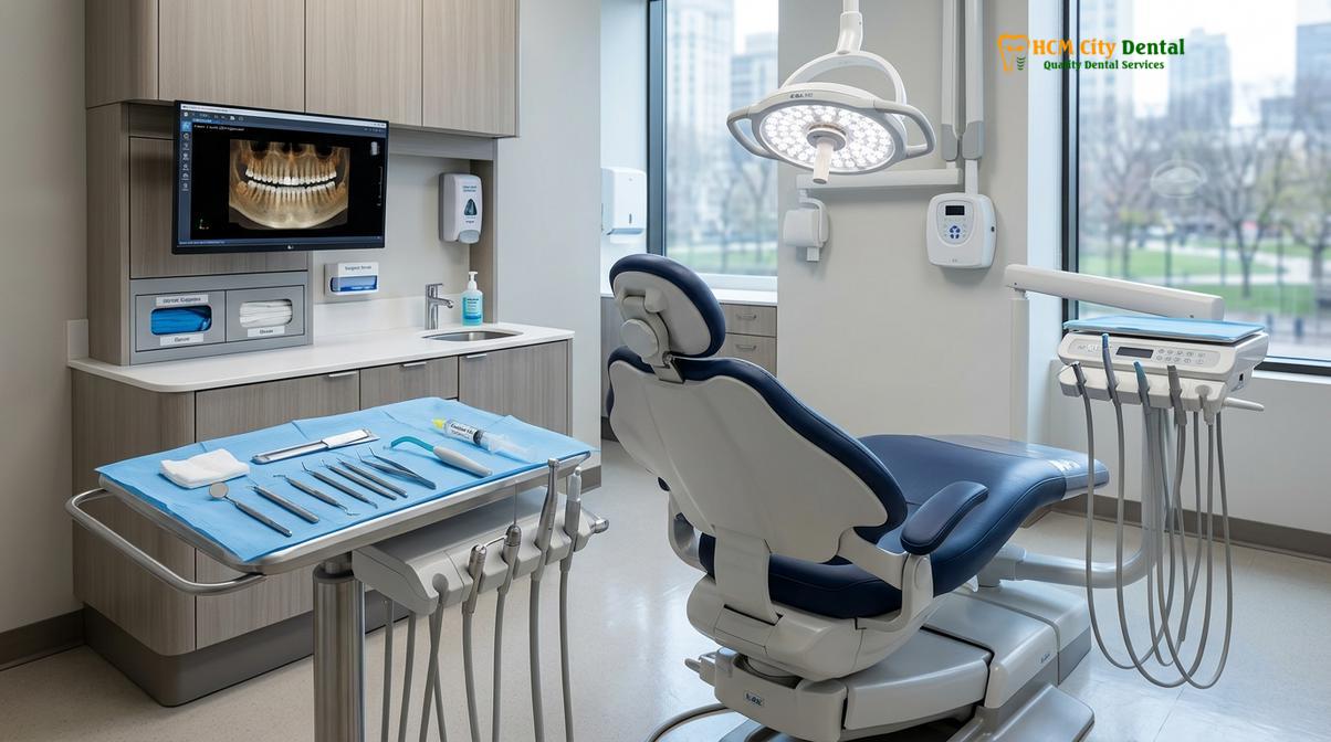
However, a 2D image has inherent limitations, particularly regarding depth perception and superimposition of structures. If the panoramic X-ray suggests that the roots of the lower wisdom tooth are in close proximity to the inferior alveolar nerve, a 3D Cone Beam Computed Tomography (CBCT) scan is strongly recommended[2]. A CBCT scan provides high-resolution, three-dimensional cross-sections of the jaw.
At HCMC Dental Clinic in Ho Chi Minh City, Dr. Nguyen Van Cuong emphasizes the critical role of 3D imaging in complex cases. “A CBCT scan eliminates the guesswork,” notes Dr. Cuong. “It allows us to view the exact trajectory of the nerve in relation to the roots from the sagittal, coronal, and axial planes. This precise mapping dictates our surgical approach, determining whether a full extraction is safe or if an alternative technique is required.”
During the pre-surgical assessment, the clinician will also evaluate the patient’s overall medical history, including any medications that might affect bleeding or bone healing (such as bisphosphonates or anticoagulants). The health of the adjacent second molar is also scrutinized, as deep impactions can cause root resorption or periodontal defects on the neighboring tooth. Patients wondering about the necessity of the procedure can explore our comprehensive analysis on should wisdom teeth be removed.
The Surgical Extraction Workflow
The extraction process involves administering local anesthesia, elevating a mucoperiosteal flap, selectively removing surrounding bone, sectioning the tooth, and carefully elevating the fragments.
The removal of an impacted bottom wisdom tooth is a highly choreographed surgical procedure. Because of the dense mandibular bone and the need to protect surrounding structures, the workflow prioritizes technique and finesse over brute force. The procedure is typically performed under profound local anesthesia, often supplemented with conscious sedation depending on patient anxiety and case complexity.
Step 1: Profound Anesthesia
The procedure begins with an inferior alveolar nerve block, which numbs the entire lower quadrant of the jaw, including the teeth, lower lip, and chin. Additional local infiltration is used to anesthetize the buccal (cheek) and lingual (tongue) soft tissues. Ensuring complete anesthesia is paramount for patient comfort and surgical cooperation.
Step 2: Flap Design and Elevation
To access the impacted tooth and the surrounding bone, the surgeon creates a precise incision in the gum tissue. A mucoperiosteal flap is then carefully elevated, pulling the gum tissue back to expose the surgical site. The design of this flap is critical; it must provide adequate visibility while ensuring a healthy blood supply for optimal post-operative healing.
Step 3: Ostectomy (Bone Removal)
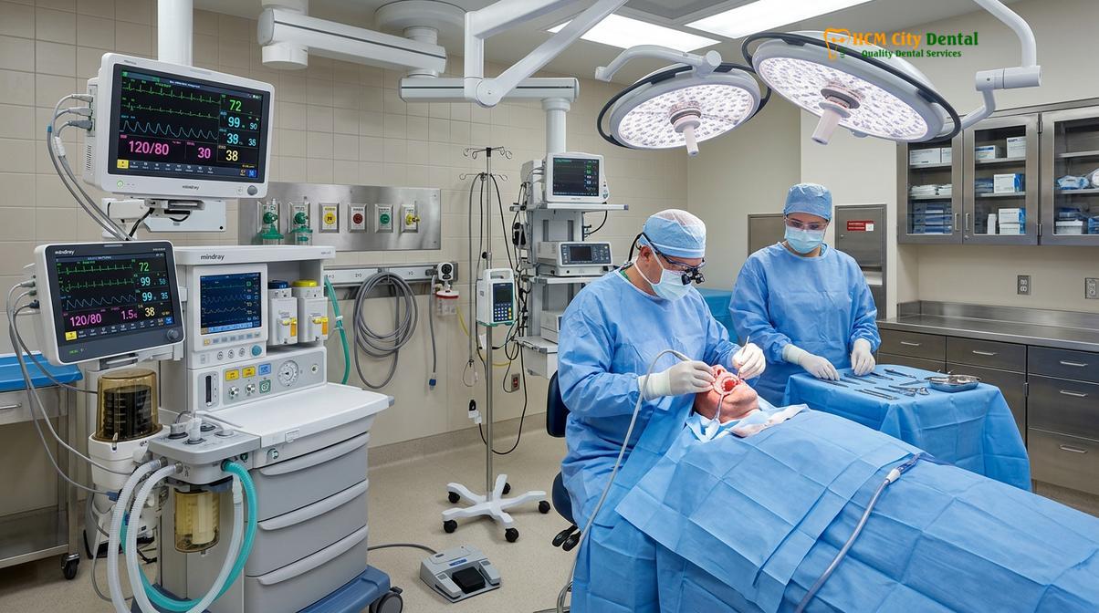
Because the lower jawbone is dense and unyielding, the surgeon often needs to remove a small amount of bone overlying or surrounding the tooth crown. This is performed using a specialized surgical handpiece with copious sterile saline irrigation to prevent the bone from overheating. The goal is to create a pathway for the tooth to exit without applying excessive pressure to the mandible.
Step 4: Odontotomy (Tooth Sectioning)
This is arguably the most crucial step in bottom wisdom teeth removal. Rather than attempting to force the entire tooth out of a small bony opening—which could fracture the jaw or damage the nerve—the surgeon uses a bur to section the tooth into smaller pieces[5]. Typically, the crown is separated from the roots, and the roots may be further divided. This allows each fragment to be gently elevated and removed individually along its path of least resistance.
Step 5: Delivery and Debridement
Once the tooth fragments are removed, the extraction socket is thoroughly inspected and debrided. Any infected tissue, remnants of the dental follicle, or bone spicules are cleaned out. The socket is irrigated with sterile saline to ensure a clean environment for blood clot formation.
Step 6: Suturing and Hemostasis
Finally, the mucoperiosteal flap is repositioned and secured with sutures. These stitches help control bleeding, protect the underlying blood clot, and promote primary intention healing of the soft tissue. Most modern surgical sutures are resorbable and will dissolve on their own within one to two weeks. For a broader understanding of extraction protocols, refer to our general tooth extraction guide.
| Impaction Type | Anatomical Position | Surgical Complexity | Common Clinical Risks |
|---|---|---|---|
| Mesioangular | Tilted forward toward the 2nd molar | Moderate to High | Decay on adjacent tooth, pericoronitis |
| Vertical | Upright but trapped under bone/gum | Moderate | Soft tissue infection, cyst formation |
| Horizontal | Lying flat, parallel to the jawbone | High | Deep bone removal required, nerve proximity |
| Distoangular | Tilted backward into the mandibular ramus | Very High | Difficult surgical access, extensive ostectomy |
Coronectomy: A Conservative Alternative for High-Risk Cases
When a lower wisdom tooth’s roots are intimately entwined with the inferior alveolar nerve, a coronectomy removes only the crown while leaving the roots intact to prevent neurological injury.
In certain clinical scenarios, 3D imaging reveals that the roots of the lower wisdom tooth are in direct contact with, or even wrapping around, the inferior alveolar nerve canal. In these high-risk cases, attempting a complete extraction carries an unacceptably high risk of causing permanent paresthesia (numbness) to the patient’s lower lip and chin.
To mitigate this risk, an alternative surgical technique known as a coronectomy (or intentional partial odontectomy) may be recommended[3]. The principle of a coronectomy is to remove the source of the infection and pathology—the crown of the tooth—while deliberately leaving the vital root structure undisturbed within the jawbone.
The procedure begins similarly to a standard extraction, with flap elevation and bone removal to expose the crown. However, instead of elevating the entire tooth, the surgeon sections the tooth horizontally, completely amputating the crown. The remaining roots are then reduced a few millimeters below the crest of the surrounding bone. The pulp chamber of the retained roots is left untreated, and the gum tissue is sutured completely closed over the site.
Clinical Case Review: High-Risk Horizontal Impaction
A 24-year-old patient presented to HCMC Dental Clinic in Ho Chi Minh City with recurrent pain in the lower right quadrant. A CBCT scan ordered by Dr. Cuong revealed a horizontally impacted wisdom tooth with roots intimately hooked around the inferior alveolar nerve. To avoid neurological damage, a coronectomy was performed. The crown was successfully sectioned and removed, alleviating the patient’s pericoronitis. Follow-up radiographs at 6 and 12 months showed healthy bone fill over the retained roots, with no signs of infection or nerve impairment, demonstrating the efficacy of this conservative approach.
While a coronectomy is a highly effective risk-management strategy, it is not suitable for all teeth. It is contraindicated if the roots themselves are infected, if the tooth is highly mobile, or if the tooth is horizontally impacted in a way that prevents safe sectioning of the crown without mobilizing the roots. In a small percentage of cases, the retained roots may slowly migrate toward the surface over time, requiring a secondary, much safer procedure to remove them once they are clear of the nerve.
Post-Operative Healing and Complication Management
Recovery from lower wisdom tooth extraction typically involves managing localized swelling and trismus, while actively preventing complications such as alveolar osteitis (dry socket) and secondary infections.
The recovery phase following bottom wisdom teeth removal is a critical period that requires strict adherence to post-operative instructions. Because the surgery involves bone removal and tissue manipulation in a highly vascular area, a certain degree of physiological response is expected. Understanding the difference between normal healing and potential complications is essential for patient peace of mind.
Normal Post-Operative Sequelae:
- Swelling (Edema): Swelling is a normal inflammatory response to surgery. It typically peaks around 48 to 72 hours post-operation before gradually subsiding. Applying ice packs to the outside of the face during the first 24 hours can help minimize this.
- Trismus (Jaw Stiffness): Difficulty opening the mouth fully is common after lower wisdom tooth surgery. This is due to inflammation of the masticatory muscles (primarily the masseter and medial pterygoid) caused by the surgical trauma and prolonged mouth opening during the procedure. Gentle jaw stretching exercises after the first few days can aid recovery.
- Mild Oozing: Slight bleeding or pink-tinged saliva is normal for the first 24 hours. Firm pressure with a sterile gauze pad is the most effective way to manage this.
Managing Complications: Alveolar Osteitis (Dry Socket)
The most notorious complication associated with lower wisdom teeth removal is alveolar osteitis, commonly known as a dry socket. This occurs when the blood clot that forms in the extraction socket is dislodged, dissolves prematurely, or fails to form entirely[4]. This leaves the underlying alveolar bone and nerve endings exposed to the oral environment, causing severe, radiating pain that typically begins 3 to 5 days after surgery.
“The incidence of alveolar osteitis is significantly higher in mandibular third molar extractions compared to any other site in the oral cavity. Prevention relies heavily on the stability of the initial coagulum and the avoidance of negative intraoral pressure, such as smoking or using straws.”
Lower wisdom teeth are particularly prone to dry sockets due to the dense bone (which has a poorer blood supply than the upper jaw) and the pooling of saliva and food debris in the lower posterior region. If a dry socket occurs, it is not an infection, but rather a delay in healing. Treatment involves the clinician gently irrigating the socket and placing a medicated dressing to soothe the exposed bone and provide immediate pain relief.
Clinical Warning: Protecting the Blood Clot
To prevent a dry socket, patients must strictly avoid smoking, vaping, using straws, or vigorous spitting for at least 72 hours post-surgery. These actions create negative pressure in the mouth that can dislodge the fragile blood clot. Additionally, maintain a soft diet and avoid chewing directly over the surgical site.
Another potential, though rare, complication is altered sensation (paresthesia) of the lip, chin, or tongue. If the inferior alveolar or lingual nerve was bruised during the procedure, the patient might experience tingling or numbness after the local anesthesia wears off. In the vast majority of cases, this is temporary and resolves as the nerve tissue heals over several weeks or months. Patients experiencing unusual referred pain should also consult our guide on can wisdom teeth cause headaches to understand the broader systemic effects of dental impactions.
When to See a Doctor
While post-operative discomfort and swelling are expected, certain symptoms indicate a deviation from normal healing and require prompt clinical evaluation. You should contact your dental provider immediately if you experience any of the following red flags:
- Uncontrolled Bleeding: Heavy, bright red bleeding that does not subside after applying firm, continuous pressure with a gauze pad for 45 minutes.
- Signs of Infection: A persistent fever, chills, or swelling that continues to worsen after the third post-operative day, rather than improving.
- Severe, Worsening Pain: Pain that becomes excruciating 3 to 5 days after the surgery and is unresponsive to prescribed analgesics (a strong indicator of a dry socket).
- Pus or Foul Discharge: A bad taste in the mouth accompanied by purulent discharge (pus) from the surgical site.
- Difficulty Breathing or Swallowing: Any swelling that extends into the neck or throat and compromises your airway is a medical emergency.
If you are experiencing any of these symptoms, or if you need to schedule a comprehensive evaluation of your wisdom teeth, do not hesitate to seek professional care. You can easily book a dentist appointment online to have your extraction sites evaluated and ensure your recovery is on track.
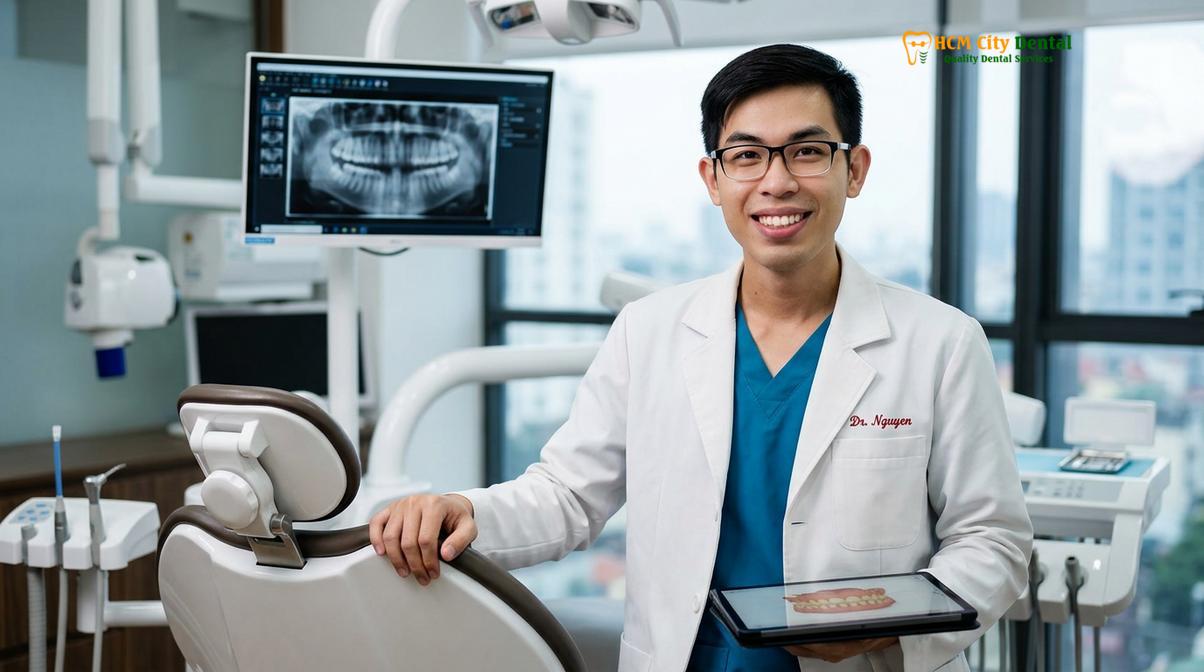
Ultimately, while bottom wisdom teeth removal is a complex surgical procedure, modern diagnostic tools and refined surgical techniques have made it highly predictable. By understanding the anatomical challenges, adhering to pre-surgical planning, and following strict post-operative care, patients can navigate this necessary procedure with confidence and minimal disruption to their daily lives. For those who are congenitally missing these teeth, you can learn more about this phenomenon in our article on having no wisdom teeth.
Frequently Asked Questions
Is bottom wisdom teeth removal more painful than the top?
Lower wisdom teeth removal often involves a more intense recovery than upper teeth due to denser jawbone and higher impaction rates. However, profound local anesthesia ensures the surgical procedure itself remains virtually painless. Post-operative discomfort is managed effectively with prescribed analgesics and anti-inflammatory medications, though you may experience more swelling and jaw stiffness compared to upper extractions.
How long does it take to recover from lower wisdom tooth surgery?
Initial healing and soft tissue closure typically take one to two weeks, while complete bone regeneration within the extraction socket can take several months. Most patients resume normal daily activities within three to five days. Adhering to a soft diet and maintaining excellent oral hygiene will significantly expedite the early phases of soft tissue recovery.
What happens if I leave an impacted bottom wisdom tooth alone?
Ignoring an impacted lower wisdom tooth can lead to recurrent pericoronitis, cyst formation, or severe decay in the adjacent second molar. Proactive clinical monitoring is essential to determine if and when extraction is necessary. Even asymptomatic impacted teeth can harbor bacteria that cause silent bone loss, making regular radiographic evaluation crucial.
Can nerve damage from bottom wisdom tooth extraction be permanent?
While temporary altered sensation (paresthesia) in the lip or tongue can occur if the nerve is bruised, permanent nerve damage is exceedingly rare. Advanced 3D imaging and alternative techniques like coronectomy significantly minimize this risk. Your oral surgeon will map the exact location of the inferior alveolar nerve prior to surgery to ensure the safest possible approach.
When can I eat solid food after the extraction?
You should stick to a soft, cool diet for the first 48 to 72 hours to protect the blood clot. Gradually reintroduce solid foods as your jaw stiffness subsides and chewing becomes comfortable. Avoid hard, crunchy, or spicy foods for at least a week, as these can irritate the surgical site or become lodged in the healing socket.
References
- Journal of Oral and Maxillofacial Surgery. Anatomical considerations and complications in third molar surgery. (2021).
- International Journal of Oral and Maxillofacial Surgery. Efficacy of CBCT in preventing inferior alveolar nerve injury during third molar extraction. (2020).
- British Dental Journal. Coronectomy of lower third molars: a clinical and radiological review. (2019).
- Journal of the American Dental Association. Management of alveolar osteitis and post-operative complications. (2022).
- Clinical Oral Investigations. Biomechanics of tooth sectioning in mandibular third molar impactions. (2018).
For more comprehensive information on dental procedures and oral health maintenance, please visit our general dental guide.