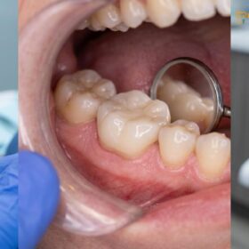
When you replace failing fillings with onlay restorations, you upgrade from a structurally compromised, patched tooth to a highly durable, biomechanically sound solution. Unlike standard direct fillings that simply plug cavities, custom-milled ceramic onlays reinforce the remaining natural tooth structure, preventing catastrophic fractures and effectively sealing out recurrent decay.
Clinical Summary:
Replacing failing large fillings with ceramic onlays represents a conservative, biomimetic approach that preserves maximum healthy tooth structure. Utilizing advanced CAD/CAM technology and precise intraoral scanners, clinicians can fabricate highly accurate, biocompatible restorations. This advanced methodology eliminates the inherent structural weaknesses of massive direct composites and the microleakage associated with aging amalgams, ensuring superior marginal adaptation, optimal occlusal stability, and long-term protection against secondary caries.
Key Takeaways:
- Ceramic onlays preserve natural tooth structure far better than aggressive full crown preparations.
- Digital intraoral scanning ensures a precise marginal fit to prevent secondary decay.
- High-strength materials like IPS e.max and Cercon HT Zirconia withstand heavy posterior biting forces.
- Resin-adhesive cementation creates a unified, fracture-resistant tooth-restoration complex.
- Replacing large amalgams with biocompatible onlays eliminates metal-induced gum irritation.
The Lifecycle of Direct Composite and Amalgam Fillings
Direct fillings have a finite lifespan; amalgams eventually corrode and expand, while composites suffer from wear and polymerization shrinkage, ultimately leading to restoration failure.
Every dental restoration placed in the human mouth is subjected to a harsh, dynamic environment. The oral cavity presents constant challenges, including extreme temperature fluctuations, acidic dietary challenges, and immense occlusal (biting) forces. Because of these factors, direct restorations—whether traditional silver amalgam or modern tooth-colored composite resin—have a limited restoration lifetime. Understanding the lifecycle of these materials is crucial for recognizing when intervention is necessary to save the underlying tooth structure.
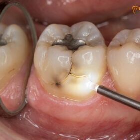
Traditional silver amalgam has been used for over a century due to its durability and ease of placement. However, amalgam is a metal alloy that undergoes physical changes over time. As it ages, it is prone to corrosion and creep (slow deformation under mechanical stress). Furthermore, metal expands and contracts at a different rate than natural tooth enamel when exposed to hot and cold foods. This continuous thermal cycling creates internal wedging forces that can induce micro-cracks in the surrounding healthy enamel. Over a period of 10 to 15 years, these micro-cracks often propagate, leading to fractured cusps or split teeth [1]. Consequently, amalgam replacement becomes a clinical necessity not just for aesthetic reasons, but to prevent catastrophic structural failure of the tooth.
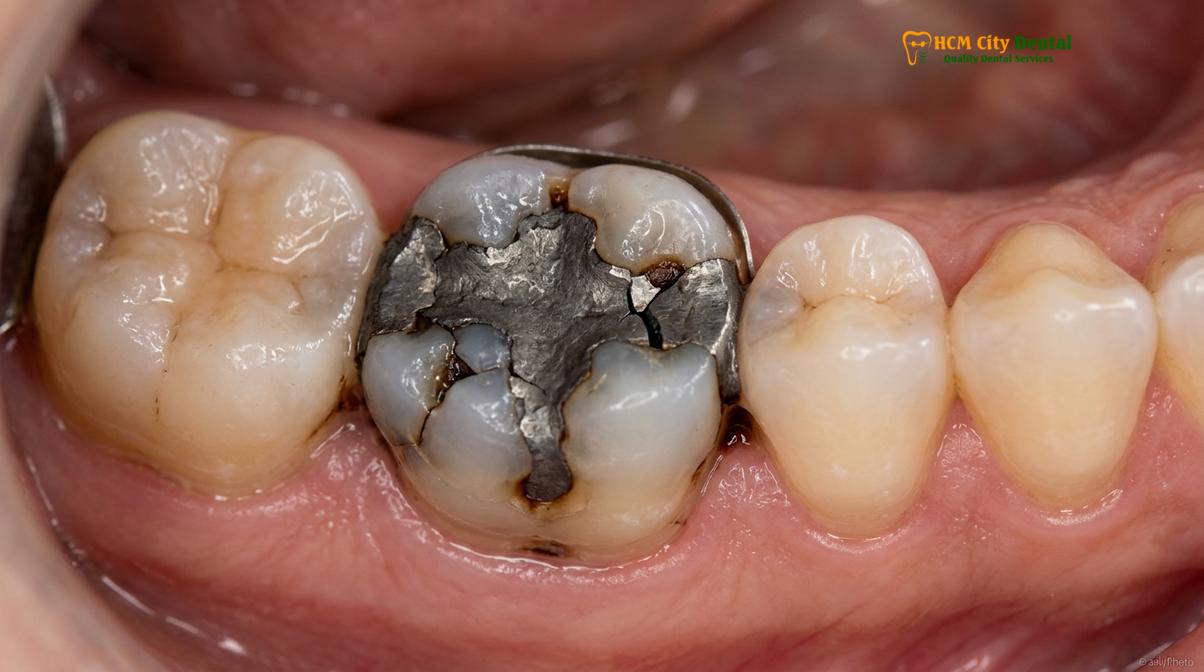
On the other hand, direct composite resin fillings offer excellent initial aesthetics and the ability to bond directly to the tooth structure. However, when used to restore massive cavities, a large composite filling faces significant biomechanical limitations. During the curing process, composite resin undergoes polymerization shrinkage—it contracts slightly as it hardens. In a large cavity preparation, this shrinkage generates immense stress on the cavity walls, potentially pulling the enamel inward and causing postoperative sensitivity or microscopic marginal gaps. Furthermore, composite resin has a lower modulus of elasticity and lower wear resistance compared to laboratory-processed ceramics. Over years of heavy chewing, the composite surface degrades, loses its anatomical contour, and the adhesive bond at the margins begins to fatigue.
What Causes a Large Filling to Fail?
Large fillings fail primarily due to structural fatigue, excessive occlusal stress, and the breakdown of the restorative material’s marginal seal, which allows bacterial infiltration.
The failure of a dental restoration is rarely a sudden event; rather, it is the culmination of progressive degradation over several years. When a cavity is exceptionally large, the remaining natural tooth structure is inherently weakened. A direct filling relies on the remaining walls of the tooth to hold it in place. If those walls are thin, the tooth is highly susceptible to structural failure. When a patient bites down, the filling acts like a wedge, driving the weakened cusps apart. This phenomenon, known as cuspal flexure, eventually leads to the fracture of the natural tooth structure surrounding the filling.
Another primary cause of failure is margins microleakage. The margin is the critical junction where the restorative material meets the natural tooth. In a healthy restoration, this margin is seamlessly sealed. However, due to the wear and tear of mastication, thermal cycling, and the chemical degradation of dental adhesives over time, this seal can break down. Once the margin opens—even microscopically—saliva, food particles, and cariogenic bacteria can percolate into the gap. Because this area is impossible to clean with a toothbrush or floss, it becomes an ideal incubator for decay [2].
Clinical Warning: Ignoring the early signs of a failing filling, such as mild sensitivity to cold or a rough edge felt with the tongue, can lead to severe complications. If a cracked tooth or deep microleakage is left untreated, the bacteria can reach the dental pulp, necessitating complex root canal therapy or resulting in tooth extraction.
Furthermore, improper bite alignment can accelerate the demise of a large filling. If a restoration is placed slightly too high, it absorbs a disproportionate amount of the occlusal load during chewing. For patients who suffer from bruxism (chronic teeth grinding or clenching), the excessive forces can quickly shatter a large composite filling or cause an old amalgam to fracture the surrounding enamel. Restoring the proper vertical dimension of occlusion (VDO) and ensuring harmonious bite alignment are critical factors in extending the lifespan of any dental work.
Recurrent Decay: The Silent Threat Under Old Restorations
Bacteria infiltrating the microscopic gaps of a degraded filling cause hidden cavities, known as recurrent decay, which silently destroy the tooth from the inside out.
One of the most insidious problems in restorative dentistry is recurrent decay (also known as secondary caries). Unlike a primary cavity that forms on the visible surface of a virgin tooth, recurrent decay develops underneath or along the margins of an existing restoration. It is a silent threat because it is often completely hidden from the patient’s view and may not cause any pain until the infection has reached the deep dentin or the nerve of the tooth.
The process begins with margins microleakage. As the old filling degrades, the adhesive bond fails, creating a microscopic pathway. Cariogenic bacteria, primarily Streptococcus mutans, colonize this gap. They feed on fermentable carbohydrates from the patient’s diet and excrete lactic acid. This acid demineralizes the tooth structure beneath the filling. Because the environment under a failing restoration is dark, moist, and protected from salivary buffering and oral hygiene efforts, the decay can progress rapidly.
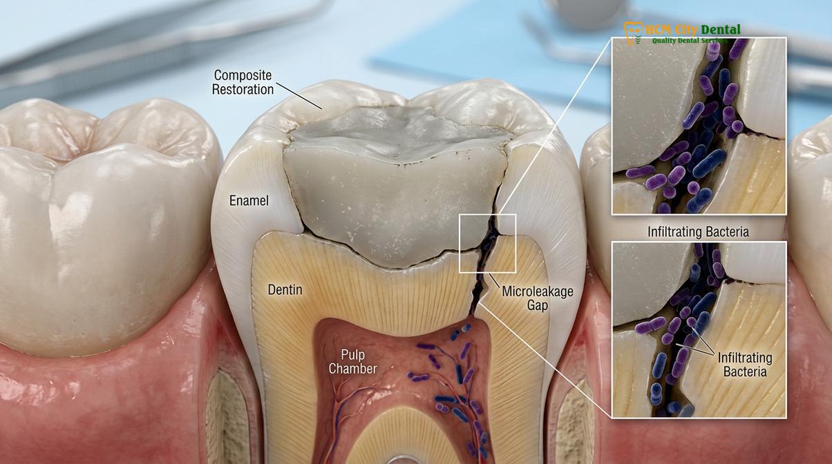
Detecting recurrent decay old composite filling or amalgam requires careful clinical and radiographic examination. While digital X-rays are invaluable for spotting radiolucencies (dark spots indicating decay) under restorations, some filling materials are highly radiopaque and can mask the early signs of secondary caries. Therefore, clinicians must also rely on tactile examination using an explorer, visual inspection under high magnification (dental loupes or microscopes), and patient symptom history. Secondary decay detection is a critical skill, as catching the problem early allows for more conservative treatment options.
If recurrent decay is left unchecked, it will eventually undermine the entire foundation of the tooth. The dentin becomes soft and necrotic, leaving the overlying enamel unsupported. This is often the point at which a patient experiences a sudden “broken tooth” while eating something relatively soft; the tooth didn’t break because the food was hard, but because the internal structure had been entirely hollowed out by recurrent decay [3].
Why Replacing a Large Filling with Another Filling is a Mistake
Repeatedly patching a massive cavity with direct filling material weakens the remaining enamel, fails to protect the cusps, and significantly increases the risk of catastrophic tooth fracture.
When a large filling fails, the instinct of many patients—and sometimes practitioners—is to simply drill out the old material and pack in a new, even larger direct filling. However, from a biomechanical standpoint, this is often a critical error. Every time a tooth is re-prepared for a new filling, more natural tooth structure is inevitably removed to ensure clean margins and eliminate decay. Eventually, the cavity outline form becomes so vast that the remaining enamel walls are too thin to withstand the forces of mastication.
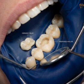
This brings us to the core preservation philosophy of modern biomimetic dentistry. The goal is to preserve maximum natural tooth structure while restoring the tooth’s original strength and biomechanical function. A massive direct filling acts as an intracoronal restoration (sitting inside the tooth). It does not protect the fragile cusps; instead, it acts as a wedge. Conversely, custom dental inlays and onlays are extracoronal restorations. An onlay covers and protects one or more cusps, distributing chewing forces evenly across the entire tooth surface, much like a helmet protects a head.
“When we evaluate a tooth with a massive, failing restoration, our primary objective is structural reinforcement. Simply placing another large composite filling is a temporary patch that invites future fractures. By transitioning to a custom-milled ceramic onlay, we utilize advanced adhesive protocols to bind the weakened cusps together, restoring the tooth to near-original strength while preserving the healthy enamel that a full crown would needlessly destroy.”
— Dr. Nguyen Van Cuong, Lead Clinician
Furthermore, the material properties of direct composite resin are simply inadequate for replacing large volumes of missing tooth structure in the posterior (back) of the mouth. The polymerization shrinkage of a massive composite filling creates immense stress, leading to immediate microleakage. In contrast, a laboratory-fabricated or CAD/CAM milled porcelain overlay is fully polymerized outside the mouth. It exhibits zero shrinkage upon cementation, ensuring a perfectly sealed, passive fit.
| Feature | Large Direct Composite Filling | Custom Ceramic Onlay |
|---|---|---|
| Structural Support | Acts as a wedge; weakens remaining cusps. | Covers and binds cusps together; reinforces tooth. |
| Material Shrinkage | High polymerization shrinkage causing stress. | Zero shrinkage; milled from solid ceramic blocks. |
| Marginal Seal | Prone to degradation and microleakage over time. | Precise CAD/CAM fit with highly durable resin seal. |
| Wear Resistance | Moderate; degrades under heavy chewing forces. | Excellent; mimics the hardness of natural enamel. |
| Longevity | Typically 5 to 7 years before replacement needed. | 15 to 20+ years with proper oral hygiene. |
Upgrading to a Ceramic Onlay: Step-by-Step Transition
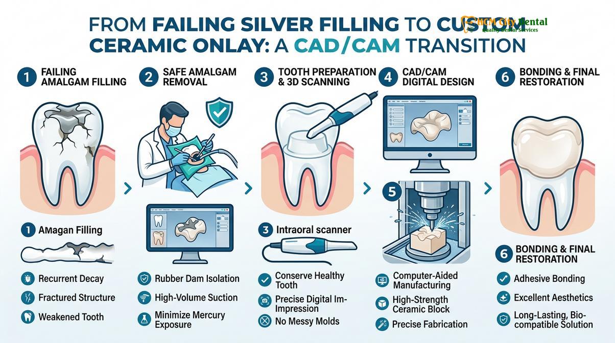
Transitioning to a ceramic onlay involves the safe removal of old material, precise digital 3D scanning, CAD/CAM milling, and meticulous resin-adhesive cementation.
The clinical workflow to replace failing fillings with onlay restorations is a highly precise, multi-step process that leverages the latest advancements in digital dentistry and material science. The procedure begins with the safe amalgam removal protocol. If the failing restoration is silver amalgam, the dentist utilizes high-volume evacuation, copious water spray, and specialized sectioning techniques to remove the metal in chunks. This minimizes the generation of heat and prevents the release of mercury vapor, ensuring a safe environment for both the patient and the clinical team.
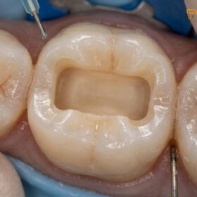
Once the old restorative material and any underlying tooth decay are completely removed, the clinician evaluates the remaining tooth structure. The cavity outline form is carefully refined. Unlike traditional crown preparations that require aggressive 360-degree shaving of the tooth, conservative restorations like inlays and onlays require minimal reduction. The dentist creates smooth, flowing chamfer margins that provide a distinct finish line for the ceramic material to seat against seamlessly [4].
At this stage, advanced biomimetic techniques such as Immediate Dentin Sealing (IDS) and Proximal Box Elevation (PBE) may be employed. IDS involves applying a dentin bonding agent to the freshly cut dentin immediately after preparation. This protects the dental pulp from bacterial invasion, prevents postoperative sensitivity, and significantly increases the final bond strength of the restoration. If the cavity extends deep below the gum line, PBE is used to build up the deep margin with a specialized composite resin, relocating the margin to a supragingival (above the gum) position where it can be easily scanned and kept clean.
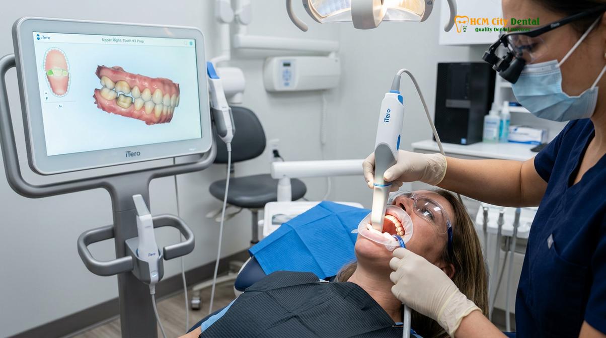
Next, the traditional, uncomfortable silicone impression putty is entirely bypassed. Instead, the clinician uses a state-of-the-art iTero scanner or Medit i700 intraoral scanner to capture a highly accurate 3D digital impression of the prepared tooth and the surrounding bite alignment. This digital data is instantly transmitted to the dental laboratory. Through a direct lab partnership & CAD/CAM technology, expert technicians design the custom restoration virtually. The design is then sent to a precision milling machine, which carves the onlay out of a solid block of high-end ceramic.
Material selection is paramount for long-term success. For teeth in the aesthetic zone or those requiring moderate strength, Ivoclar IPS e.max (lithium disilicate) is often chosen for its exceptional translucency and strong bonding capabilities. For posterior molars subjected to heavy bruxism or intense chewing forces, Dentsply Sirona Cercon HT Zirconia may be utilized due to its unparalleled flexural strength and fracture toughness.
The final step is the delivery and bonding protocol. The ceramic onlay is tried in to verify the marginal fit and occlusal contacts. Once confirmed, the internal surface of the ceramic is treated (e.g., etched with hydrofluoric acid and silanated for e.max, or sandblasted and primed for Zirconia). The tooth enamel is etched, and a sophisticated enamel bonding protocol is executed. The onlay is then permanently affixed using a dual-cure resin-adhesive cementation system. This creates a monoblock effect—a unified, incredibly strong complex where the ceramic and the natural tooth function as a single biomechanical unit [5].
Clinical Case Studies: Silver to Ceramic Makeovers
Real-world clinical outcomes demonstrate how replacing massive, failing metal fillings with custom ceramic onlays restores both biomechanical function and natural aesthetics.
The transformative power of upgrading from failing direct restorations to precision-milled ceramics is best illustrated through clinical outcomes. Patients frequently present with a history of chronic sensitivity, food impaction, and aesthetic dissatisfaction due to aging, dark restorations. By applying the principles of biomimetic dentistry, clinicians can predictably resolve these issues while safeguarding the tooth’s long-term prognosis.
Consider the scenario of replacing large amalgam with onlay saigon. A patient may present with a massive silver filling on a lower first molar that was placed two decades ago. Clinical examination reveals marginal ditching, recurrent decay, and a visible hairline crack propagating down the mesial cusp. If left untreated, this tooth is destined for a catastrophic split that would likely require extraction and a dental implant.
Clinical Case Highlight: HCMC Dental Clinic
A 45-year-old patient visited HCMC Dental Clinic in Ho Chi Minh City complaining of sharp pain when chewing hard foods. Diagnostics revealed a failing silver filling restoration ceramic upgrade was necessary due to severe microleakage and a weakened lingual cusp. Dr. Nguyen Van Cuong carefully removed the amalgam, performed Immediate Dentin Sealing, and utilized the Medit i700 scanner to design a custom IPS e.max porcelain overlay. The conservative preparation saved the remaining healthy buccal enamel. Post-treatment, the patient reported immediate relief from chewing pain, and the tooth was beautifully restored to its natural anatomical contour and strength.
In cases involving multiple failing restorations, the treatment plan may also involve restoring the vertical dimension of occlusion (VDO). Over years of wear, large composite fillings can flatten out, causing the patient’s bite to collapse slightly. By carefully designing multiple ceramic onlays and partial crowns, the dental team can rebuild the proper anatomical height of the teeth, alleviating stress on the temporomandibular joint (TMJ) and providing a more comfortable, stable bite alignment.
The aesthetic improvement is also profound. 100% metal-free, biocompatible ceramic prevents the unsightly gray shadowing often seen around amalgam fillings and eliminates the risk of metal-induced gum line irritation or allergic reactions. The result is a holistic dentistry choice that looks, feels, and functions exactly like a healthy natural tooth.
When to See a Doctor
Recognizing the early warning signs of a failing dental restoration can save you from experiencing severe pain, undergoing complex root canal therapy, or losing the tooth entirely. You should schedule a comprehensive clinical evaluation if you experience any of the following symptoms associated with an old filling:
- Localized Sensitivity: A sharp, fleeting pain when consuming hot, cold, or sweet foods and beverages, indicating potential microleakage or recurrent decay.
- Pain on Chewing: Specifically, a sharp twinge of pain when you release biting pressure. This is a classic symptom of a cracked tooth syndrome, often caused by the wedging effect of a large failing filling.
- Visible Defects: If you can see dark shadows underneath the enamel, noticeable ditching or gaps around the edges of the filling, or if a piece of the filling or tooth has chipped off.
- Food Impaction: Constantly getting food stuck between teeth that have large fillings, which suggests the contact point has worn away or the margin has failed.
- Roughness: Feeling a sharp or rough edge with your tongue where the filling meets the tooth.
“Patients often wait until a tooth physically breaks or causes unbearable pain before seeking help. However, through careful clinical diagnostics and digital scanning, we can identify failing margins and hidden decay long before a catastrophic failure occurs. Early intervention with a conservative ceramic onlay is the most predictable way to save the natural tooth.”
— Dr. Nguyen Van Cuong

If you suspect your old restorations are reaching the end of their lifespan, seeking expert care is essential. At HCMC Dental Clinic in Ho Chi Minh City, our team specializes in conservative, biomimetic restorative dentistry. We utilize advanced digital workflows to provide precise, long-lasting solutions.
Understanding the investment in your dental health is important. According to our current pricing structure, which incorporates a special 40% pre-arrival discount for WhatsApp bookings, upgrading your smile is highly accessible:
- Inlay / Onlay (Lab-made ceramic): Walk-in: ~$385 (10.0M VND) | WhatsApp pre-booking discount: From $231 (6.0M VND).
- Overlay / Partial Crown: Walk-in: ~$446 (11.6M VND) | WhatsApp pre-booking discount: From $269 (7.0M VND).
Furthermore, international and local patients alike can proceed with complete peace of mind, knowing that our restorations are backed by a global warranty and remote WhatsApp checkup support, ensuring your investment is protected long after you leave the clinic.
Frequently Asked Questions
Why do fillings fail after a few years?
Fillings fail due to continuous occlusal stress, material degradation, and thermal expansion. Over time, amalgams corrode and expand, while composites suffer from wear and polymerization shrinkage, leading to marginal breakdown and microleakage. The constant harsh environment of the mouth accelerates this fatigue.
What is recurrent decay?
Recurrent decay is a new cavity that forms underneath or around the margins of an existing dental restoration. It occurs when the seal between the filling and the tooth breaks down, allowing cariogenic bacteria to infiltrate and destroy the hidden dentin.
Is it safe to replace silver fillings with ceramic onlays?
Yes, it is highly safe and beneficial when performed using strict isolation protocols. Dentists use specialized techniques, including high-volume suction and water spray, to safely remove the old amalgam, preventing mercury exposure, before bonding a biocompatible ceramic onlay.
Will an onlay prevent future cavities?
While an onlay provides a superior, tightly sealed margin that significantly reduces the risk of secondary caries, it does not make the tooth immune to decay. Excellent daily oral hygiene, proper diet, and regular dental checkups remain essential to maintain the restoration.
How long does a ceramic onlay last compared to a large filling?
A well-maintained ceramic onlay can last 15 to 20 years or more, whereas a massive direct filling typically fails within 5 to 7 years. The longevity of an onlay is due to its superior material strength, zero polymerization shrinkage, and precise CAD/CAM fit.
References
- Journal of Esthetic and Restorative Dentistry. Longevity of direct and indirect restorations in posterior teeth. (2021).
- International Journal of Prosthodontics. Biomechanical behavior of teeth restored with large composite resins vs ceramic onlays. (2020).
- Journal of Dentistry. Microleakage and marginal adaptation of CAD/CAM ceramic restorations. (2022).
- American Dental Association. Clinical guidelines on the replacement of amalgam restorations. (2019).
- Journal of Prosthodontic Research. Fracture resistance of lithium disilicate and zirconia partial crowns. (2023).