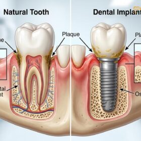
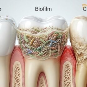
Dental plaque is a sticky, colorless biofilm of bacteria that constantly forms on teeth. If not removed through daily brushing and flossing, it absorbs minerals from saliva and hardens into tartar (dental calculus), a porous, yellow-brown deposit that requires professional dental scaling for safe removal.
Clinical Summary:
The transition from dental plaque to tartar represents a critical shift from a reversible bacterial accumulation to an irreversible calcified deposit. Plaque is a dynamic biofilm that continuously forms on the acquired enamel pellicle, feeding on dietary carbohydrates to produce enamel-demineralizing acids. Within 24 to 48 hours of inadequate oral hygiene, this soft biofilm undergoes mineralization, absorbing calcium and phosphate from saliva to become dental calculus (tartar). Unlike plaque, tartar cannot be removed by at-home brushing. Its porous surface acts as a retentive scaffold for further bacterial colonization, driving the progression of plaque-induced gingivitis into destructive periodontitis. Effective management requires professional intervention utilizing advanced ultrasonic scaling and Guided Biofilm Therapy (GBT) to safely eradicate subgingival deposits, preserve enamel integrity, and halt periodontal attachment loss.
Key Takeaways:
- Plaque is Soft: A sticky, colorless bacterial biofilm that can be disrupted and removed with proper daily brushing and flossing.
- Tartar is Hard: Calcified plaque that has absorbed salivary minerals, requiring professional dental instruments for safe removal.
- Rapid Calcification: The mineralization process from soft plaque to hardened calculus can begin in as little as 24 to 48 hours.
- Periodontal Risk: Tartar buildup below the gumline triggers chronic inflammation, leading to gingival recession and alveolar bone loss.
- Advanced Removal: Modern clinics utilize Guided Biofilm Therapy (GBT) and dual ultrasonic scalers for painless, enamel-safe debridement.
What is Plaque?
Plaque is a dynamic, bacteria-laden biofilm that adheres to the acquired enamel pellicle within minutes after brushing, feeding on dietary carbohydrates to produce enamel-eroding acids.
To understand the fundamental differences between dental plaque vs tartar, one must first examine the microscopic origins of oral biofilms. Dental plaque is not merely a collection of food debris; it is a highly organized, complex ecological community of microorganisms. The formation of plaque begins almost immediately after a comprehensive dental cleaning. Salivary glycoproteins precipitate onto the clean tooth surface, forming a thin, acellular layer known as the acquired pellicle. While this pellicle protects the enamel from acid exposure, it also provides receptor sites for initial bacterial adhesion.
Within hours, early colonizers—primarily Gram-positive facultative anaerobes such as Streptococcus mutans and Streptococcus sanguinis—attach to the pellicle via van der Waals forces and specific molecular interactions. As these bacteria metabolize fermentable carbohydrates from the host’s diet, they excrete extracellular polymeric substances (EPS). This sticky, protective matrix encapsulates the bacterial colonies, forming the structural backbone of dental plaque. The EPS matrix makes the biofilm highly resistant to antimicrobial agents and host immune responses [1].
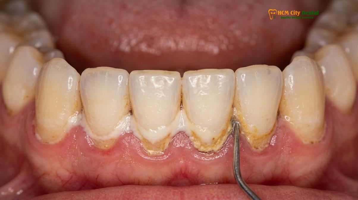
Clinically, plaque presents as a soft, whitish or pale yellow film that accumulates primarily along the gingival margin, in the interproximal spaces between teeth, and within the deep fissures of molars. Because it is soft and structurally vulnerable to mechanical disruption, plaque can be effectively managed and removed through diligent at-home oral hygiene practices, including twice-daily brushing with a fluoridated toothpaste and daily interdental cleaning. However, if left undisturbed, the microbial composition of the plaque shifts. The environment becomes increasingly anaerobic, favoring the proliferation of pathogenic Gram-negative bacteria associated with periodontal disease.
How Plaque Calcifies in 48h
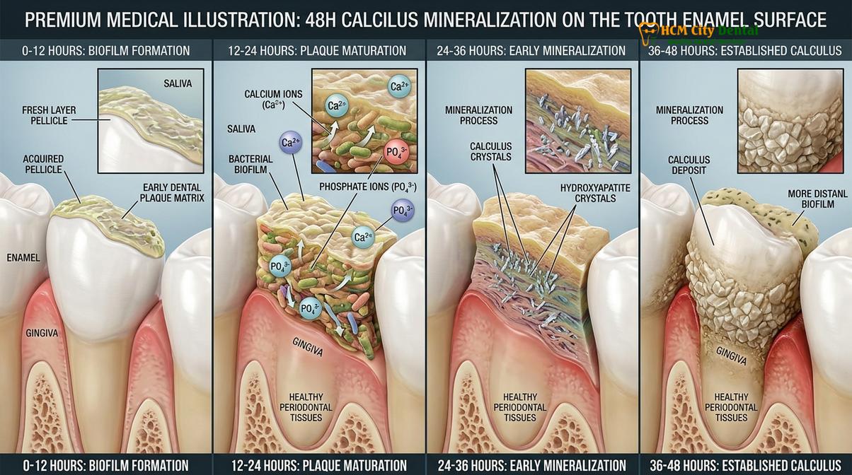
Within 24 to 48 hours, undisturbed plaque undergoes calculus mineralization, absorbing calcium phosphate from saliva to form a rigid, porous structure known as tartar.
The transformation of soft biofilm into hard dental calculus is a fascinating yet detrimental biochemical process known as calculus mineralization. Human saliva is naturally supersaturated with calcium and phosphate ions, an evolutionary mechanism designed to facilitate continuous enamel remineralization. However, when a mature plaque biofilm is allowed to persist on the tooth surface, it acts as a nucleating matrix for these minerals.
The mineralization process can initiate in as little as 24 to 48 hours of inadequate oral hygiene. As the pH within the deeper layers of the plaque fluctuates, calcium and phosphate ions precipitate out of the saliva and crystallize within the extracellular polymeric matrix of the biofilm. The initial mineral phases, such as brushite and octacalcium phosphate, gradually mature into highly stable hydroxyapatite crystals, cementing the bacterial mass into a rigid, stone-like deposit [2].
“The critical danger of calculus is not just its physical presence, but its microscopic topography. The highly porous surface of tartar acts as an ideal, un-cleanable scaffold for fresh, highly pathogenic biofilm to accumulate, driving the inflammatory cascade deeper into the periodontium.”
— Dr. Nguyen Van Cuong, Lead Periodontal Specialist
Clinically, tartar is classified into two distinct categories based on its location relative to the gingival margin:
- Supragingival Calculus: Located above the gumline, this type of tartar is typically yellow or tan. It derives its mineral content primarily from saliva and is most commonly found near the orifices of the major salivary glands, such as the lingual surfaces of the lower anterior teeth and the buccal surfaces of the upper molars.
- Subgingival Calculus: Located below the gumline within the periodontal pocket, this tartar is often dark brown, green, or black. Its dark coloration is due to the incorporation of blood pigments (hemosiderin) from chronically inflamed, bleeding gums. Subgingival calculus derives its minerals from gingival crevicular fluid and is a primary driver of advanced periodontitis.
Comparison Table: Plaque vs Tartar
Understanding the clinical distinctions between plaque and tartar is essential for maintaining optimal oral hygiene and recognizing when professional intervention is required.
While plaque and tartar are stages of the same pathological continuum, their physical properties, clinical implications, and removal protocols differ drastically. The following table outlines the primary distinctions between the two substances.
| Clinical Parameter | Dental Plaque (Biofilm) | Dental Tartar (Calculus) |
|---|---|---|
| Composition | Living bacteria, food debris, and extracellular polymeric substances (EPS). | Calcified dead bacteria, calcium phosphate, and hydroxyapatite crystals. |
| Texture & Consistency | Soft, sticky, and easily disrupted. Feels “fuzzy” to the tongue. | Hard, rigid, and stone-like. Feels rough and porous. |
| Coloration | Colorless, pale yellow, or whitish. Often invisible without disclosing solution. | Yellow, tan, brown, or black (especially subgingival deposits). |
| Formation Time | Begins forming within minutes to hours after brushing. | Mineralization begins within 24 to 48 hours of plaque accumulation. |
| Removal Method | At-home mechanical disruption (brushing, flossing, water flossers). | Strictly requires professional dental scaling and debridement. |
| Primary Pathology | Acid production leading to enamel demineralization and dental caries. | Gingival irritation, periodontal pocket formation, and alveolar bone loss. |
Recognizing these differences empowers patients to take proactive control of their oral health. While diligent home care can entirely prevent the accumulation of plaque, once the calculus mineralization threshold is crossed, the patient must rely on professional clinical protocols to restore periodontal health.
Can You Remove Tartar at Home?
Attempting to remove hardened calculus at home using DIY tools poses a severe tartar scraping hazard, risking irreversible enamel damage, gingival lacerations, and localized infections.
With the proliferation of online tutorials and the availability of consumer-grade dental picks, many individuals wonder if they can bypass the dentist and remove tartar at home. From a clinical perspective, the answer is an unequivocal no. Attempting to scale your own teeth introduces a significant tartar scraping hazard that far outweighs any perceived convenience or cost savings.
Dental calculus is bonded to the tooth surface with a strength that rivals the tooth structure itself. Professional dental hygienists and periodontists undergo years of rigorous training to develop the tactile sensitivity, precise angulation, and fulcrum techniques required to fracture calculus away from the tooth without damaging the underlying enamel or cementum. When untrained individuals attempt to use sharp, non-sterile metal instruments in their own mouths, several severe complications frequently occur [3].
Clinical Warning: The Dangers of DIY Tartar Removal
- Enamel Micro-Scratches: Improper angulation of metal picks creates microscopic gouges in the enamel. These scratches permanently dull the tooth’s appearance and create highly retentive areas where future plaque and tartar will accumulate even faster.
- Gingival Trauma: Slipping with a sharp instrument can cause severe lacerations to the delicate gingival tissues, leading to profuse bleeding, painful ulcerations, and permanent gum recession.
- Subgingival Impaction: Amateurs often inadvertently push calculus fragments deeper into the periodontal pocket, exacerbating inflammation and triggering acute periodontal abscesses.
- Infection Risk: Using non-autoclaved tools introduces foreign pathogens directly into the bloodstream via bleeding gums, posing a risk for systemic bacteremia.
Furthermore, DIY methods are entirely ineffective against subgingival calculus—the dark, hidden tartar that causes the most severe periodontal destruction. Safe and comprehensive removal of these deposits requires specialized ultrasonic technology, proper illumination, and clinical expertise.
Professional Scaling and Advanced Removal Protocols
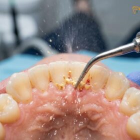
Professional dental scaling utilizes advanced ultrasonic technology and targeted air-polishing to safely eradicate calcified deposits and subgingival biofilm without compromising the underlying tooth structure.
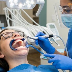
When plaque has calcified into tartar, professional dental scaling is the only safe and effective method for removal. At HCMC Dental Clinic, we have moved beyond traditional, aggressive hand-scraping techniques. Instead, we employ a sophisticated, technology-driven approach to periodontal maintenance that prioritizes patient comfort, enamel preservation, and comprehensive biofilm eradication.
The Dual Scaler Advantage
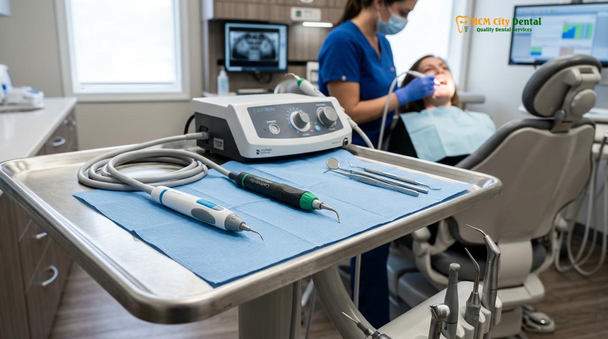
Not all calculus deposits are identical, and therefore, a one-size-fits-all approach to scaling is clinically suboptimal. Our clinic utilizes a “Dual Scaler Advantage,” equipping our operatories with both Piezoelectric and Magnetostrictive ultrasonic technologies to customize the treatment to the patient’s specific anatomical needs.
- Piezoelectric Scalers (e.g., Acteon/Satelec): These advanced devices utilize ceramic discs to generate high-frequency vibrations operating up to 32 kHz. The tip moves in a precise, linear back-and-forth motion, meaning only the lateral sides of the tip are active. This technology generates significantly less heat and requires less water coolant. It is exceptionally gentle, making it the preferred choice for patients with dentin hypersensitivity, pediatric patients, and those undergoing orthodontic treatment.
- Magnetostrictive Scalers (e.g., Dentsply Cavitron): These units utilize a stack of metal strips to generate an elliptical, figure-eight motion at the tip. Because all surfaces of the tip (front, back, and sides) are active, magnetostrictive scalers are incredibly efficient at breaking down heavy, tenacious calculus bridges, eradicating deep subgingival tartar, and performing comprehensive full mouth debridement.
Both technologies rely on the physical principles of cavitation and acoustic microstreaming. As the ultrasonic tip vibrates against the calculus, it shatters the calcified matrix. Simultaneously, the rapid movement of the water coolant creates millions of microscopic vacuum bubbles. When these bubbles collapse (cavitation), they release shockwaves that lyse (burst) bacterial cell walls, effectively disinfecting the periodontal pocket [4].
Guided Biofilm Therapy (GBT): The Swiss Standard
For patients seeking the ultimate in preventive care and comfort, HCMC Dental Clinic features the premium Swiss EMS Guided Biofilm Therapy (GBT) protocol. GBT represents a paradigm shift in dental prophylaxis, moving away from blind scaling toward a targeted, minimally invasive approach.
Dr. Nguyen Van Cuong emphasizes the clinical superiority of the GBT protocol: “By utilizing disclosing agents to visualize the biofilm before we begin, we eliminate the guesswork. We only instrument the areas that require it, preserving the pristine enamel and ensuring a virtually painless experience for the patient.”
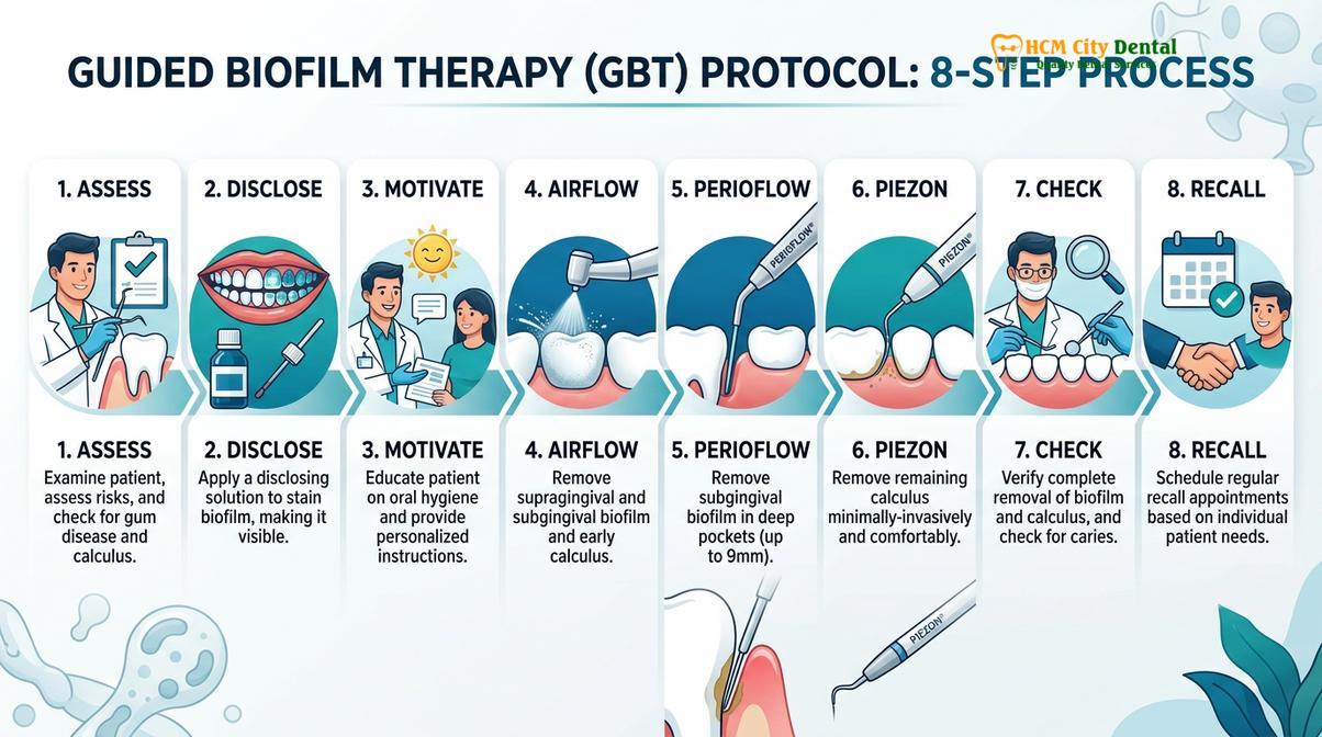
The GBT protocol follows eight systematic steps:
- Assess: Comprehensive clinical examination, probing depths, and caries risk assessment.
- Disclose: Application of a specialized dye that stains fresh plaque pink and mature plaque blue, making the invisible biofilm visible to both the clinician and the patient.
- Motivate: Personalized oral hygiene instruction based on the disclosed areas, empowering the patient to improve their home care.
- AirFlow: Utilizing warm water and low-abrasive erythritol powder (with an ultra-fine 25μm grain size) to gently and painlessly remove biofilm, stains, and early calculus from enamel, dentin, soft tissues, and orthodontic appliances. Erythritol is safe and will not scratch titanium implants, porcelain crowns, or delicate veneers.
- PerioFlow: For patients with deep periodontal pockets (>4mm), a specialized subgingival nozzle is used to deliver the erythritol powder deep into the pocket, eradicating the anaerobic biofilm.
- Piezon: Targeted use of the Piezoelectric ultrasonic scaler to remove any remaining hard calculus. Because the biofilm has already been removed by the AirFlow, the use of metal instruments is minimized by up to 80%.
- Check: Final clinical inspection to ensure all deposits are removed and the application of topical fluoride varnish for enamel remineralization.
- Recall: Establishing a customized maintenance schedule based on the patient’s specific risk profile.
Clinical Pricing and Treatment Options at HCMC Dental Clinic
HCMC Dental Clinic offers transparent, tiered pricing for dental hygiene services, ranging from standard ultrasonic scaling to comprehensive full mouth debridement and premium GBT protocols.
Understanding the cost of periodontal care is crucial for our international and local patients. At HCMC Dental Clinic in Ho Chi Minh City, we maintain a transparent pricing structure that reflects the advanced technology and clinical expertise utilized in our treatments. We offer a tiered approach based on the severity of the calculus accumulation and the specific technology requested by the patient.
According to the latest clinic fee schedule, our professional dental cleaning services are priced as follows:
- Standard Ultrasonic Scaling & Polishing: 500,000 to 800,000 VND (~$20 to $32 USD). This is ideal for patients with mild to moderate supragingival calculus who maintain a regular 6-month recall schedule. (Walk-in rate: 800,000 to 1,300,000 VND).
- Airflow Prophylaxis / Guided Biofilm Therapy (GBT): 1,500,000 to 2,000,000 VND (~$60 to $80 USD). The premium, minimally invasive Swiss EMS protocol utilizing erythritol powder for maximum comfort and stain removal. (Walk-in rate: 2,500,000 to 3,300,000 VND).
- Scaling & Root Planing (SRP deep cleaning per quadrant): 1,000,000 to 1,500,000 VND (~$40 to $60 USD). A therapeutic procedure requiring local anesthesia, designed for patients with active periodontitis and subgingival calculus in pockets deeper than 4mm. (Walk-in rate: 1,600,000 to 2,500,000 VND).
- Full Mouth Debridement (Heavy Tartar Removal): 2,000,000 to 3,000,000 VND (~$80 to $120 USD). A comprehensive initial therapy for patients who have not received dental care in several years and present with massive calculus bridges obscuring the clinical crown. (Walk-in rate: 3,300,000 to 5,000,000 VND).
To make premium dental care more accessible, patients who book their appointments in advance via our official WhatsApp channel are eligible for a -40% WhatsApp booking discount off the standard walk-in rates.
Case Study: Full Mouth Debridement
Severe calculus accumulation requires a phased clinical approach, combining magnetostrictive scaling and deep root planing to restore periodontal health and halt alveolar bone loss.
To illustrate the clinical impact of professional intervention, consider the following documented case managed by our periodontal team.
Clinical Case Review: Severe Calculus Accumulation
Patient Profile: A 45-year-old male presented to HCMC Dental Clinic in Ho Chi Minh City with a chief complaint of severe halitosis, spontaneous gingival bleeding, and visible dark deposits on his lower anterior teeth. The patient reported avoiding dental visits for over eight years due to dental anxiety.
Clinical Findings: Periodontal probing revealed generalized pocket depths of 5-7mm. Massive supragingival calculus bridges were present on the lingual surfaces of teeth #22 through #27. Subgingival exploration confirmed dense, tenacious calculus deposits extending to the base of the periodontal pockets. Diagnosis: Generalized Stage III Periodontitis.
Treatment Protocol: Dr. Nguyen Van Cuong initiated a phased treatment plan.
Phase 1: Full Mouth Debridement using the Magnetostrictive Cavitron scaler to fracture and remove the gross supragingival calculus bridges, allowing the swollen gingival tissues to begin healing.
Phase 2: Two weeks later, the patient returned for Scaling and Root Planing (SRP) under local anesthesia. The Piezoelectric scaler and specialized Gracey curettes were utilized to meticulously debride the subgingival root surfaces, smoothing the cementum to facilitate tissue reattachment.
Phase 3: Application of targeted antimicrobial irrigation (Chlorhexidine 0.12%) and implementation of a strict 3-month periodontal maintenance recall utilizing the GBT protocol.
Outcome: At the 3-month re-evaluation, the patient exhibited a dramatic reduction in gingival inflammation. Bleeding on probing was reduced by 85%, and pocket depths had decreased to a manageable 3-4mm. The patient’s halitosis was entirely resolved, and his dental anxiety was significantly alleviated due to the gentle nature of the ultrasonic technology.
When to See a Doctor
While understanding the difference between plaque and tartar is educational, recognizing the clinical signs of periodontal deterioration is critical for preserving your natural dentition. You should schedule a comprehensive periodontal evaluation if you experience any of the following symptoms:
- Bleeding Gums: Healthy gums do not bleed. Bleeding during brushing, flossing, or eating hard foods is a primary indicator of plaque-induced gingivitis or active periodontitis.
- Persistent Halitosis: Chronic bad breath or a metallic taste in the mouth that does not resolve with brushing is often caused by the volatile sulfur compounds produced by anaerobic bacteria trapped within porous tartar deposits.
- Visible Deposits: The appearance of hard, yellow, brown, or black crusts along the gumline or between the teeth indicates that calculus mineralization has occurred and professional scaling is required.
- Gingival Recession: If your teeth appear longer than they used to, or if you experience sudden sensitivity to hot and cold temperatures, tartar buildup may be causing the gingival tissues to pull away from the tooth, exposing the sensitive root surface.
- Tooth Mobility: Any shifting, looseness, or changes in the way your teeth fit together when you bite down is a late-stage sign of severe alveolar bone loss caused by untreated subgingival calculus.
Early intervention is the cornerstone of conservative dentistry. By addressing tartar accumulation promptly, you can prevent the irreversible destruction of the periodontal ligaments and alveolar bone.
Frequently Asked Questions
Why does plaque turn into tartar so fast?
Plaque turns into tartar rapidly because saliva is naturally supersaturated with calcium and phosphate ions. When oral hygiene is inadequate, these minerals precipitate into the sticky bacterial biofilm within 24 to 48 hours, initiating the calcification process. Factors such as a high-carbohydrate diet, reduced salivary flow (dry mouth), and individual variations in salivary pH can further accelerate this mineralization, turning soft plaque into rigid calculus in a matter of days.
Is DIY tartar scraping safe?
No, DIY tartar scraping is highly dangerous. Using non-sterile, sharp instruments at home lacks the necessary tactile feedback and angulation control, leading to severe enamel scratches, irreversible gingival lacerations, and a high risk of localized bacterial infections. Professional dental hygienists undergo extensive training to safely fracture calculus away from the tooth without damaging the underlying cementum or triggering acute periodontal abscesses.
How often should I get professional scaling?
Most patients require professional scaling every six months to maintain optimal periodontal health. However, individuals with a history of periodontitis, heavy calculus accumulation, or systemic conditions like diabetes may need maintenance visits every three to four months. Your dentist will determine the most appropriate recall interval based on your specific rate of calculus formation, probing depths, and overall caries risk profile [5].
Does removing tartar damage tooth enamel?
When performed by a licensed dental professional using modern ultrasonic scalers and low-abrasive powders, tartar removal is entirely safe for tooth enamel. Advanced technologies like Guided Biofilm Therapy specifically target deposits without scratching the underlying tooth structure. The ultrasonic vibrations are calibrated to shatter the brittle calculus matrix while leaving the resilient enamel completely unharmed.
What is the difference between standard scaling and GBT?
Standard scaling primarily uses ultrasonic and hand instruments to remove hard calculus, followed by gritty paste polishing. Guided Biofilm Therapy (GBT) uses warm water and ultrafine erythritol powder to painlessly eradicate biofilm and early calculus before targeted ultrasonic scaling. GBT is considered a more comfortable, minimally invasive approach that is highly effective for stain removal and is exceptionally safe for delicate restorations, implants, and orthodontic brackets.
References
- Journal of Clinical Periodontology. Guided Biofilm Therapy clinical efficacy. (2021).
- International Journal of Dental Hygiene. Ultrasonic scaling vs hand scaling. (2020).
- Journal of Periodontology. Scaling and root planing for periodontitis. (2019).
- Clinical Oral Investigations. Erythritol air-polishing powder safety. (2022).
- Journal of the American Dental Association. Professional cleaning recall intervals. (2018).