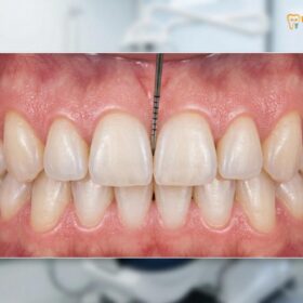
Altered passive eruption gums occur when the gum tissue fails to recede normally during tooth development, leaving excess tissue covering the enamel. This results in a gummy smile and short, square-looking teeth, which can be corrected clinically using precision laser contouring to reveal the natural anatomical crown.
Clinical Summary:
Altered passive eruption (APE) is a developmental dental anomaly where the gingival margin remains positioned too far coronally on the anatomic crown in adulthood. Unlike active eruption, where the tooth emerges into the oral cavity, passive eruption involves the apical migration of the dentogingival junction. When this process is delayed or halted, it creates the illusion of short clinical crowns and excessive gingival display. Diagnosis relies on periodontal probing, bone sounding, and radiographic evaluation to determine the relationship between the cementoenamel junction (CEJ) and the alveolar bone crest. Treatment protocols at advanced centers like HCMC Dental Clinic utilize minimally invasive AMD Picasso Lasers for precise gingivectomy, or osseous resective surgery (osteoplasty/osteotomy) when the bone crest is too close to the CEJ. Restoring the proper supracrestal tissue attachment ensures long-term periodontal health and optimal aesthetic symmetry.
Key Takeaways:
- Altered passive eruption masks the true length of the anatomical crown, causing a disproportionately “gummy” appearance.
- Coslet’s classification system categorizes the condition based on the amount of keratinized gingiva and the alveolar bone crest position.
- Accurate diagnosis requires careful measurement of the biological width to prevent chronic inflammation post-treatment.
- Minimally invasive diode lasers offer bloodless, sutureless correction with rapid 48-hour recovery times.
- Severe skeletal cases may require orthognathic surgery in collaboration with maxillofacial specialists.
Active vs. Passive Tooth Eruption Stages
Tooth eruption is a continuous biological process involving active movement toward the occlusal plane and passive apical migration of the surrounding gum tissue. Understanding these distinct stages is critical for diagnosing gingival abnormalities.
The development of a mature, healthy dentition is not a single event but a complex, multi-phased physiological journey. In clinical dentistry, the eruption of teeth is divided into two distinct but overlapping phases: active eruption and passive eruption. Active eruption refers to the physical movement of the tooth crown through the jawbone and oral mucosa until it meets its antagonist in the opposing arch. This phase establishes the foundational bite alignment and occlusal vertical dimension.
However, the visual length of the tooth is heavily dictated by the second phase. Passive eruption is the gradual apical (downward, toward the root) migration of the gingival tissues, which progressively exposes more of the clinical crown. Gottlieb and Orban originally classified this passive physiological process into four distinct stages. In Stage 1, the teeth reach the line of occlusion, and the junctional epithelium lies entirely on the tooth enamel. As the patient ages, the tissue transitions into Stage 2, where the epithelial attachment rests partially on the enamel and partially on the cementum, though the base of the gingival sulcus remains on the enamel.
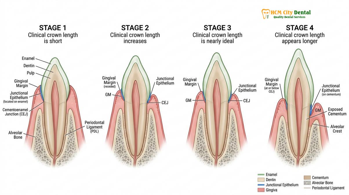
By the time a patient reaches early adulthood, the dentogingival complex should ideally reach Stage 3. In this optimal stage, the entire junctional epithelium lies on the cementum, with the base of the sulcus perfectly aligned at the cementoenamel junction (CEJ). This exposes the full anatomical crown, providing the natural, elongated appearance of adult teeth. Stage 4 is generally considered a pathological state, characterized by gingival recession where the epithelial attachment migrates further apically, exposing the sensitive root surface[1].
“Passive eruption is not merely the receding of gums, but a complex physiological shift of the dentogingival junction that, when interrupted, fundamentally alters the aesthetic and functional harmony of the smile.”
When this biological migration fails to progress past Stage 1 or Stage 2, the condition is clinically diagnosed as delayed passive eruption. The gingival margin remains anchored too high on the enamel, effectively hiding a significant portion of the healthy tooth structure. This developmental pause is not a disease in the traditional sense—it does not inherently cause pain or tooth decay—but it creates a structural imbalance that traps plaque and severely compromises dental aesthetics.
What is Altered Passive Eruption (APE) and Classification?
Altered passive eruption is clinically classified by evaluating the width of keratinized tissue and the distance from the alveolar bone crest to the cementoenamel junction. This diagnostic matrix dictates the appropriate surgical intervention.
To effectively treat altered passive eruption gums, periodontal specialists rely on a highly specific diagnostic framework known as Coslet’s Classification. Introduced in 1977, this system remains the gold standard in periodontology for categorizing the relationship between the gingiva, the underlying alveolar bone, and the tooth structure. The classification is divided into two main Types based on soft tissue dimensions, which are further divided into two Subtypes based on hard tissue (bone) positioning.
Type 1 APE is characterized by a noticeably wide band of keratinized gingiva. The mucogingival junction (the line where the tough, pink keratinized gum meets the loose, red alveolar mucosa) is positioned far apically, meaning there is more than enough robust tissue present. Conversely, Type 2 APE presents with a narrow, inadequate band of keratinized tissue, meaning the mucogingival junction is located very close to the free gingival margin. Differentiating between Type 1 and Type 2 is crucial because excising tissue in a Type 2 case could completely remove the protective keratinized layer, leading to severe mucosal complications.
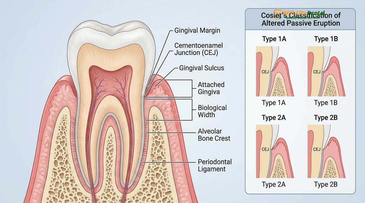
The Subtypes (A and B) evaluate the underlying bone. In Subtype A, the alveolar bone crest is located in its normal physiological position, approximately 1.5 to 2.0 millimeters apical to the CEJ. This space is vital because it houses the supracrestal tissue attachment (formerly known as the biological width)—the connective tissue and epithelial attachment that seal the tooth against bacterial invasion. In Subtype B, the alveolar bone crest is abnormally high, sitting directly at or near the CEJ. This leaves no room for the supracrestal tissue attachment on the root surface, forcing it to attach to the enamel instead[2].
| Classification | Keratinized Gingiva Width | Alveolar Crest Position | Indicated Surgical Treatment |
|---|---|---|---|
| Type 1A | Wide / Adequate | 1.5 – 2.0 mm apical to CEJ | Laser Gingivectomy |
| Type 1B | Wide / Adequate | At or near the CEJ | Gingivectomy + Osseous Resection |
| Type 2A | Narrow / Inadequate | 1.5 – 2.0 mm apical to CEJ | Apically Positioned Flap |
| Type 2B | Narrow / Inadequate | At or near the CEJ | Apically Positioned Flap + Osseous Resection |
Understanding these classifications is paramount. If a dentist attempts a simple soft-tissue excision on a Subtype B case without addressing the bone, the tissue will inevitably rebound and grow back, or worse, trigger chronic, painful inflammation as the body fights to re-establish its biological width.
Why Delayed Eruption Makes Teeth Look Worn and Square
When excess gingival tissue covers the cervical third of the enamel, it distorts the natural width-to-length ratio of the teeth. This results in a boxy, artificially worn appearance commonly referred to as short teeth syndrome.
The aesthetic impact of altered passive eruption is profound. In a naturally developed adult smile, the central incisors exhibit a specific mathematical proportion, often aligning with the principles of the Golden Ratio. The ideal width-to-length ratio of a maxillary central incisor is approximately 75% to 80%. This means the tooth should be visibly taller than it is wide, creating an elegant, rectangular appearance that conveys youth and vitality.
When a patient suffers from delayed passive eruption, the gingival margin fails to retreat, covering the cervical (top) third of the tooth. This drastically reduces the clinical crown height—the portion of the tooth visible above the gum line—while the width remains unchanged. The resulting ratio often approaches 100%, creating teeth that look perfectly square. This visual distortion is frequently misdiagnosed by laypersons as severe bruxism (teeth grinding) or age-related wear, leading to the colloquial term “short teeth syndrome.”
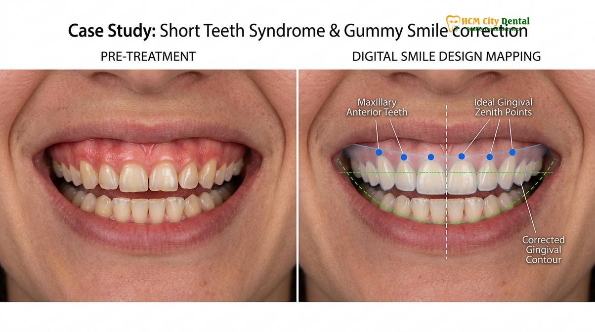
Furthermore, this condition is a primary contributor to a gummy smile. When the patient smiles, the upper lip elevates to reveal not only the shortened teeth but also a massive expanse of pink gingival tissue. The lack of proper gingival zenith points—the highest point of the gum scallop, which should be displaced slightly distal to the long axis of the tooth—leaves the gum line looking flat, blunt, and asymmetrical. Correcting this requires a meticulous eye for facial symmetry and a deep understanding of dental anatomy.
Clinical Case Study: Correcting Short Teeth Syndrome
A 26-year-old female patient presented to HCMC Dental Clinic in Ho Chi Minh City with complaints of a “gummy smile” and square-looking teeth. Clinical examination by Dr. Nguyen Van Cuong revealed a classic case of Type 1B altered passive eruption. The anatomical crowns were fully developed but hidden beneath 3mm of excess gingival tissue, and the alveolar bone crest was situated directly at the CEJ. Utilizing digital smile design (DSD) and picasso laser bone sounding, Dr. Cuong mapped the ideal gingival zenith points. The treatment involved a combined laser gingivectomy and minor osteoplasty to re-establish a healthy 2mm supracrestal tissue attachment. Within two weeks, the patient exhibited perfectly proportioned clinical crowns with zero signs of gingival inflammation.
Correcting Altered Passive Eruption: Laser Gingivectomy Solutions
Modern treatment for altered passive eruption relies on advanced diode lasers to precisely sculpt the gingival margin. This offers a minimally invasive alternative to traditional scalpel surgery with vastly improved recovery times.
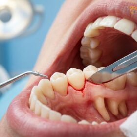
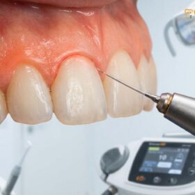
The clinical management of altered passive eruption has been revolutionized by the integration of laser technology. At the forefront of this evolution are AMD Picasso Lasers, which utilize an 810nm diode soft tissue laser. This specific wavelength is highly absorbed by melanin and hemoglobin, making it exceptionally effective for precise soft tissue ablation. Unlike traditional scalpel gingivectomies that result in significant bleeding and require sutures, the diode laser provides bloodless, sutureless cuts. As the laser vaporizes the excess tissue, it simultaneously provides instant coagulation and cauterization of the blood vessels and nerve endings.
Before initiating the aesthetic gum contouring procedure, the clinician must perform a critical diagnostic step known as picasso laser bone sounding. Under profound local anesthesia, a specialized periodontal probe is gently pushed through the gingival sulcus until it contacts the alveolar bone crest. This tactile measurement confirms whether the patient is a Subtype A (requiring only soft tissue removal) or a Subtype B (requiring bone modification). By utilizing digital smile design (DSD), the clinician can pre-plan the exact architectural changes needed to achieve perfect symmetry.
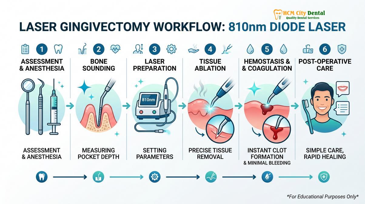
During the procedure, Dr. Nguyen Van Cuong emphasizes the importance of calibrating the laser settings to match the patient’s specific tissue biotype. The sterile beam of the laser not only sculpts the new gingival zenith points but also decontaminates the surgical site, drastically minimizing the risk of postoperative infection. Because the laser seals the tissue as it cuts, the inflammatory response is blunted, leading to a rapid 48-hour recovery period where patients experience minimal to no swelling[3].
For patients considering this transformative procedure, understanding the clinical pricing structure is essential. According to the latest clinic fee schedule, the costs are structured based on the complexity of the intervention:
- Laser Gingivectomy (per tooth): 1,500,000 VND (~$60 USD) (Walk-in: 2,500,000 VND).
- Full-Arch Laser Gum Contouring (up to 10 teeth): 12,000,000 VND (~$480 USD) (Walk-in: 20,000,000 VND).
- Functional/Esthetic Crown Lengthening (per tooth, including bone contouring): 3,000,000 VND (~$120 USD) (Walk-in: 5,000,000 VND).
Patients booking their consultation via WhatsApp can take advantage of a -40% discount, making this advanced laser therapy highly accessible for those seeking to permanently resolve their delayed passive eruption.
Advanced Surgical Interventions for Complex Cases
When excessive gingival display is caused by severe skeletal discrepancies or hypermobile lip muscles rather than isolated passive eruption, multidisciplinary surgical interventions become necessary to achieve facial harmony.
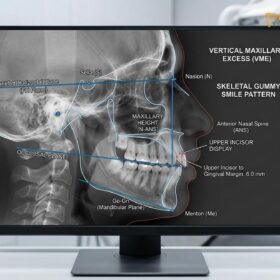
While laser gingivectomy is highly effective for isolated cases of altered passive eruption gums, a comprehensive clinical evaluation may reveal that the gummy smile is multifactorial. In some instances, the excess gum display is driven by Vertical Maxillary Excess (VME)—a skeletal condition where the upper jawbone grows too far downward. For these severe skeletal cases requiring a Le Fort I osteotomy (orthognathic surgery to reposition the entire upper jaw), HCMC Dental Clinic works in collaboration with Surgical Jaw Partners. These complex procedures are coordinated with elite maxillofacial specialists at the National Hospital of Odonto-Stomatology in Ho Chi Minh City (Bệnh viện Răng Hàm Mặt Trung Ương TP.HCM), ensuring the highest level of clinical authority and patient safety[4].
Another common contributing factor is a hyperactive levator muscle, which causes the upper lip to retract excessively during a smile. If the anatomical crown is already fully exposed and the bone levels are normal, cutting more gum tissue would be disastrous. Instead, the appropriate treatment is Surgical Lip Repositioning (Lip Lowering). This procedure involves removing a strip of mucosa from the inside of the upper lip and suturing it lower, physically restricting the lip’s upward mobility. The cost for this surgical intervention is 15,000,000 VND (~$600 USD) (Walk-in: 25,000,000 VND).
For patients seeking a non-surgical alternative to manage a hyperactive lip, Botox Gummy Smile Correction offers a temporary but highly effective solution. By injecting botulinum toxin into the levator labii superioris muscles, the upward pull is relaxed. This treatment is priced at 4,500,000 VND (~$180 USD) for both sides (Walk-in: 7,500,000 VND) and typically lasts for three to four months before requiring maintenance.
Clinical Warning: Biological Width Violation
Performing a simple gingivectomy on a Type 1B or 2B altered passive eruption case without addressing the underlying alveolar bone position will violate the supracrestal tissue attachment (biological width). This inevitably leads to chronic gingival inflammation, rebound tissue overgrowth, and localized periodontal bone loss. Accurate bone sounding is mandatory prior to any soft tissue excision.
When to See a Doctor
Patients should seek a professional periodontal evaluation if they experience a persistently gummy smile, disproportionately short teeth, or chronic gingival inflammation that does not resolve despite rigorous oral hygiene and regular dental scaling. Because altered passive eruption creates deep pseudopockets—areas where the gum tissue overlaps the enamel without actually attaching to it—these zones become highly susceptible to plaque accumulation and localized gingivitis.
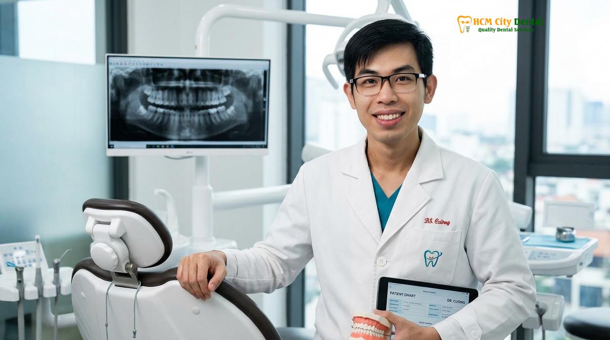
International patients engaging in dental tourism frequently visit Ho Chi Minh City for comprehensive smile makeovers, drawn by the advanced diagnostic capabilities available. A qualified specialist can accurately differentiate between simple delayed passive eruption, skeletal vertical maxillary excess, or a hyperactive lip muscle. Early intervention is highly recommended, as correcting the gingival architecture not only improves aesthetics but also facilitates easier home care and better long-term oral health[5].
“Early diagnostic intervention for altered passive eruption not only transforms the aesthetic proportions of the smile but also creates a self-cleansing gingival architecture that prevents future periodontal complications.”
If you suspect your teeth are hidden beneath excess gum tissue, scheduling a comprehensive evaluation with the General & Preventive Dentistry team is the first step toward uncovering your natural smile.
Frequently Asked Questions
Is altered passive eruption common?
Yes, altered passive eruption is a relatively common developmental condition, estimated to affect approximately 12% of the adult population. While it is not a disease, it is a frequent primary complaint among patients seeking aesthetic dental consultations for a gummy smile or disproportionately short teeth. Because it often presents without pain or infection, many individuals remain undiagnosed until they seek cosmetic improvements.
Will APE resolve on its own?
No, altered passive eruption will not resolve on its own once a patient reaches adulthood. Because the active and passive phases of tooth eruption are biologically complete by the early twenties, any remaining excess gingival tissue covering the anatomical crown requires professional surgical intervention to correct. Waiting will not cause the gums to recede naturally to the proper cementoenamel junction level.
How does a dentist diagnose altered passive eruption?
A dentist diagnoses altered passive eruption through a combination of periodontal probing, radiographic imaging, and bone sounding. These clinical measurements allow the specialist to locate the exact position of the cementoenamel junction (CEJ) and the alveolar bone crest beneath the gum line. This data is essential for classifying the condition according to Coslet’s criteria and determining whether bone recontouring is necessary.
Does laser gum contouring for APE hurt?
Laser gum contouring is highly tolerable and virtually painless, as it is performed under local anesthesia. The use of advanced diode lasers instantly cauterizes nerve endings and blood vessels during the procedure, significantly reducing postoperative discomfort and swelling compared to traditional scalpel surgery. Most patients manage any mild post-procedure sensitivity with standard over-the-counter analgesics.
How long is the recovery after treating delayed passive eruption?
Recovery after laser treatment for delayed passive eruption is remarkably fast, typically taking only 48 to 72 hours for the initial soft tissue healing. Patients can usually resume normal daily activities immediately, though full tissue maturation and stabilization of the gingival margin may take several weeks. If osseous resection (bone contouring) was required, the underlying bone remodeling process will continue for several months, though this is generally not felt by the patient.
References
- Journal of Clinical Periodontology. Esthetic crown lengthening outcomes and biological width. (2021).
- International Journal of Periodontics & Restorative Dentistry. Classification and management of altered passive eruption. (2020).
- Journal of Aesthetic and Restorative Dentistry. Diode laser applications in gingival contouring. (2019).
- Journal of Oral and Maxillofacial Surgery. Le Fort I osteotomy stability in vertical maxillary excess. (2022).
- Clinical Oral Investigations. Surgical lip repositioning and botulinum toxin efficacy. (2018).