Vertical Maxillary Excess (VME) is a complex skeletal deformity characterized by the disproportionate downward overgrowth of the upper jaw bone, resulting in an elongated lower face and a severe gummy smile. Clinical management requires precise cephalometric diagnosis to determine whether biomechanical orthodontic intrusion or orthognathic surgery is the most appropriate corrective pathway.
Clinical Summary:
Vertical Maxillary Excess manifests as an excessive vertical dimension of the maxilla, leading to lip incompetence, a hyperdivergent facial profile, and significant gingival display. Treatment protocols are dictated by the severity of the skeletal discrepancy. Mild to moderate cases may be managed via orthodontic intrusion using temporary anchorage devices (TADs). Severe presentations necessitate a Le Fort I osteotomy to physically impact the maxilla superiorly. Following skeletal correction, soft tissue refinement using advanced 810nm diode lasers is routinely performed to calibrate gingival zenith points, respect the supracrestal tissue attachment, and finalize the esthetic architecture of the smile.
Key Takeaways:
- VME is a structural skeletal anomaly, distinct from purely dental or muscular causes of excessive gingival display.
- Accurate diagnosis relies on lateral cephalometric radiographs to evaluate the lower anterior facial height and occlusal plane angulation.
- Orthodontic intrusion utilizing TADs offers a minimally invasive alternative for moderate vertical discrepancies.
- Severe skeletal cases require collaborative orthognathic surgery, specifically superior repositioning via Le Fort I osteotomy.
- Post-surgical soft tissue management with diode lasers ensures optimal gingival contouring and long-term esthetic stability.
Understanding the Skeletal Framework: Upper Jaw Overgrowth
Vertical Maxillary Excess is a developmental skeletal anomaly where the upper jaw grows excessively in the vertical dimension, fundamentally altering facial proportions, lip dynamics, and dental occlusion.
The human facial skeleton develops through a complex interplay of genetic predispositions and environmental functional matrices. In a normodivergent facial growth pattern, the maxilla and mandible grow in harmony, maintaining balanced proportions between the upper, middle, and lower thirds of the face. However, in cases of Vertical Maxillary Excess, the maxilla exhibits an exaggerated downward and forward growth vector. This VME jaw bone overgrowth significantly increases the lower anterior facial height (LAFH), leading to what is clinically recognized as “long face syndrome.”[1]
The pathophysiology of this condition involves the excessive vertical development of the dentoalveolar process. As the maxilla grows downward, it carries the maxillary dentition with it. To maintain an occlusal relationship, the mandible often rotates downward and backward (clockwise rotation), which further exacerbates the appearance of an elongated face and often results in a retrognathic (recessed) chin profile. This skeletal gummy smile is fundamentally different from a dental gummy smile caused by delayed passive eruption, where the bone position is normal but the gum tissue has failed to recede properly during tooth eruption.
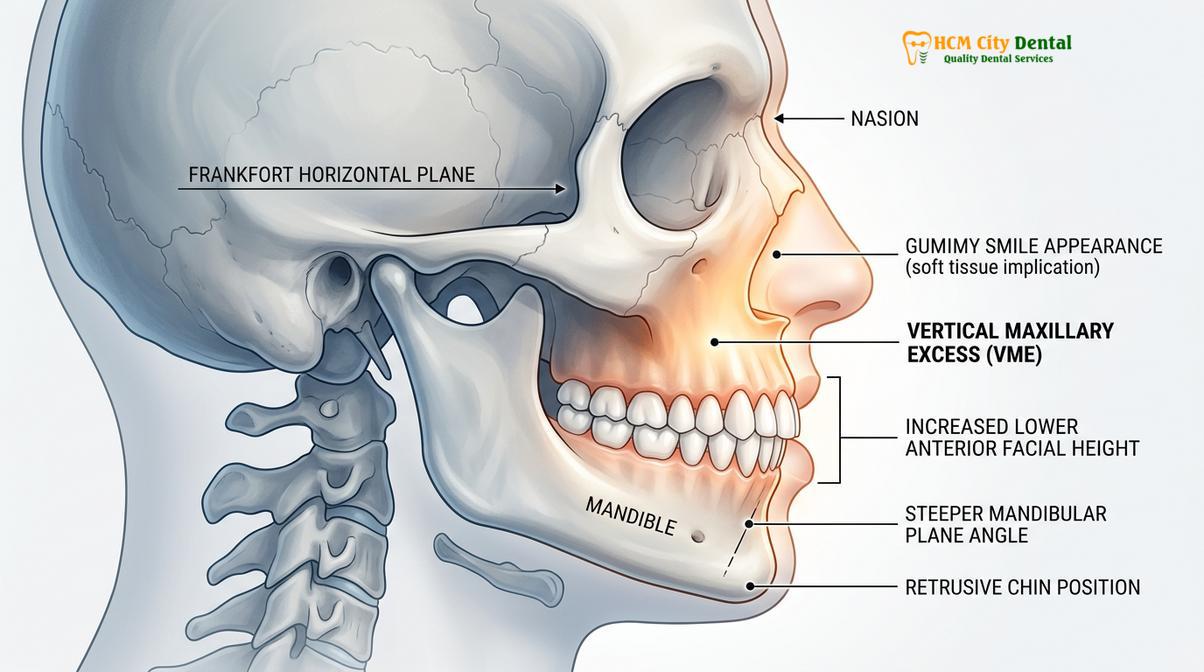
Clinically, patients with upper jaw overgrowth present with several hallmark features. At rest, there is typically lip incompetence—an inability to close the lips without straining the mentalis muscle in the chin. The interlabial gap (the space between the upper and lower lips at rest) is significantly increased, often exposing more than 3 to 4 millimeters of the maxillary incisors. During a dynamic smile, the excessive vertical dimension becomes starkly apparent, revealing a wide band of gingival tissue. Furthermore, the altered skeletal framework often impacts the airway and masticatory function, making this not merely an esthetic concern but a comprehensive functional discrepancy.
Understanding the distinction between skeletal and dentoalveolar components is crucial. While the basal bone of the maxilla may be vertically elongated, the alveolar bone housing the teeth also undergoes compensatory vertical growth. This dual-layered overgrowth requires a treatment approach that addresses both the skeletal base and the dentoalveolar complex to achieve lasting stability and functional harmony.
Diagnosing Vertical Maxillary Excess (VME) via Cephalometric X-Rays
Accurate diagnosis of VME relies on detailed lateral cephalometric radiography to quantify the spatial relationship of the maxilla to the cranial base and evaluate the extent of vertical elongation.
The cornerstone of diagnosing a skeletal gummy smile is the lateral cephalometric radiograph. This standardized imaging modality allows clinicians to perform precise angular and linear measurements, comparing the patient’s skeletal architecture against established normative data. The primary objective of cephalometric analysis in these cases is to isolate the maxilla’s vertical position and determine its contribution to the overall facial disharmony.[2]
Key cephalometric indicators include the evaluation of the lower anterior facial height (LAFH), measured from the anterior nasal spine (ANS) to the menton (Me). In patients with VME, this measurement is significantly increased. Additionally, the distance from the palatal plane to the incisal edge of the maxillary central incisors is evaluated to quantify the dentoalveolar extrusion. The mandibular plane angle (SN-MP or FMA) is typically steep, reflecting the downward and backward rotation of the mandible caused by the vertically excessive maxilla.
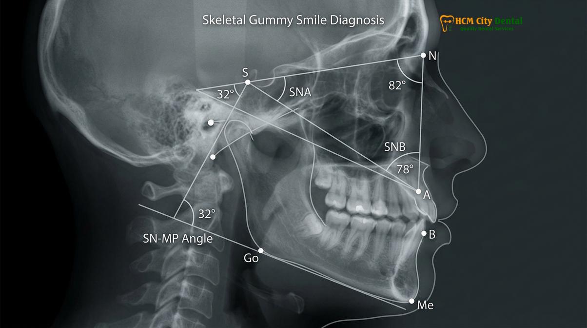
Beyond skeletal measurements, modern diagnostic protocols incorporate comprehensive soft tissue analysis. The length of the upper lip (measured from subnasale to stomion superius) is critical; a normal upper lip length is approximately 20-22mm in females and 22-24mm in males. If the lip length is normal but gingival display is excessive, the diagnosis points strongly toward skeletal VME. Conversely, a short upper lip may compound the visual impact of a mild skeletal discrepancy.
| Diagnostic Parameter | Normative Value (Approximate) | Typical VME Presentation | Clinical Implication |
|---|---|---|---|
| Lower Anterior Facial Height (ANS-Me) | 60 – 65 mm | > 70 mm | Elongated lower face, “long face syndrome” |
| Maxillary Incisor Display at Rest | 2 – 3 mm | > 4 – 5 mm | Lip incompetence, chronic mouth breathing |
| Mandibular Plane Angle (SN-MP) | 32 degrees | > 38 degrees | Clockwise mandibular rotation, retrognathic profile |
| Palatal Plane to Maxillary Incisor Edge | 28 – 30 mm | > 34 mm | Severe dentoalveolar extrusion requiring intrusion |
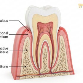
During the diagnostic phase, it is also imperative to evaluate the periodontal phenotype and the supracrestal tissue attachment (formerly known as the biological width). In cases where compensatory eruption has occurred, the relationship between the alveolar crest, the cementoenamel junction (CEJ), and the gingival margin must be mapped via bone sounding. This ensures that any subsequent treatment—whether orthodontic or surgical—respects the minimum 2mm attachment required to prevent chronic inflammation or iatrogenic bone resorption.
The Three Grades of VME and Esthetic Impacts
VME is clinically categorized into three distinct grades based on the millimeter extent of gingival display and the severity of lip incompetence, guiding the appropriate therapeutic intervention.
To standardize treatment protocols and facilitate clear communication between orthodontists, oral surgeons, and periodontists, the severity of a skeletal gummy smile is classified into specific grades. The grade of maxillary excess is determined by measuring the amount of gingival tissue exposed during a full, uninhibited Duchenne smile, as well as evaluating the resting posture of the lips.
Grade I (Mild VME): In this category, the patient displays approximately 2 to 4 millimeters of gingival tissue upon smiling. At rest, lip incompetence is minimal or absent, and the patient can achieve lip seal without noticeable mentalis strain. Grade I cases are often borderline and may be successfully managed with non-surgical approaches, such as orthodontic intrusion or minor soft tissue contouring.
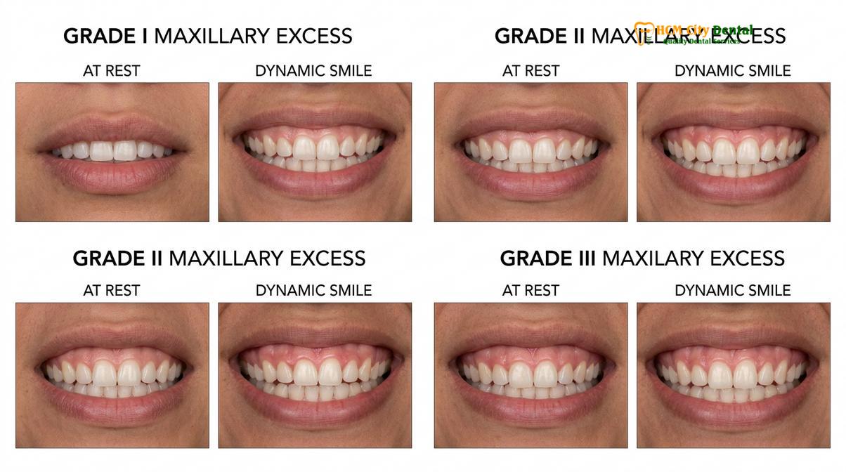
Grade II (Moderate VME): Patients with Grade II excess exhibit 4 to 8 millimeters of gingival display. Lip incompetence becomes a prominent clinical feature, with an interlabial gap of 4 to 6 millimeters at rest. The patient must actively contract the perioral musculature to close their lips, which can lead to a flattened chin profile. These cases require significant biomechanical intervention, often utilizing temporary anchorage devices (TADs) to achieve meaningful skeletal intrusion.
Grade III (Severe VME): This is the most extreme presentation, characterized by more than 8 millimeters of gingival exposure during a smile. The upper jaw overgrowth is so pronounced that the entire clinical crown and a vast expanse of alveolar mucosa are visible. Lip incompetence is severe, often resulting in chronic mouth breathing and secondary gingival inflammation due to desiccation. Grade III VME is almost exclusively a surgical indication, requiring a Le Fort I osteotomy for comprehensive correction.
“The accurate grading of vertical maxillary excess is not merely an academic exercise; it is the fundamental blueprint that dictates whether a patient will achieve stability through orthodontics alone or requires the transformative intervention of orthognathic surgery.”
It is also vital to assess the role of the facial musculature. In some instances, a patient may have a moderate skeletal discrepancy compounded by a hyperactive levator muscle (levator labii superioris alaeque nasi). This hypermobility causes the upper lip to retract excessively during smiling, mimicking a higher grade of VME. Differentiating between skeletal overgrowth and muscular hypermobility is essential, as the latter may benefit from adjunctive neuromodulator therapies (such as Botox) rather than aggressive bone reduction.
Treatment Paths: Skeletal Intrusion vs. Maxillofacial Surgery
Correcting VME requires either biomechanical orthodontic intrusion using TADs for moderate cases or surgical repositioning of the maxilla via Le Fort I osteotomy for severe skeletal discrepancies.
The management of VME represents one of the most challenging frontiers in dentofacial orthopedics. The chosen treatment pathway depends entirely on the severity of the skeletal dysplasia, the patient’s age, and their specific esthetic and functional goals. The two primary modalities are non-surgical orthodontic intrusion and orthognathic surgery.
Orthodontic Intrusion VME (Non-Surgical Approach)
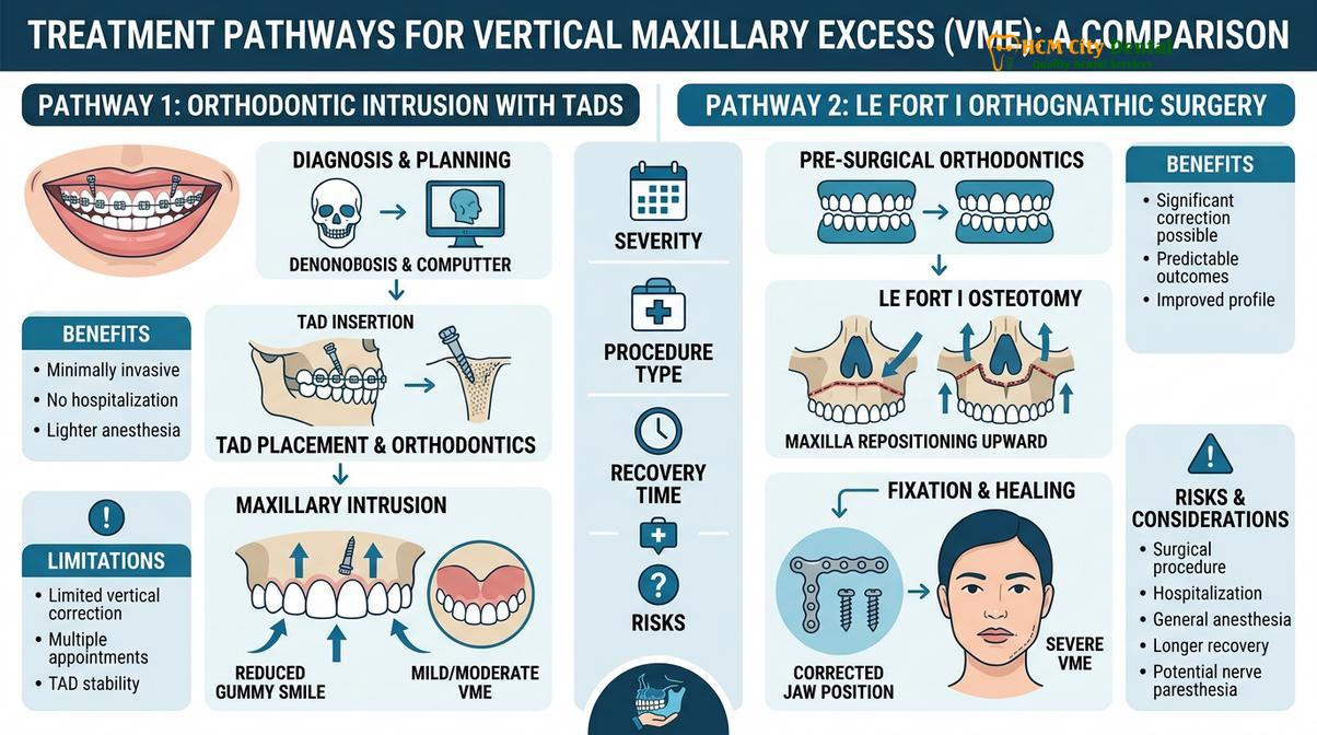
For patients with Grade I or moderate Grade II VME who decline surgery, orthodontic intrusion VME utilizing Temporary Anchorage Devices (TADs) offers a powerful alternative. TADs are biocompatible titanium micro-screws temporarily placed into the dense cortical bone of the maxilla—typically in the infrazygomatic crest or the interradicular spaces. These devices provide absolute anchorage, allowing the orthodontist to apply continuous, controlled upward forces to the entire maxillary dentoalveolar complex.[3]
By utilizing TADs in conjunction with rigid archwires and elastomeric chains or NiTi coil springs, the maxillary teeth and their surrounding alveolar bone can be intruded by 3 to 5 millimeters. This true bodily intrusion reduces the lower anterior facial height, allows the mandible to autorotate counterclockwise (improving chin projection), and significantly diminishes the gummy smile. However, this process requires meticulous biomechanical control to prevent root resorption and ensure the occlusal plane remains level.
Le Fort I Osteotomy (Surgical Approach)
For severe Grade II and Grade III cases, orthognathic surgery is the gold standard. The procedure of choice is the Le Fort I osteotomy with superior impaction. During this surgery, the maxillofacial surgeon makes an incision high in the maxillary vestibule, exposing the facial skeleton. A horizontal osteotomy is performed above the roots of the upper teeth, completely separating the maxilla from the cranial base.
Once mobilized, a predetermined segment of bone is removed from the superior aspect of the maxilla (osteoplasty). The entire upper jaw is then repositioned upward (impacted) and secured in its new, anatomically correct position using rigid titanium mini-plates and screws. This superior repositioning directly eliminates the excessive vertical dimension, allows the lips to close naturally without strain, and dramatically transforms the facial profile.
For severe skeletal cases requiring Le Fort I osteotomy orthognathic surgery, HCMC Dental Clinic works in close collaboration with Surgical Jaw Partners—specifically the highly esteemed maxillofacial specialists at the National Hospital of Odonto-Stomatology in Ho Chi Minh City (Bệnh viện Răng Hàm Mặt Trung Ương TP.HCM), recognized as the highest clinical authority in the region. Dr. Nguyen Van Cuong emphasizes that this collaborative approach ensures patients receive world-class surgical care combined with meticulous pre- and post-surgical orthodontic detailing.
Soft Tissue Management and Esthetic Refinement
Following skeletal correction, precise soft tissue contouring using advanced diode lasers is essential to establish ideal gingival architecture and harmonize the final digital smile design.
Whether a patient undergoes orthodontic intrusion or orthognathic surgery, the final phase of treating a skeletal gummy smile almost always involves soft tissue refinement. As the bone is repositioned or intruded, the overlying gingival tissues may bunch up or heal with irregular margins. To achieve a truly flawless result, the gingival zenith points—the highest point of the gum scallop, located slightly distal to the long axis of each tooth—must be meticulously calibrated.
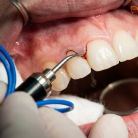
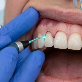
At HCMC Dental Clinic, Dr. Nguyen Van Cuong utilizes state-of-the-art AMD Picasso Lasers (Diode soft tissue laser, 810nm) for this critical finishing stage. The 810nm wavelength is highly absorbed by melanin and hemoglobin, making it the ideal tool for soft tissue microsurgery. The laser provides bloodless, sutureless cuts with instant coagulation and cauterization. Because the procedure is minimally invasive, patients experience significantly reduced postoperative pain and swelling, enjoying a rapid 48-hour recovery. Furthermore, the sterile beam of the diode laser minimizes any risk of postoperative infection.[4]
Clinical Case Review: Comprehensive VME Management
A 24-year-old patient presented to HCMC Dental Clinic in Ho Chi Minh City with Grade II VME, exhibiting 6mm of gingival display and lip incompetence. After a thorough cephalometric evaluation, the patient opted for a combined approach. Initial orthodontic alignment was followed by TAD-supported maxillary intrusion, achieving 4mm of skeletal lift. To perfect the result, Dr. Cuong performed a laser gingivectomy using the AMD Picasso Laser to adjust the gingival zenith points and lengthen the clinical crowns. The synergistic treatment resolved the lip incompetence and delivered a perfectly proportioned, highly esthetic smile without the need for a Le Fort I osteotomy.
Prior to any surgical or laser intervention, thorough dental scaling and root planing are mandatory to ensure the periodontium is entirely free of inflammation. When planning the final gingival contours, the clinical team employs Digital Smile Design (DSD) software to map the exact millimeter adjustments required, ensuring the final soft tissue drape perfectly frames the newly positioned dentition.[5]
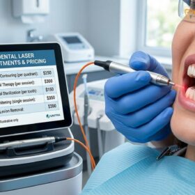
For patients exploring advanced gum contouring and gummy smile correction, understanding the financial investment is crucial. According to the latest clinic fee schedule, the Pricing Structure for soft tissue and adjunctive procedures is highly transparent:
- Laser Gingivectomy (per tooth): 1,500,000 VND (~$60 USD) (Walk-in: 2,500,000 VND).
- Full-Arch Laser Gum Contouring (up to 10 teeth): 12,000,000 VND (~$480 USD) (Walk-in: 20,000,000 VND).
- Functional/Esthetic Crown Lengthening (per tooth, including bone contouring): 3,000,000 VND (~$120 USD) (Walk-in: 5,000,000 VND).
- Surgical Lip Repositioning / Lip Lowering: 15,000,000 VND (~$600 USD) (Walk-in: 25,000,000 VND).
- Botox Gummy Smile Correction (both sides): 4,500,000 VND (~$180 USD) (Walk-in: 7,500,000 VND).
Note: International patients engaging in dental tourism can take advantage of a -40% WhatsApp booking discount prior to arrival.
When to See a Doctor
Early clinical evaluation is critical for patients exhibiting chronic lip incompetence, excessive gingival display, or difficulty chewing, as these may indicate underlying skeletal discrepancies.
Identifying Vertical Maxillary Excess early can significantly broaden the available treatment options. Patients should seek a comprehensive evaluation from a qualified orthodontist or maxillofacial specialist if they experience persistent difficulty closing their lips at rest, chronic dry mouth or gingivitis due to mouth breathing, or if they feel their smile is dominated by gum tissue rather than teeth.
For international patients, dental tourism in Ho Chi Minh City offers access to world-class diagnostics and multidisciplinary care. A thorough consultation will involve 3D Cone Beam Computed Tomography (CBCT), digital intraoral scanning, and a detailed facial analysis to differentiate between a simple soft tissue issue and a complex skeletal deformity.
Important Clinical Considerations
Attempting to treat a severe skeletal gummy smile (Grade III VME) solely with soft tissue laser gingivectomy is a critical clinical error. Removing too much gum tissue without addressing the underlying bone overgrowth will violate the supracrestal tissue attachment, leading to severe chronic inflammation, bone loss, and eventual relapse. A personalized clinical examination is mandatory to determine the correct anatomical diagnosis before any irreversible treatment is initiated.
“The successful management of vertical maxillary excess relies on a profound respect for facial biomechanics. By accurately diagnosing the skeletal foundation, we can tailor interventions that not only enhance esthetics but fundamentally restore optimal oral function.”
Frequently Asked Questions
Find evidence-based answers to the most common patient inquiries regarding the diagnosis, treatment, and recovery associated with Vertical Maxillary Excess.
Can VME cause bite issues?
Yes, Vertical Maxillary Excess frequently causes severe bite issues, including anterior open bites, Class II malocclusions, and transverse discrepancies. The excessive downward growth of the upper jaw alters the occlusal plane, preventing the upper and lower teeth from meeting correctly during function. This skeletal imbalance often forces the mandible to rotate backward, further complicating the bite and potentially leading to temporomandibular joint (TMJ) strain and inefficient mastication.
Is jaw intrusion with TADs effective for VME?
Orthodontic intrusion using Temporary Anchorage Devices (TADs) is highly effective for mild to moderate Vertical Maxillary Excess. By applying continuous biomechanical force, TADs can intrude the maxillary dentoalveolar complex by several millimeters, reducing gingival display without invasive surgery. However, this approach requires healthy periodontal support, adequate root length, and meticulous force application to prevent root resorption. It is generally not sufficient for severe Grade III skeletal discrepancies.
What is the difference between dental and skeletal gummy smiles?
A dental gummy smile is caused by altered passive eruption or short clinical crowns, whereas a skeletal gummy smile results from the physical overgrowth of the upper jaw bone (VME). In dental cases, the jaw bone is in a normal position, and the issue is resolved with soft tissue contouring or crown lengthening. In skeletal cases, the entire maxilla is elongated, requiring bone repositioning (orthognathic surgery) or orthodontic intrusion to correct the foundational discrepancy.
How long is the recovery after Le Fort I osteotomy for VME?
Initial recovery from a Le Fort I osteotomy takes approximately two to three weeks, during which swelling subsides and patients adhere to a strict soft diet. Complete bone healing and final stabilization of the skeletal and soft tissue changes require three to six months. During the initial phase, patients are closely monitored by their surgical team to ensure proper rigid fixation stability and to manage postoperative edema, while orthodontic detailing continues to perfect the bite.
Can laser gum contouring fix VME on its own?
No, laser gum contouring cannot resolve the underlying bone discrepancy of true Vertical Maxillary Excess. However, it is frequently used as a finishing procedure after skeletal correction to perfect the gingival margins and establish ideal zenith points for optimal esthetics. If laser contouring is performed aggressively on a skeletal VME case without bone reduction, it risks violating the biological width, leading to chronic periodontal inflammation and tissue rebound.
References
- American Journal of Orthodontics and Dentofacial Orthopedics. Cephalometric evaluation of the long face syndrome. (2020).
- Journal of Clinical Periodontology. Soft tissue management and biological width following orthognathic surgery. (2019).
- International Journal of Periodontics & Restorative Dentistry. Biomechanics of orthodontic intrusion using TADs. (2021).
- Journal of Aesthetic and Restorative Dentistry. Diode laser applications in gingival zenith calibration. (2023).
- Journal of Oral and Maxillofacial Surgery. Stability of Le Fort I osteotomy in vertical maxillary excess. (2022).