The biological width of gums is the natural protective seal of soft tissue attached to the tooth directly above the alveolar bone. Measuring approximately 2mm, it defends the underlying bone from bacterial infection and must be strictly preserved during restorative dental procedures to prevent chronic inflammation.
Clinical Summary:
Biological width is a fundamental periodontal concept dictating the health and stability of the dentogingival junction. It comprises the junctional epithelium and supracrestal connective tissue attachment, forming a vital immunological barrier against oral pathogens. In restorative dentistry, encroaching upon this critical 2mm zone with crowns, veneers, or fillings triggers severe inflammatory responses, localized bone loss, and chronic discomfort. Correcting a biological width violation typically requires functional crown lengthening, utilizing advanced diode lasers or osseous recontouring to re-establish healthy tissue margins. Precision diagnostics, including bone sounded probing, ensure that restorative margins respect this anatomical boundary, preserving both functional stability and optimal gingival aesthetics.
Key Takeaways:
- The biological width averages 2.04mm, comprising 0.97mm of epithelial attachment and 1.07mm of connective tissue.
- Restorative margins must remain at least 2.5mm to 3mm away from the alveolar crest to prevent soft tissue encroachment.
- Violating this space leads to chronic gingival inflammation, bleeding on probing, and unpredictable alveolar bone resorption.
- Functional crown lengthening surgically relocates the alveolar crest apically to restore a healthy supracrestal attachment.
- Modern interventions utilize 810nm diode lasers for bloodless, minimally invasive tissue contouring with rapid 48-hour recovery.
What is the Biological Width? (Junctional Epithelium and Connective Tissue)
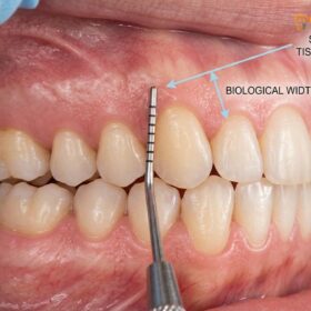
Biological width is the precise dimension of soft tissue attached to the tooth coronal to the alveolar crest, functioning as an essential immunological and physical barrier.
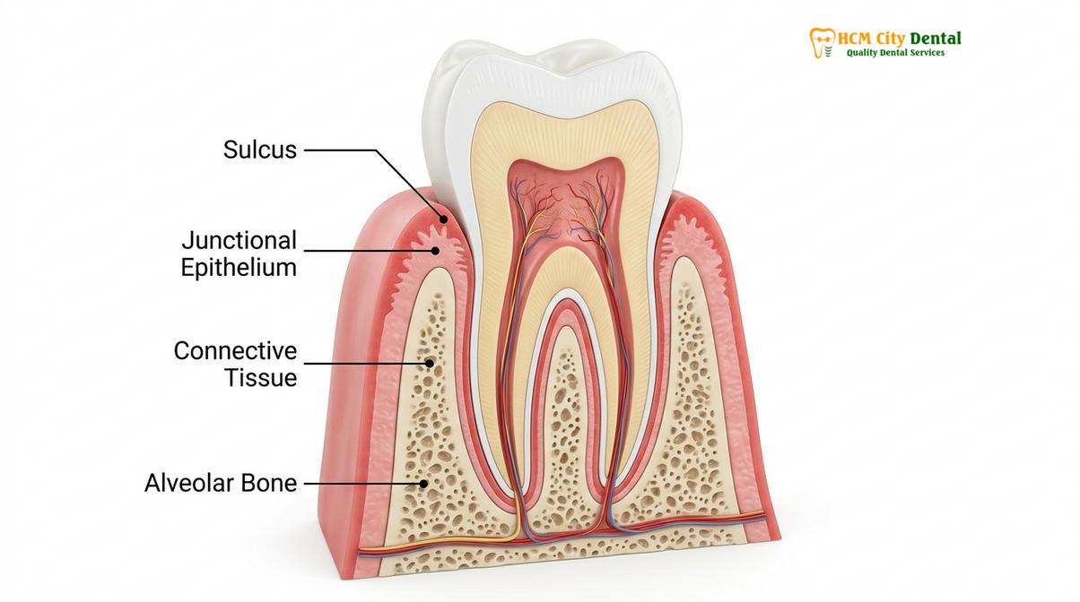
In the human body, ectodermal tissues serve as the primary defense mechanism against the invasion of bacteria, foreign materials, and environmental pathogens. The oral cavity presents a unique anatomical challenge: teeth and dental implants must physically penetrate this defensive epithelial barrier to function. The natural, highly specialized seal that develops around the cervical portion of the tooth to protect the underlying alveolar bone from disease is known clinically as the biological width[1].
Histologically, this dimension is not merely empty space; it is a complex, highly organized structure composed of two distinct layers. Based on foundational cadaveric studies, the dentogingival junction exhibits a remarkably consistent proportional relationship. The total biological width averages 2.04mm, which is the sum of two specific attachments:
- The Junctional Epithelium (0.97mm): This layer attaches to the enamel or cementum via hemidesmosomes. It is highly permeable, allowing gingival crevicular fluid and immune cells to pass through and combat bacterial plaque, but it is physically delicate.
- The Connective Tissue Attachment (1.07mm): Located immediately apical to the junctional epithelium, this layer consists of dense bundles of collagen fibers (dentogingival, alveologingival, and circular fibers) that insert directly into the root cementum. This supracrestal tissue attachment provides the robust mechanical seal that protects the bone.
Above these two attached layers lies the histological gingival sulcus, which averages 0.69mm in depth. Therefore, the total distance from the crest of the alveolar bone to the free gingival margin is typically around 3.0mm in a healthy periodontium. Understanding this anatomy is the cornerstone of both periodontics and restorative dentistry. If this barrier is compromised, the underlying bone becomes highly susceptible to osteoclastic activity and rapid resorption[2].
The 2mm Rule: Why Crowns and Veneers Must Avoid Deep Tissue Encroachment
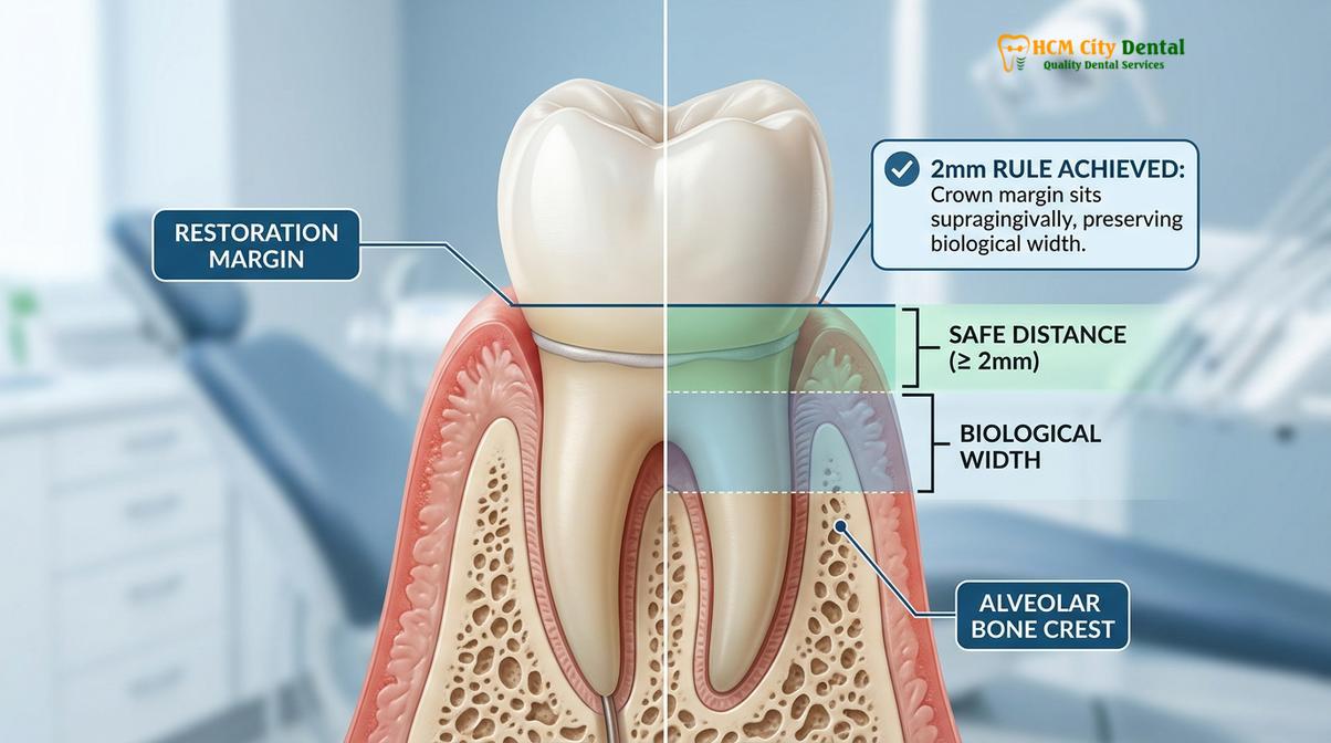
Dental restorations must maintain a safe distance of at least 2mm from the bone crest to prevent destructive inflammatory responses and ensure long-term periodontal health.
The relationship between periodontal health and the restoration of teeth is intimate and inseparable. When a tooth requires a crown, veneer, or large filling, the clinician must decide where to place the edge of the restoration, known as the restorative margin. There are three primary options for margin placement: supragingival (above the gums), equigingival (even with the gums), and subgingival (below the gums). While supragingival margins are the healthiest for the periodontium, aesthetic demands in the anterior aesthetic zone often necessitate subgingival placement to hide the transition line between the restoration and the natural tooth.
When subgingival margins are indicated, the clinician must strictly adhere to the “2mm Rule.” The restorative margin must never disrupt the junctional epithelium attachment or the connective tissue apparatus. Because the biological width occupies approximately 2mm of space above the bone, and the sulcus occupies about 1mm, the restorative margin should ideally be placed no deeper than 0.5mm into the sulcus. This leaves a safe buffer of 2.5mm between the margin and the alveolar bone crest[3].
“In modern aesthetic dentistry, respecting the periodontium is non-negotiable. When we calibrate gingival zenith points for a smile makeover, we must simultaneously perform biological width sounding. A beautiful ceramic veneer will ultimately fail if it acts as a chronic irritant to the supracrestal attachment.”
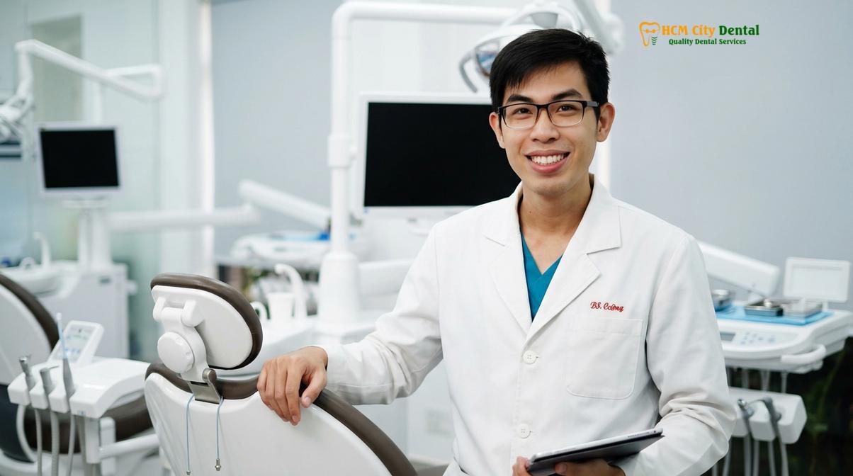
— Dr. Nguyen Van Cuong, Lead Clinician at HCMC Dental Clinic
The risk of encroachment varies depending on the patient’s periodontal biotype. Individuals with a thick, flat biotype generally have more dense fibrotic tissue that is somewhat resistant to recession but highly prone to pocket formation if violated. Conversely, patients with a thin, highly scalloped biotype have delicate tissue and thin underlying buccal bone. In these cases, even minor subgingival trauma during tooth preparation or impression taking can lead to immediate and irreversible gingival recession, resulting in the dreaded “black triangle” disease and exposed crown margins.
Surgical Violations of Biological Width: Chronic Bleeding, Bone Loss, and Pain
Encroaching on the supracrestal tissue attachment triggers a chronic localized inflammatory cascade, leading to unpredictable bone resorption and persistent gingival bleeding.
A biological width violation occurs when a restorative margin is placed too deeply beneath the gingival tissue crest, physically impinging on the junctional epithelium and connective tissue. The body perceives this deep margin not as a helpful medical device, but as a foreign body and a constant source of plaque retention. Because the restorative material is occupying the space that the biological width needs to exist, a severe and chronic inflammatory response is initiated.
The physiological response to this violation is highly destructive. The body attempts to recreate the necessary 2mm of space between the alveolar bone and the restorative margin to allow for tissue reattachment. To achieve this, osteoclasts are recruited to the area to resorb the alveolar bone. This bone loss is often unpredictable and accompanied by severe clinical symptoms.
Clinical Signs of a Biological Width Violation:
- Chronic Progressive Inflammation: The gums around the specific crown or veneer remain persistently red, swollen, and tender, regardless of the patient’s oral hygiene efforts.
- Bleeding on Probing (BOP): The tissue bleeds easily upon gentle brushing, flossing, or clinical examination.
- Localized Gingival Hyperplasia: The gums may overgrow and become bulbous in an attempt to wall off the irritant.
- Alveolar Bone Loss: Radiographic evidence of localized bone resorption, often leading to increased pocket depth and clinical attachment loss.
- Unexplained Pain: Patients frequently report a dull, chronic ache or pressure originating from the gums surrounding the restored tooth.
It is crucial to differentiate between a biological width violation and standard plaque-induced gingivitis. While gingivitis can be resolved with professional dental scaling and improved home care, inflammation caused by a width violation will never resolve with hygiene alone. The physical irritant (the deep margin) must be addressed. If left untreated, the chronic inflammation can lead to premature failure of the restoration, severe periodontal disease, and eventual tooth loss[4].
Restoring biological width through Functional Crown Lengthening
Functional crown lengthening surgically reshapes the gingiva and underlying bone to re-establish a healthy 2mm supracrestal attachment zone for restorative procedures.
When a tooth has decayed or fractured below the gum line, or when an existing crown has violated the biological width, the clinician cannot simply place a new restoration in the same compromised position. The anatomical foundation must be corrected first. This is achieved through a surgical procedure known as functional crown lengthening. The goal of this procedure is to surgically relocate the alveolar bone crest apically (downward), thereby recreating the necessary space for the biological width to re-establish itself on healthy tooth structure.
The workflow for functional crown lengthening is highly precise. Following profound local anesthesia, a full-thickness mucoperiosteal flap is elevated to expose the underlying bone. The clinician then performs an osteotomy (removal of supporting bone) and osteoplasty (reshaping of non-supporting bone) using specialized surgical burs and hand instruments. The bone is carefully contoured to mimic the natural scalloped architecture of the periodontium, ensuring that the new bone crest is at least 3mm apical to the planned restorative margin.
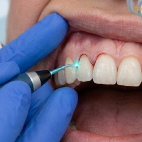
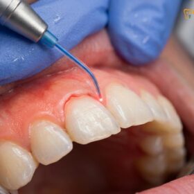
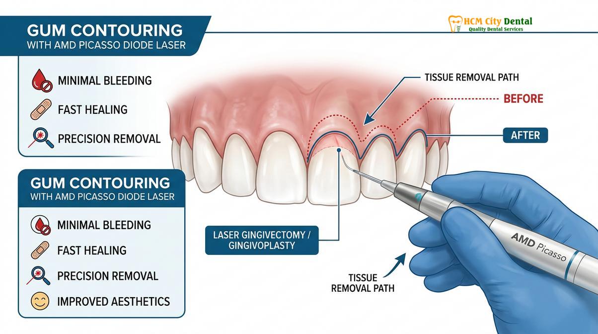
In modern clinical settings, the management of the soft tissue component of this procedure has been revolutionized by laser technology. At HCMC Dental Clinic, specialists utilize AMD Picasso Lasers (Diode soft tissue laser, 810nm). The 810nm wavelength is highly absorbed by melanin and hemoglobin, making it exceptionally effective for soft tissue surgery. The benefits of this technology include:
- Bloodless and Sutureless Cuts: The laser provides instant coagulation and cauterization, maintaining a clear, dry surgical field.
- Minimally Invasive: The precision of the laser minimizes collateral thermal damage to surrounding healthy tissues.
- Sterile Beam: The high-energy light sterilizes the surgical pocket, significantly reducing the risk of postoperative infection.
- Rapid Recovery: Patients experience vastly reduced postoperative pain and swelling, often achieving a rapid 48-hour soft tissue recovery compared to traditional scalpel surgery.
For patients seeking these advanced periodontal interventions, understanding the financial investment is important. The clinic offers transparent pricing for both local residents and international patients engaging in dental tourism. Note that a -40% WhatsApp booking discount is currently available for scheduled appointments.
| Periodontal Procedure | Standard Walk-in Price (VND) | Discounted Price (VND) | Estimated USD |
|---|---|---|---|
| Laser Gingivectomy (per tooth) | 2,500,000 VND | 1,500,000 VND | ~$60 USD |
| Functional/Esthetic Crown Lengthening (per tooth, includes bone contouring) | 5,000,000 VND | 3,000,000 VND | ~$120 USD |
| Full-Arch Laser Gum Contouring (up to 10 teeth) | 20,000,000 VND | 12,000,000 VND | ~$480 USD |
| Botox Gummy Smile Correction (both sides) | 7,500,000 VND | 4,500,000 VND | ~$180 USD |
| Surgical Lip Repositioning / Lip Lowering | 25,000,000 VND | 15,000,000 VND | ~$600 USD |
Advanced Clinical Workflows: Managing Severe Cases and Gummy Smiles
Complex cases involving vertical maxillary excess or hyperactive lip muscles require multidisciplinary approaches, combining orthognathic surgery, laser contouring, and neuromodulators.
While biological width violations and altered passive eruption can often be managed with laser gingivectomy and crown lengthening, some patients present with severe skeletal or muscular discrepancies that expose excessive gingival tissue. These cases require a comprehensive, multidisciplinary approach guided by Digital Smile Design (DSD) principles to ensure that the final zenith points align perfectly with the patient’s facial aesthetics.
For patients with a hyperactive levator muscle—where the upper lip pulls up excessively during a smile—minimally invasive neuromodulator therapy is highly effective. Botox injections temporarily relax the specific elevator muscles of the upper lip, preventing it from rising too high and exposing the gums. For a more permanent soft tissue solution, Surgical Lip Repositioning (Lip Lowering) is performed. This procedure involves removing a strip of mucosa from the maxillary vestibule and suturing the lip in a lower, more restrictive position, effectively masking the gingival display.
However, in severe skeletal cases diagnosed as Vertical Maxillary Excess (VME), where the upper jaw bone itself has grown too far downward, soft tissue procedures alone are insufficient. For these complex orthognathic cases, HCMC Dental Clinic works in collaboration with Surgical Jaw Partners at the National Hospital of Odonto-Stomatology in Ho Chi Minh City (Bệnh viện Răng Hàm Mặt Trung Ương TP.HCM). As the highest clinical authority in the region, their maxillofacial specialists perform the Le Fort I osteotomy. This major surgery involves sectioning the entire upper jaw, impacting it upward to reduce the vertical height, and securing it with titanium plates, thereby definitively resolving the skeletal gummy smile[5].
Clinical Case Study: Multidisciplinary Smile Rehabilitation
A 32-year-old patient visited HCMC Dental Clinic in Ho Chi Minh City complaining of short, square teeth and chronically inflamed gums around old porcelain veneers. Clinical examination and bone sounded probing revealed a severe biological width violation and altered passive eruption. Dr. Cuong initiated a comprehensive treatment plan. First, the old veneers were removed, and the inflamed tissue was allowed to heal. Next, functional crown lengthening was performed using the AMD Picasso Diode laser to re-establish a healthy 2mm supracrestal attachment and calibrate the gingival zenith points. After a 12-week osseous healing period, new lithium disilicate veneers were fabricated using Digital Smile Design. The result was a harmonious, inflammation-free smile with perfectly proportioned teeth and healthy, stippled gingiva.
When to Consult a Periodontal Specialist
If you are experiencing chronic redness, bleeding, or pain around a dental crown, bridge, or veneer, it is imperative to seek a professional periodontal evaluation. These symptoms are strong indicators that the restorative margins may be encroaching upon your biological width. Delaying treatment can lead to irreversible alveolar bone loss and the eventual failure of the restoration.
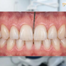
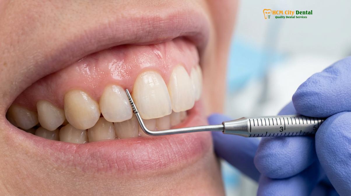
During your consultation, the specialist will perform a comprehensive diagnostic workup. This includes a visual assessment of the tissue biotype, radiographic evaluation to check for interproximal bone loss, and the critical technique of bone sounded probing. Under local anesthesia, the dentist will gently measure the exact distance from the gingival margin to the bone crest to definitively diagnose any attachment violations.
“Early diagnosis of a biological width violation can save both the tooth and the restoration. We strongly advise patients not to ignore chronic localized bleeding around crowns. A simple diagnostic probing can determine if the issue is hygiene-related or if surgical margin correction is required.”
— Dr. Nguyen Van Cuong
Whether you require a simple General & Preventive Dentistry intervention like deep scaling, or complex aesthetic gum contouring procedures, a personalized clinical examination is essential to determine your candidacy and outline the safest, most effective treatment workflow.
Frequently Asked Questions
How does a dentist measure biological width?
A dentist measures biological width using a technique called bone sounded probing. Under local anesthesia, a sterile periodontal probe is gently pushed through the attachment tissues until it contacts the alveolar bone crest, allowing the clinician to calculate the exact tissue dimensions. This measurement is compared against the depth of the restorative margin to diagnose any potential violations accurately.
What happens if a crown violates biological width?
If a crown violates the biological width, it triggers a chronic inflammatory response. The body attempts to recreate the necessary space by resorbing the underlying alveolar bone, leading to persistent bleeding, localized pain, gingival hyperplasia, and eventual periodontal attachment loss. The tissue will remain chronically inflamed until the physical irritant is removed or surgically corrected.
Does laser gingivectomy violate biological width?
No, a properly executed laser gingivectomy does not violate biological width. Advanced diode lasers precisely remove excess unattached gingiva while strictly preserving the 2mm supracrestal tissue attachment, ensuring the underlying bone and junctional epithelium remain healthy and undisturbed. The laser’s precision allows for exact calibration of the gingival zenith points without encroaching on the bone.
Can a biological width violation heal on its own?
A biological width violation cannot heal on its own without clinical intervention. The chronic inflammation will persist until the restorative margin is corrected or surgical crown lengthening is performed to surgically relocate the alveolar bone and re-establish the natural attachment zone. Improved oral hygiene cannot resolve the inflammation caused by a physical margin impingement.
How long does it take to recover from functional crown lengthening?
Initial soft tissue recovery from functional crown lengthening takes approximately two to three weeks. However, complete osseous healing and stabilization of the gingival margins require about three to six months before final restorative impressions for crowns or veneers can be safely taken. Rushing the final restoration before the tissue has fully stabilized can lead to aesthetic failures and margin exposure.
References
- Journal of Clinical Periodontology. Dimensions of the dentogingival junction in humans. (1961).
- International Journal of Periodontics & Restorative Dentistry. Biologic width and its relation to periodontal biotypes. (1994).
- Journal of Esthetic and Restorative Dentistry. The perio-restorative interface: biologic width implications. (2018).
- Clinical Oral Investigations. Efficacy of 810nm diode lasers in soft tissue surgery. (2020).
- Journal of Oral and Maxillofacial Surgery. Stability of Le Fort I osteotomy in vertical maxillary excess. (2022).