Full mouth reconstruction is a comprehensive clinical approach that rebuilds all teeth in both the upper and lower jaws. It combines restorative and cosmetic dentistry to resolve severe decay, missing teeth, and bite dysfunction, ultimately restoring optimal oral health, chewing capacity, and facial aesthetics.
Clinical Summary:
Full mouth rehabilitation integrates multiple dental disciplines to address complex oral pathologies. By combining implantology, prosthodontics, and cosmetic dentistry, clinicians can rebuild compromised dentition. This process requires meticulous diagnostic planning, 3D imaging, and precise execution to ensure long-term functional stability and aesthetic harmony for patients suffering from extensive tooth loss, severe occlusal wear, or congenital dental anomalies. The ultimate goal is to establish a biologically sound, functionally efficient, and visually pleasing stomatognathic system.
Key Takeaways:
- Comprehensive reconstruction addresses both functional chewing mechanics and facial aesthetics simultaneously.
- Treatment plans often integrate titanium implants, ceramic crowns, and ultra-thin porcelain veneers.
- Precise bite calibration is essential to prevent future temporomandibular joint (TMJ) disorders and ceramic fractures.
- Advanced 3D CBCT imaging ensures accurate surgical and prosthetic planning for predictable outcomes.
- Phased treatment timelines allow for proper tissue healing, provisional testing, and manageable financial planning.
The Smile Makeover Challenge: Structural and Aesthetic Restoration
A comprehensive restoration goes beyond simple aesthetics, requiring a robust structural foundation that supports long-term oral health, proper phonetics, and a balanced facial profile.
When a patient presents with widespread dental deterioration, a standard cosmetic approach is rarely sufficient. The challenge of a true full mouth reconstruction lies in the necessity to rebuild the underlying architecture of the mouth before addressing the visible aesthetics. A superficial cosmetic smile makeover might temporarily improve the appearance of the anterior teeth, but without a stable posterior foundation and a harmonious bite, those aesthetic restorations are highly susceptible to premature failure, chipping, or debonding. Therefore, the clinical workflow must prioritize disease control, structural integrity, and biomechanical balance.
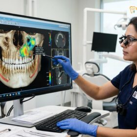
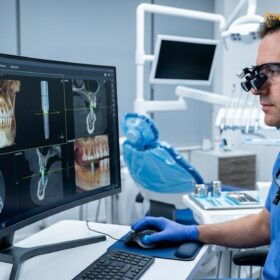
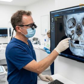
The initial diagnostic phase is arguably the most critical component of the entire reconstructive process. Clinicians must gather extensive data, including full-mouth series radiographs, 3D Cone Beam Computed Tomography (CBCT) scans, intraoral digital impressions, and detailed photographic records. This data allows the dental team to evaluate the periodontal health, endodontic status, and structural viability of every remaining tooth. Teeth that are deemed structurally compromised beyond repair are slated for extraction, while those that can be salvaged are prepared for core build-ups, root canal therapy, or crown lengthening procedures.
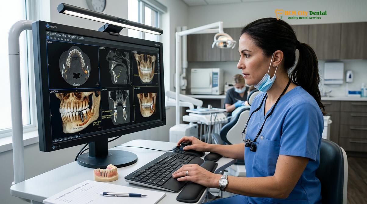
Dr. Nguyen Van Cuong frequently emphasizes that a successful full mouth smile design is an engineering feat as much as it is an artistic endeavor. The biological width of the gums, the density of the alveolar bone, and the dynamic movements of the temporomandibular joint must all be factored into the blueprint. If the foundation is weak—due to untreated periodontitis or insufficient bone volume—any restorative work placed on top will inevitably fail. Consequently, the first phase of treatment often involves deep periodontal scaling, bone grafting, and the stabilization of active dental caries [1].
“Rebuilding a smile is akin to constructing a high-rise building. You cannot focus on the glass facade until the steel framework and the deep foundation are perfectly engineered to withstand the daily forces of nature. In dentistry, those forces are the immense pressures of mastication.”
Furthermore, the aesthetic parameters must be customized to the patient’s unique facial features. The interpupillary line, the facial midline, and the curvature of the lower lip during a natural smile dictate the incisal edge position of the new upper teeth. By utilizing digital smile design software, clinicians can simulate the final outcome, allowing the patient to visualize the proposed changes before any irreversible procedures are initiated. This collaborative approach ensures that the final structural and aesthetic restoration aligns with both the clinical requirements and the patient’s personal expectations.
Restoring Posterior Chewing Power with Dental Implants
Titanium implants serve as the gold standard for replacing missing posterior teeth, providing the necessary root-level support to withstand heavy occlusal forces and preserve jawbone density.
The posterior teeth—the molars and premolars—are the workhorses of the human dentition. They are responsible for the grinding and crushing of food, bearing the brunt of the muscular forces generated by the masseter and temporalis muscles. When these teeth are lost due to decay, fracture, or periodontal disease, the entire dynamic of the bite shifts. The anterior teeth, which are designed primarily for tearing and aesthetics, are suddenly forced to take on the heavy loads of chewing. This abnormal stress leads to rapid wear, flaring, and eventual loss of the front teeth—a phenomenon known as posterior bite collapse.
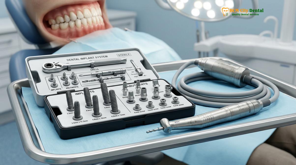
To prevent or correct this collapse, restoring posterior chewing power is paramount. While traditional removable partial dentures can fill the gaps, they do not stimulate the underlying alveolar bone, leading to progressive bone resorption over time. Furthermore, they often lack the stability required for efficient mastication. This is where implantology becomes indispensable. By surgically placing titanium fixtures into the jawbone, clinicians can create artificial roots that integrate biologically with the patient’s own bone tissue through a process called osseointegration [2].
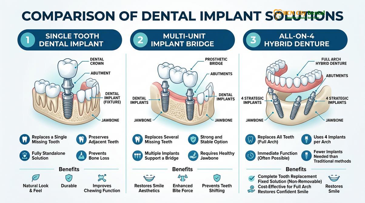
For patients missing multiple posterior teeth, there are several implant-based strategies available. Single implants can be used to replace individual teeth, while implant-supported bridges can span larger edentulous spaces using fewer surgical fixtures. In cases of complete edentulism (total tooth loss) in an arch, a full arch implant prosthetics approach, such as the All-on-4 hybrid denture, can provide a fixed, highly durable solution that restores near-natural chewing capacity. When comparing fixed implants to traditional removable options, the biomechanical advantages of implants are clear: they preserve bone, offer superior stability, and eliminate the discomfort associated with acrylic flanges resting on sensitive gum tissue.
| Restoration Type | Bone Preservation | Chewing Efficiency | Longevity | Maintenance Requirement |
|---|---|---|---|---|
| Single Dental Implants | Excellent (stimulates bone) | High (mimics natural teeth) | 15+ years to lifetime | Standard brushing and flossing |
| Implant-Supported Bridge | Excellent in implant sites | High | 10-15+ years | Requires specialized floss threaders |
| Traditional Fixed Bridge | Poor (bone loss under pontic) | Moderate to High | 7-15 years | Requires specialized floss threaders |
| Removable Partial Denture | None (accelerates bone loss) | Low to Moderate | 5-8 years | Daily removal and soaking required |
The surgical placement of posterior implants requires precise planning, particularly in the upper jaw where the maxillary sinus cavity resides, and in the lower jaw where the inferior alveolar nerve runs. Advanced 3D imaging allows the implantologist to measure bone volume accurately and, if necessary, plan for supplementary procedures such as sinus lifts or guided bone regeneration. By utilizing premium implant systems, clinicians can ensure high primary stability and predictable long-term integration, laying a rock-solid foundation for the final ceramic crowns.
Achieving Front Smile Aesthetics with Porcelain Veneers
Anterior restorations focus on mimicking natural enamel translucency and shape, utilizing ultra-thin ceramic layers to correct discoloration, chips, and minor misalignments.
Once the posterior foundation is secured and the bite is stabilized, the focus of the full mouth reconstruction shifts to the anterior segment—the teeth that are most visible when smiling and speaking. The aesthetic demands in this region are exceptionally high. The restorations must not only function correctly during the incisive movements of the jaw but must also interact harmoniously with the lips, the gingival architecture, and the patient’s overall facial proportions.
For anterior teeth that are structurally sound but aesthetically compromised, porcelain veneers are often the treatment of choice. These ultra-thin shells of custom-milled or hand-layered ceramic are bonded to the facial surfaces of the teeth. They are highly resistant to staining and possess optical properties that closely mimic natural enamel, including fluorescence and opalescence. Veneers can dramatically alter the size, shape, and color of the teeth, closing diastemas (gaps), masking severe tetracycline stains, and correcting mild orthodontic misalignments without the need for braces.
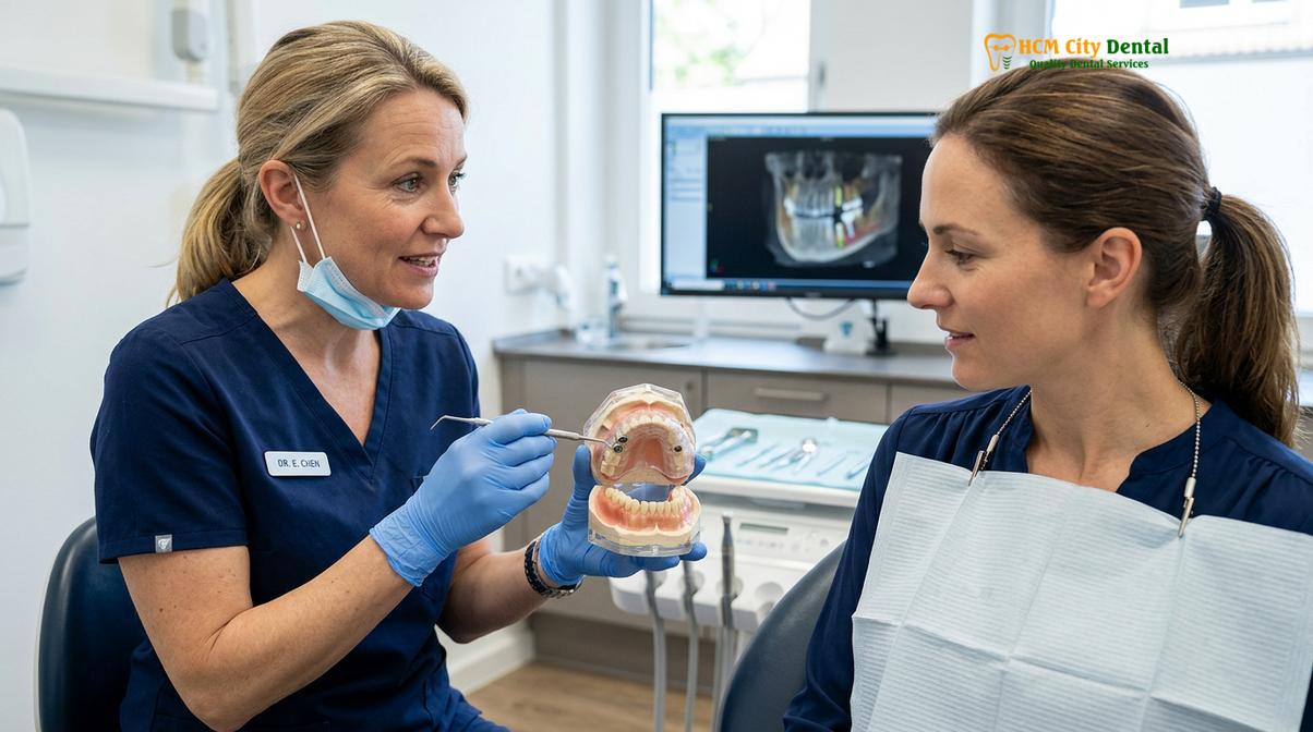
However, in a full mouth reconstruction, it is common to encounter a mix of natural teeth and missing teeth in the anterior zone. This requires the seamless integration of dental implants and porcelain veneers. Achieving a uniform appearance across different restorative materials—such as matching a veneer bonded to natural dentin with a crown cemented over a titanium implant abutment—is one of the most challenging aspects of cosmetic dentistry. It requires a master ceramist who understands how different underlying substrates affect the final color and translucency of the ceramic [3].
Clinical Case Review: A 55-year-old patient presented to HCMC Dental Clinic in Ho Chi Minh City with severe anterior wear and two missing upper incisors. The treatment plan utilized advanced implantology techniques to replace the missing incisors with titanium fixtures and custom zirconia abutments. Concurrently, the adjacent natural teeth were minimally prepared for lithium disilicate (E.max) veneers. By utilizing a single master ceramist for both the implant crowns and the veneers, the clinical team achieved a virtually seamless aesthetic integration, restoring both the patient’s confidence and anterior guidance.
The soft tissue management around anterior restorations is equally critical. The gums must frame the teeth symmetrically, with healthy, stippled papillae filling the embrasure spaces between the teeth. If the gingival margins are uneven, a periodontist may perform a gingivectomy or crown lengthening procedure to establish a harmonious gum line before the final impressions for the veneers and crowns are taken. This meticulous attention to the “pink aesthetics” (the gums) is just as important as the “white aesthetics” (the teeth) in achieving a truly natural-looking smile makeover.
Bite Calibration and Occlusion Alignment
Establishing a harmonious bite is the most critical step in complex rehabilitations, ensuring that upper and lower teeth meet evenly without destructive friction or joint strain.
The success of a full mouth reconstruction hinges entirely on the management of occlusion—the way the upper and lower teeth come together. If the bite is misaligned, the resulting interferences can cause catastrophic failures, including the fracturing of expensive porcelain restorations, the loosening of dental implants, and the development of severe temporomandibular joint (TMJ) disorders. Therefore, the ability to accurately restore bite alignment is the hallmark of an expert restorative clinician.
In cases of severe tooth wear (attrition) caused by bruxism (teeth grinding) or acid erosion, patients often lose their Vertical Dimension of Occlusion (VDO). This means the lower jaw over-closes, leading to a collapsed facial appearance, deep wrinkles around the mouth, and strain on the TMJ complex. Re-establishing the correct VDO is a delicate process. The clinician must determine the optimal resting position of the jaw muscles—known as Centric Relation—and build the new bite to support this physiologically relaxed state [4].
To achieve this, the dentist will often use a facebow transfer and a semi-adjustable articulator. These tools allow the clinician to mount the patient’s dental models in a way that accurately replicates the exact hinge axis and movements of their jaw. In modern practices, this analog process is frequently supplemented or replaced by digital jaw tracking and T-Scan technology, which measures the precise timing and force of every tooth contact dynamically in real-time.
Clinical Warning: Altering a patient’s Vertical Dimension of Occlusion (VDO) without proper neuromuscular testing and a provisional trial phase can lead to severe chronic pain, muscle spasms, and the inability to chew properly. A new bite must always be tested with temporary restorations before final ceramics are fabricated.
Once the ideal bite is determined, the patient is placed in provisional (temporary) restorations. This “test drive” phase is crucial. It allows the patient to evaluate the aesthetics, phonetics, and comfort of the new bite over several weeks or months. If the patient experiences any muscle tenderness or difficulty speaking, the provisionals can be easily adjusted. Only when the patient is completely asymptomatic and satisfied with the function are the final ceramic restorations fabricated to replicate the exact contours of the successful provisionals.
Timeline and Scheduling for Combined Treatments in Saigon
Complex dental rehabilitations are executed in carefully planned phases over several months, allowing for surgical healing, provisional testing, and final ceramic placement.
A full mouth reconstruction is not a single event but a journey. Patients must be prepared for a phased approach that respects the biological healing times of the human body. Rushing the process, particularly when bone grafting or implant osseointegration is involved, significantly increases the risk of complications and implant failure. For international patients traveling to Ho Chi Minh City, understanding this timeline is essential for proper travel and financial planning.
The typical timeline is divided into several distinct phases. Phase 1 involves comprehensive diagnostics, 3D imaging, and the formulation of the treatment plan. Phase 2 is the surgical and preparatory phase, which may include extractions, implant placement, bone grafting, and the preparation of natural teeth. During this phase, the patient is fitted with high-quality provisional restorations, ensuring they never leave the clinic without a functional and aesthetic smile. If implants are placed, a healing period of three to six months is generally required for osseointegration to occur [5].
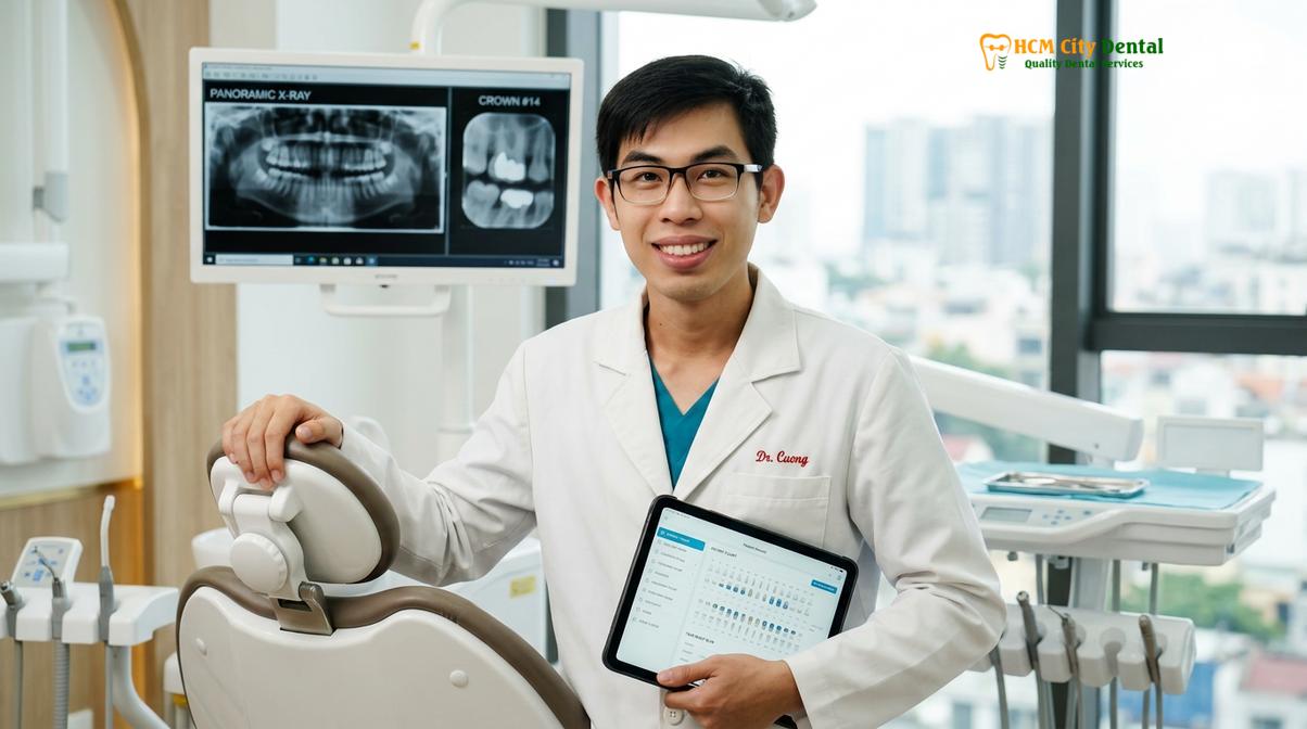
“Patience is a clinical virtue in full mouth rehabilitation. The provisional phase is not just about waiting for implants to heal; it is a critical diagnostic period where we fine-tune the patient’s new bite, speech, and aesthetics. The final ceramics are only as good as the blueprint we perfect during this temporary phase,” notes Dr. Cuong.
Phase 3 involves the final impressions or digital scans of the healed implant sites and prepared teeth. The master ceramists at the dental laboratory then spend several weeks meticulously crafting the final zirconia or lithium disilicate restorations. Finally, Phase 4 is the delivery and final calibration, where the permanent crowns, bridges, and veneers are bonded into place, and the occlusion is fine-tuned to perfection.
Many patients choose to undergo these extensive procedures at HCMC Dental Clinic due to the availability of world-class technology and highly specialized clinicians. When evaluating the comprehensive treatment pricing in Vietnam, patients often find that they can access premium materials and expert care at a fraction of the cost compared to Western countries. This makes complex rehabilitations, including restorative solutions for older adults, highly accessible without compromising on clinical excellence or safety standards.
When to See a Doctor for Full Mouth Rehabilitation
Determining whether you are a candidate for a full mouth reconstruction requires a comprehensive clinical evaluation. You should seek a professional consultation if you experience multiple signs of advanced dental deterioration. Common indications include the loss of several teeth across both arches, which severely impairs your ability to chew solid foods. Additionally, if you notice significant wear on your remaining teeth—often presenting as flattened, shortened, or chipped enamel—this may indicate a collapsing bite or severe nocturnal bruxism that requires intervention.
Chronic pain in the jaw joints (TMJ), frequent tension headaches, or a noticeable change in your facial profile (such as a sunken appearance around the mouth) are also strong indicators that your occlusal foundation needs rebuilding. It is important to note that full mouth reconstruction is a highly personalized medical procedure. There are no absolute guarantees of success, and candidacy depends heavily on your systemic health, bone density, and commitment to long-term oral hygiene. A thorough examination by a qualified prosthodontist or restorative specialist is the only way to determine the safest and most effective treatment plan for your specific anatomical needs.
Frequently Asked Questions
Can I get veneers and implants at the same time?
Yes, veneers and implants can be integrated into a single comprehensive treatment plan. While the surgical placement of implants and the preparation for veneers may occur during the same phase, the final ceramic restorations are usually delivered together after the implants have fully integrated with the jawbone to ensure perfect color matching and occlusal harmony. This synchronized delivery ensures that the translucency and shade of the veneers perfectly match the crowns placed over the titanium implants.
How long does a full mouth rehabilitation take in Saigon?
A complete full mouth rehabilitation typically requires three to six months, depending on the complexity of the case. If bone grafting or extensive implant surgeries are necessary, the timeline extends to allow for proper biological healing, whereas cases involving only crowns and veneers may be completed in a matter of weeks. International patients often make two separate trips: one for the surgical phase and provisionalization, and a second trip months later for the final ceramic delivery.
What is the cost of full mouth reconstruction in Vietnam?
The total investment for a full mouth reconstruction varies significantly based on the number of implants, the materials chosen, and the required preparatory procedures. Following a detailed clinical assessment and 3D imaging, patients receive a customized fee schedule that reflects the specific surgical and prosthodontic interventions required for their unique anatomy. Generally, the costs in Vietnam are highly competitive, allowing patients to access premium Swiss or German implant systems and master-crafted ceramics at a substantial value.
Is the full mouth reconstruction process painful?
Modern full mouth reconstruction is performed under profound local anesthesia, and often with conscious sedation, ensuring the procedures are highly comfortable. Post-operative discomfort is generally manageable with prescribed analgesics, and patients are closely monitored throughout the healing phases to minimize inflammation and ensure a smooth recovery. Advanced techniques, such as computer-guided surgery, also minimize tissue trauma, leading to faster healing times and less post-operative swelling.
How do I maintain my new teeth after a full mouth restoration?
Maintaining a reconstructed mouth requires strict daily oral hygiene, including the use of water flossers, interdental brushes, and non-abrasive toothpaste. Additionally, wearing a custom occlusal night guard is highly recommended to protect the ceramics from nocturnal grinding, alongside biannual professional cleanings and clinical evaluations. Regular radiographic monitoring of the implant sites is also crucial to ensure the long-term health of the surrounding bone and gingival tissues.
References
- Journal of Prosthetic Dentistry. Biomechanical considerations in full mouth rehabilitation. (2021).
- International Journal of Oral and Maxillofacial Implants. Posterior implant restorations and occlusal load distribution. (2020).
- Journal of Esthetic and Restorative Dentistry. Integrating anterior veneers with posterior implant prosthetics. (2019).
- Clinical Oral Investigations. Vertical dimension of occlusion and temporomandibular joint harmony. (2022).
- British Dental Journal. Phased treatment planning in complex restorative dentistry. (2018).