An All on 4 failure occurs when one or more titanium implants fail to integrate with the jawbone or when the prosthetic bridge fractures. While rare, these complications require immediate clinical intervention to prevent further bone loss and restore full masticatory function.
Clinical Summary:
While full-arch implant rehabilitations boast exceptionally high success rates, an All on 4 failure remains a complex clinical reality that patients and practitioners must navigate. Failures are generally categorized into early biological non-integration—often due to surgical trauma or poor primary stability—and late complications, which are typically driven by peri-implantitis or mechanical overload on the prosthetic bridge. The cornerstone of managing these complications lies in early detection, precise diagnostic imaging, and a structured clinical retreatment protocol. By understanding the biomechanical limits of cantilever extensions and the critical importance of meticulous oral hygiene, patients can significantly mitigate the risks associated with full-mouth restorations. Advanced retreatment strategies now allow skilled implantologists to salvage failing cases through targeted bone grafting and strategic re-implantation, ensuring long-term functional and aesthetic stability.
Key Takeaways:
- Full-arch implant failures are divided into early biological rejection and late mechanical or infectious complications.
- Smoking and poor bone density are the leading risk factors for disrupted osseointegration.
- Peri-implantitis is a progressive bacterial infection that causes severe bone loss around the titanium fixtures.
- Mechanical failures often involve the fracture of the prosthetic bridge due to excessive bite forces or improper design.
- Retreatment is highly viable but requires advanced surgical techniques, including bone grafting and guided re-implantation.
Understanding All-on-4 Success Rates and Failure Realities
While full-arch implant rehabilitation boasts high long-term survival rates, failures can occur due to biological rejection or mechanical stress. Understanding these realities helps patients and clinicians manage expectations and mitigate risks effectively.
The All-on-4 implant protocol has revolutionized the management of complete edentulism, offering patients a fixed, immediate-load solution that bypasses the need for extensive bone grafting in many cases. By tilting the posterior implants, clinicians can maximize the use of available bone and avoid vital anatomical structures such as the maxillary sinus and the inferior alveolar nerve. Statistically, this treatment modality demonstrates survival rates exceeding 95% over a ten-year period [1]. However, the term “success” in implantology is multifaceted, encompassing not only the survival of the titanium fixtures but also the integrity of the prosthetic bridge and the health of the surrounding soft tissues. When an All on 4 failure occurs, it represents a significant clinical challenge that requires a comprehensive understanding of biomechanics, microbiology, and patient-specific physiological factors.
It is crucial to differentiate between the failure of the implant fixture itself and the failure of the prosthesis. An implant fixture failure means the titanium post has lost its structural connection to the jawbone—a process known as a loss of osseointegration. Conversely, a prosthetic failure involves damage to the visible teeth, such as the chipping of acrylic, the fracture of a zirconia framework, or the loosening of the prosthetic screws that hold the bridge in place. Both scenarios are distressing for the patient, but they require vastly different clinical interventions. A fractured bridge can often be repaired or replaced without surgical intervention, whereas a failed implant necessitates a complex surgical retreatment.
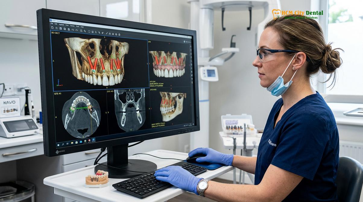
The biomechanical foundation of the full-arch concept relies heavily on the Antero-Posterior (AP) spread. The AP spread is the distance between the center of the most anterior implant and the distal aspect of the most posterior implant. A wider AP spread allows for a longer cantilever—the portion of the bridge that extends backward beyond the last implant. If the cantilever is too long relative to the AP spread, the posterior implants are subjected to immense leverage forces during chewing. Over time, this mechanical overload can lead to crestal bone loss, screw loosening, or catastrophic fracture of the metal framework. Therefore, meticulous preoperative planning using 3D imaging is non-negotiable to ensure optimal implant distribution.
Dr. Nguyen Van Cuong, a leading specialist in complex oral rehabilitations, frequently emphasizes that patient selection is the most critical determinant of long-term success. Patients presenting with severe bruxism (teeth grinding), uncontrolled systemic conditions like diabetes, or a history of aggressive periodontitis require modified treatment protocols. In some instances, placing five or six implants (All-on-5 or All-on-6) may be recommended to distribute occlusal forces more evenly, particularly in the softer bone of the upper jaw. Understanding these nuanced realities allows patients to make informed decisions when considering Dental Implants as a permanent solution for tooth loss.
Early Failures vs. Late Failures: Biological vs. Mechanical Issues
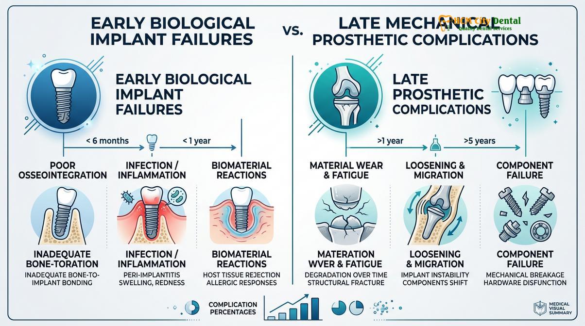
Early failures typically stem from disrupted osseointegration or surgical trauma, whereas late failures are usually driven by progressive bacterial infections or cumulative biomechanical overload on the prosthesis.
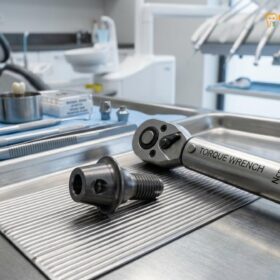
In the realm of implant dentistry, complications are generally stratified by their timeline of onset. Early failures occur within the first few months following surgery, prior to the definitive loading of the final prosthesis. These failures are almost exclusively biological in nature, representing an inability of the jawbone to heal and fuse to the titanium surface. The primary culprit for early failure is a lack of primary stability. When an implant is placed, it must achieve a specific torque value (typically >35 Ncm for immediate loading) to ensure it does not move during the initial healing phase [2]. If micromotion exceeds 150 microns, the body will form a fibrous scar tissue capsule around the implant instead of rigid bone, leading to immediate mobility and failure.
Surgical trauma is another significant factor in early complications. The drilling sequence must be performed with copious saline irrigation to prevent the bone from overheating. If the bone temperature exceeds 47°C for more than one minute, thermal necrosis occurs, causing the surrounding bone cells to die. This necrotic bone cannot support osseointegration, and the implant will inevitably fail. Furthermore, postoperative infections introduced during the surgical phase can rapidly destroy the delicate blood clot and early cellular matrix required for bone formation. Patients experiencing early complications often report acute implant rejection signs, such as localized throbbing pain, persistent swelling, and a noticeable mobility of the temporary bridge.
“The critical threshold for immediate loading relies entirely on mechanical primary stability. If micromotion exceeds the biological tolerance during the first eight weeks, fibrous encapsulation is inevitable, rendering the implant clinically failed.”
Conversely, late failures manifest years after the initial surgery and are typically the result of cumulative factors. Late biological failures are predominantly caused by peri-implantitis, a destructive inflammatory condition triggered by bacterial biofilm. Similar to severe gum disease, peri-implantitis causes the progressive loss of the supporting bone around a fully integrated implant. If left untreated, the bone loss will advance until the implant loses its structural support and falls out. The progression of this disease is often silent in its early stages, making routine radiographic monitoring essential.
Late mechanical failures are equally concerning and are directly related to the immense forces generated by the human jaw. The materials used to fabricate the full-arch bridge—whether acrylic, composite, or monolithic zirconia—are subjected to thousands of chewing cycles daily. Material fatigue can lead to micro-cracks that eventually propagate into full fractures. Additionally, the tiny retaining screws that connect the bridge to the multi-unit abutments can stretch and loosen over time. When a screw loosens, the bridge begins to micro-move, placing asymmetrical stress on the remaining implants. This highlights why choosing the right restorative material and adhering to a strict maintenance schedule is just as important as the surgery itself when comparing Dental Implants vs Dentures: Which Is the Better Investment?.
Major Causes: Poor Bone Quality, Smoking, and Peri-Implantitis
The primary catalysts for full-arch implant complications include insufficient bone density, the vasoconstrictive effects of tobacco use, and destructive bacterial inflammation surrounding the titanium fixtures.
The success of any implant-supported restoration is fundamentally dependent on the quality and quantity of the host bone. Bone density is clinically categorized into four types, ranging from Type I (dense, oak-like cortical bone) to Type IV (soft, styrofoam-like trabecular bone). The posterior maxilla (upper jaw) frequently presents with Type IV bone, which poses a significant challenge for achieving the high insertion torque required for the All-on-4 protocol. When implants are placed in poor-quality bone without adequate cortical engagement, the risk of early micromotion and subsequent failure increases dramatically. In such cases, clinicians may need to utilize longer implants, under-prepare the osteotomy site to compress the soft bone, or delay the loading of the prosthesis until full osseointegration has occurred [3].
Smoking remains one of the most detrimental lifestyle factors affecting implant survival. The nicotine and carbon monoxide present in cigarette smoke induce severe peripheral vasoconstriction, drastically reducing the blood supply to the oral tissues. Blood carries the essential oxygen, nutrients, and immune cells required for wound healing and bone regeneration. By compromising this vascular network, smoking delays angiogenesis (the formation of new blood vessels) and impairs the function of osteoblasts (bone-forming cells). Furthermore, smoking alters the local immune response, making the patient significantly more susceptible to postoperative infections. Clinical studies consistently show that smokers experience a failure rate nearly twice as high as non-smokers, particularly in complex full-arch rehabilitations.
Clinical Warning: Tobacco use severely compromises the microvascular blood supply to the jawbone. Patients who continue to smoke during the critical 3-to-6 month healing phase face an exponentially higher risk of early implant failure and severe postoperative infections.
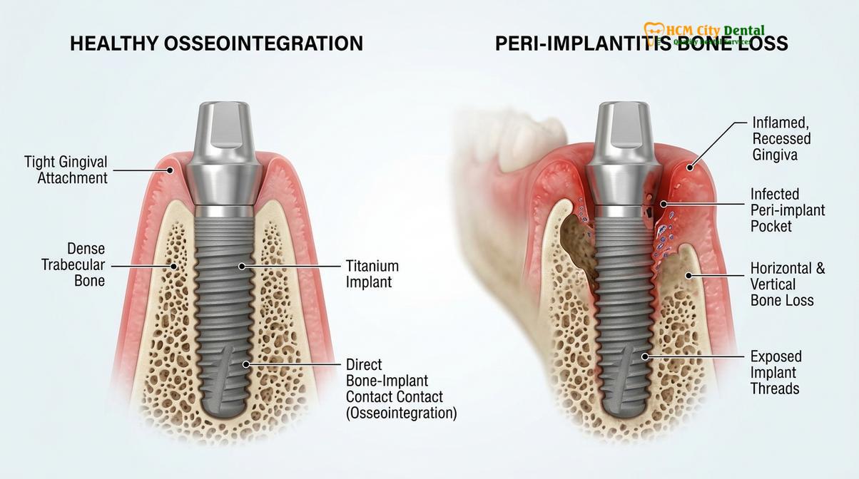
Perhaps the most insidious threat to long-term implant survival is peri-implantitis. This condition is characterized by an aggressive inflammatory response to bacterial biofilm accumulating at the implant-abutment junction. Unlike natural teeth, implants lack the protective periodontal ligament and its associated vascular supply, making them more vulnerable to rapid bacterial invasion. The pathogens involved in peri-implantitis are predominantly gram-negative anaerobes, which thrive in the deep, oxygen-deprived pockets around the implant. As the immune system attempts to fight the infection, it inadvertently releases enzymes that destroy the surrounding crestal bone.
The clinical manifestation of peri-implantitis bone loss includes deep probing depths, bleeding upon probing, purulent exudate (pus), and radiographic evidence of crater-like bone defects. Managing this condition is notoriously difficult because the rough, porous surface of the titanium implant—which is designed to promote bone ingrowth—also provides an ideal sanctuary for bacteria. Treatment often requires surgical intervention to decontaminate the implant surface using lasers, air-abrasion, or chemical agents, followed by guided bone regeneration. Understanding these risks is vital when evaluating different implant systems, such as Straumann vs Nobel Biocare Implants: What Is the Difference?, as different surface topographies may influence bacterial adherence.
| Complication Type | Primary Causes | Clinical Symptoms | Typical Timeframe |
|---|---|---|---|
| Early Biological Failure | Poor primary stability, surgical trauma, infection, smoking | Acute pain, mobility, swelling, lack of integration | 0 – 6 Months |
| Late Biological Failure | Peri-implantitis, poor oral hygiene, systemic disease | Bleeding gums, deep pockets, progressive bone loss | 1 – 10+ Years |
| Mechanical Failure | Bruxism, cantilever overload, material fatigue | Fractured acrylic/zirconia, loose screws, clicking sounds | 2 – 10+ Years |
Prevention Strategies: Guided Surgery and Meticulous Maintenance
Preventing complications relies on precise 3D computer-guided surgical planning to optimize implant positioning, combined with rigorous daily oral hygiene and routine professional maintenance.
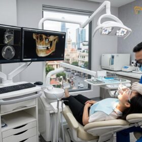
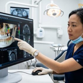
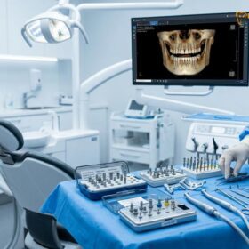
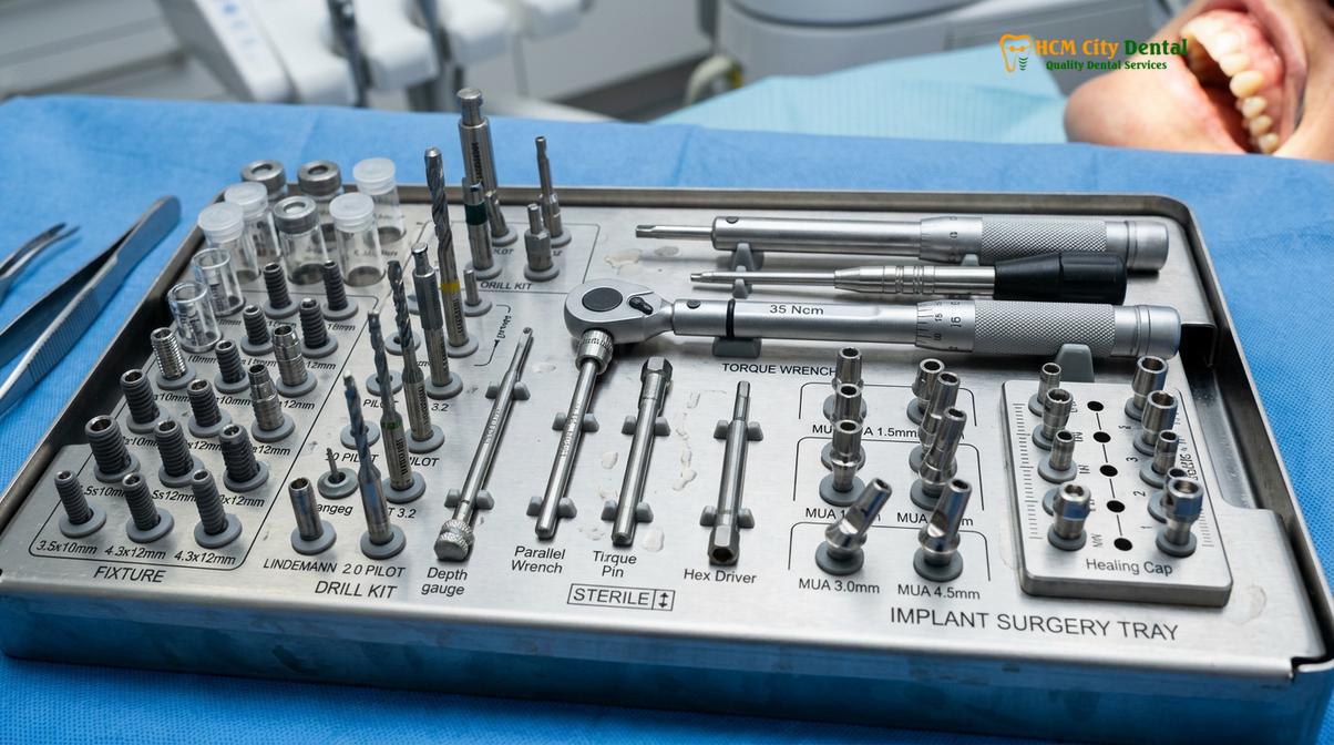
The paradigm of implant dentistry has shifted from a surgically driven approach to a prosthetically driven one. This means that the final position of the teeth dictates exactly where the implants must be placed in the bone. To achieve this level of precision, advanced digital workflows are employed. A Cone Beam Computed Tomography (CBCT) scan provides a high-resolution 3D view of the patient’s jawbone, allowing the surgeon to evaluate bone volume, locate the sinus cavities, and map the trajectory of the inferior alveolar nerve. This data is then merged with an intraoral optical scan of the soft tissues to create a comprehensive virtual model.
Using specialized software, the surgeon virtually places the implants, optimizing the AP spread and ensuring that the screw access holes will emerge in ideal locations on the final bridge. Once the virtual plan is finalized, a custom 3D-printed surgical guide is fabricated. This guide fits securely over the patient’s gums or remaining bone, directing the surgical drills with sub-millimeter accuracy. Guided surgery significantly reduces the risk of surgical errors, minimizes tissue trauma, and ensures that the implants are placed in the densest available bone, thereby maximizing primary stability and reducing the risk of early failure [4].
However, surgical precision is only half the battle; the longevity of the restoration depends heavily on the patient’s commitment to maintenance. A full-arch bridge is a complex prosthetic device that requires specialized cleaning techniques. Because the bridge is fixed in place, patients cannot simply floss between the teeth as they would with natural dentition. Instead, they must use water flossers (oral irrigators) to flush out debris trapped beneath the prosthesis. Interdental brushes and specialized superfloss are also required to clean the critical junction where the titanium abutments emerge from the gums.
Clinical Case Review: A 62-year-old patient presented to HCMC Dental Clinic in Ho Chi Minh City with a failing hybrid bridge placed at an overseas facility. The prosthesis exhibited severe mobility due to fractured retaining screws and localized peri-implantitis. Dr. Cuong utilized a 3D-guided approach to safely remove the compromised fixtures, perform targeted bone grafting, and successfully deliver a new, biomechanically optimized zirconia restoration.
Professional maintenance is equally critical. Patients with full-arch restorations should undergo professional prophylaxis every four to six months. During these visits, the dental hygienist will use specialized titanium or plastic scalers to remove calculus without scratching the implant surface. The clinician will also verify the occlusion (bite) to ensure that no excessive forces are being concentrated on a single area of the bridge. If a patient exhibits signs of bruxism, a custom night guard is mandatory to protect the porcelain or zirconia from fracturing. For older adults considering this treatment, understanding these maintenance requirements is a key topic discussed in Dental Implants for Seniors: Am I Too Old for Implants?.
Clinical Retreatment: Replacing Failed Posts Safely
When an implant fails, the clinical retreatment protocol involves safely extracting the compromised fixture, grafting the bony defect, and strategically placing a new implant to support the existing or a newly fabricated bridge.
Despite the best preventive measures, failures can still occur. When a patient presents with a failing implant, the first step is a thorough diagnostic evaluation using CBCT imaging to assess the extent of the bone defect. The clinical retreatment protocol is highly systematic, prioritizing the complete eradication of infection and the preservation of the remaining healthy bone. If the implant is mobile, it can often be removed easily using reverse torque instruments. However, if the implant is partially integrated but suffering from severe peri-implantitis, specialized trephine burs or piezoelectric surgical units may be required to carefully cut the fixture out of the bone with minimal trauma.
Once the failed implant is explanted, the surgical site is meticulously debrided. All granulation tissue and bacterial biofilm must be completely removed using surgical curettes and chemical decontamination agents, such as chlorhexidine or hydrogen peroxide. Leaving any infected tissue behind will almost certainly doom the subsequent re-implantation effort. Following debridement, the bony defect is typically grafted using a combination of particulate allograft or xenograft material, covered by a resorbable collagen membrane. This Guided Bone Regeneration (GBR) procedure rebuilds the foundation necessary to support a new implant.
The timing of re-implantation depends on the size of the defect and the quality of the surrounding bone. In some cases, a wider “rescue” implant can be placed immediately into the explantation site, engaging the deeper, healthy bone beyond the defect. However, in cases of severe bone loss, a staged approach is required. The graft is allowed to heal and consolidate for four to six months before a new implant is placed [5]. During this healing phase, the patient’s existing bridge must be modified. This failed fixed bridge repair often involves removing the section of the bridge that connected to the failed implant, allowing the prosthesis to function temporarily on the remaining healthy fixtures.
“Retreatment of a failed full-arch case is not merely a repetition of the initial surgery; it is a complex reconstructive effort that demands profound anatomical knowledge and advanced bone grafting techniques to restore biomechanical equilibrium.”
In extreme cases where the maxillary bone is completely resorbed due to prolonged infection, traditional re-implantation may not be possible. In these scenarios, advanced techniques such as the placement of zygomatic (cheekbone) or pterygoid implants may be utilized. These specialized implants anchor into dense, remote bones of the skull, entirely bypassing the compromised jawbone. While retreatment procedures are complex and require a highly skilled surgical team, they offer a highly predictable pathway to restoring the patient’s smile and function. Patients concerned about the financial implications of retreatment can review comprehensive resources such as How Much Do Dental Implants Cost in Vietnam? (Comprehensive Guide).
When to See a Doctor
Early detection is the most critical factor in salvaging a compromised implant and preventing extensive bone loss. Patients who have undergone full-arch rehabilitation must remain vigilant and monitor their oral health closely. You should schedule an immediate clinical evaluation if you experience any of the following symptoms:
- Persistent Pain or Swelling: While mild discomfort is normal immediately after surgery, pain that intensifies or returns months or years later is a primary indicator of infection or biomechanical overload.
- Mobility of the Bridge: A full-arch prosthesis should feel completely rigid. If you notice any clicking, shifting, or movement when chewing, it may indicate a loose retaining screw, a fractured framework, or a failing implant fixture.
- Bleeding or Purulent Exudate: Spontaneous bleeding from the gums around the implants, or the presence of pus (a foul-tasting discharge), is a definitive sign of active peri-implantitis that requires urgent decontamination.
- Exposed Metal Threads: If you can see the grey titanium threads of the implant fixture above the gumline, it indicates significant soft tissue recession and underlying bone loss.
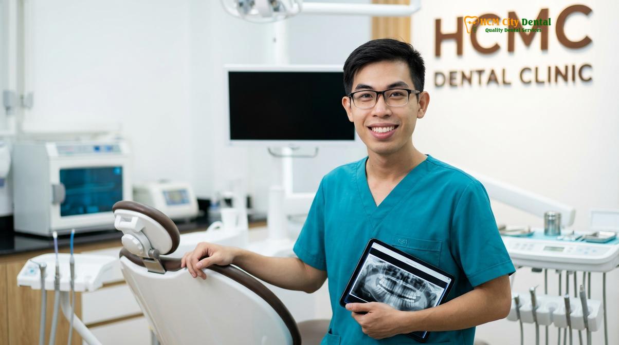
Do not wait for the symptoms to worsen. Ignoring these warning signs can lead to the rapid destruction of the jawbone, making retreatment significantly more difficult and invasive. If you suspect an issue with your restoration, seek a professional diagnostic review. For expert guidance and personalized care, consider exploring Dental Implants: Transform Your Smile Today! [Expert Tips] | Dr. Cuong to understand the comprehensive diagnostic protocols utilized at HCMC Dental Clinic.
Frequently Asked Questions
What does a failed All-on-4 implant feel like?
A failed All-on-4 implant typically feels loose, painful when chewing, or exhibits swelling and bleeding around the gumline. You may notice the entire prosthetic bridge shifting slightly, persistent discomfort, or a foul taste in your mouth indicating a localized bacterial infection. In some cases, the failure is painless but presents as a visible recession of the gums, exposing the grey titanium threads of the implant.
Can you redo an All-on-4 if it fails?
Yes, an All-on-4 procedure can often be redone if a failure occurs, though it requires a specialized clinical approach. The retreatment usually involves removing the failed fixture, grafting new bone to repair the defect, and placing a new implant once the site has adequately healed. In cases of severe bone loss, advanced techniques like zygomatic implants may be utilized to bypass the damaged areas entirely.
How does smoking affect All-on-4 success?
Smoking significantly increases the risk of All-on-4 failure by constricting blood vessels, which reduces oxygen and nutrient flow to the healing bone. This delayed healing process compromises osseointegration and drastically elevates the risk of postoperative infections and long-term peri-implantitis. Clinicians strongly advise complete smoking cessation for several weeks before and after surgery to optimize clinical outcomes.
How long does it take to recover from an implant failure retreatment?
Recovery from an implant retreatment typically takes between three to six months, depending on the extent of bone grafting required. While the soft tissue heals within a few weeks, the underlying jawbone needs several months to fully integrate with the new titanium fixture. During this period, your dentist will modify your existing bridge or provide a temporary prosthesis to ensure you maintain functional aesthetics.
Is it normal for an All-on-4 bridge to feel bulky?
It is entirely normal for a new All-on-4 bridge to feel slightly bulky during the initial adaptation period, as it replaces missing teeth, gum tissue, and bone volume. The prosthesis requires a certain thickness of acrylic or zirconia to prevent mechanical fracture under chewing forces. However, if the bulkiness interferes with speech or causes persistent biting issues after a few weeks, the prosthesis may require clinical adjustment.
References
- Journal of Oral and Maxillofacial Surgery. Biomechanical principles and tissue response in full-arch implant prosthetics. (2021).
- International Journal of Oral and Maxillofacial Implants. Survival rates and complications of All-on-4 treatments: A systematic review. (2020).
- Clinical Implant Dentistry and Related Research. The impact of smoking and poor bone quality on immediate loading protocols. (2019).
- Journal of Periodontology. Diagnosis and management of peri-implantitis in full-arch rehabilitations. (2022).
- British Dental Journal. Retreatment strategies for failed osseointegrated implants in the edentulous maxilla. (2018).