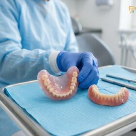
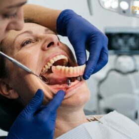
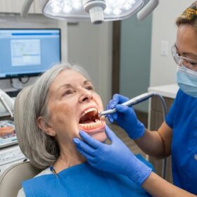
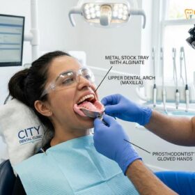
Proper denture adhesive application requires precise placement on clean, dry prosthetics to create a secure, comfortable seal without excess overflow. Understanding the correct techniques, appropriate adhesive types, and safe removal protocols ensures optimal retention while protecting delicate oral tissues from irritation and potential systemic toxicity.
Clinical Summary:
Denture adhesives function by enhancing interfacial surface tension between the prosthetic base and the oral mucosa through polymer hydration. Proper application demands a strict “less is more” approach to prevent tissue displacement, altered occlusion, and the risk of zinc toxicity. Continuous reliance on heavy adhesive application often masks underlying alveolar bone resorption, necessitating professional clinical evaluation for relining, rebasing, or transitioning to implant-supported alternatives to maintain long-term stomatognathic health.
Key Takeaways:
- Adhesives rely on natural salivary moisture to activate polymer hydration and create a cohesive retention matrix.
- Applying more than three to four small, pea-sized dots of adhesive can compromise the prosthetic fit and alter the bite.
- Zinc-based adhesive products pose severe neurological risks if used excessively over prolonged periods.
- Thorough daily removal of residue from both the gums and the denture base is mandatory to prevent bacterial colonization.
- Chronic dependency on adhesives strongly indicates a need for a professional prosthetic reline to address bone loss.
How Denture Adhesives Work: Enhancing Mucosa Surface Tension
Denture adhesives create a secure seal by absorbing saliva to form a viscous, cohesive layer that maximizes interfacial surface tension between the prosthetic base and the oral mucosa.
The biomechanical foundation of a custom removable prosthetic relies heavily on the intimate adaptation between the acrylic base and the underlying soft tissues. However, even with a masterfully crafted prosthetic, physiological changes, minor anatomical discrepancies, or heavy occlusal forces during mastication can challenge this retention. This is where the science of denture adhesive application becomes clinically relevant. Adhesives do not function as traditional “glues” that permanently bond two dry surfaces together; rather, they act as dynamic, moisture-activated interfacial agents that dramatically enhance the natural physical forces of retention, specifically cohesion, adhesion, and surface tension[1].
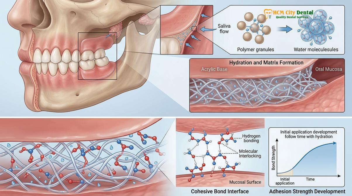
When a patient applies an adhesive to the intaglio (tissue-bearing) surface of their denture and places it into the oral cavity, the material immediately interacts with the patient’s saliva. The primary active ingredients in most modern adhesives are water-soluble polymers, such as carboxymethylcellulose (CMC) and polymethyl vinyl ether-maleic anhydride (PVM-MA) copolymers. As these polymers absorb salivary moisture, they hydrate, swell, and cross-link, transforming from a dry or semi-viscous state into a thick, elastic, and highly cohesive gel matrix. This gel fills any microscopic voids or macroscopic gaps between the rigid polymethyl methacrylate (PMMA) denture base and the compressible oral mucosa, effectively eliminating air pockets that would otherwise break the suction seal.
The creation of a secure denture seal is heavily dependent on the principle of interfacial surface tension. In a well-fitting denture without adhesive, a thin film of saliva provides a baseline level of surface tension that holds the prosthetic against the palate or alveolar ridge. However, natural saliva has a relatively low viscosity. By introducing an adhesive, the viscosity of this interfacial layer is exponentially increased. The swollen polymer chains entangle with the mucin proteins naturally present in saliva, creating a robust, viscoelastic bond that resists the vertical and lateral dislodging forces generated during chewing and speaking. Furthermore, this gel layer acts as a shock absorber, distributing occlusal loads more evenly across the mucosal surface and reducing localized pressure points that could lead to ulceration or erythema.
It is crucial to understand that the efficacy of this mechanism is finite. The adhesive matrix is designed to be temporary and water-soluble. Over the course of 8 to 12 hours, continuous exposure to excess saliva, dietary liquids, and the mechanical shearing forces of mastication will gradually dilute and break down the polymer cross-links. As the gel loses its cohesive strength, the retention of the denture decreases, signaling the need for removal, cleaning, and reapplication. Attempting to bypass this natural breakdown by applying excessive amounts of adhesive initially does not prolong the hold; instead, it disrupts the optimal thickness of the interfacial layer, leading to a loss of surface tension and a compromised fit.
Types of Adhesives: Creams, Powders, and Pre-cut Strips
Selecting the optimal adhesive involves choosing between the robust hold of creams, the thin profile of powders, and the highly controlled application of pre-cut strips based on individual clinical needs.
The dental market offers a variety of adhesive formulations, each engineered to address specific patient needs, anatomical challenges, and manual dexterity levels. Understanding the distinct properties, advantages, and limitations of each type is essential for achieving optimal prosthetic stability without compromising oral hygiene or tissue health. The three primary modalities utilized in modern prosthodontics are creams, powders, and pre-cut strips[2].
Denture Adhesive Cream: Creams, or pastes, are the most widely utilized form of adhesive globally. They are typically dispensed from a tube and offer the highest degree of initial tack and long-term cohesive strength. Creams are particularly beneficial for patients with moderate alveolar ridge resorption or those who require maximum stability for demanding masticatory function. The thick viscosity of the cream allows it to act as a volumetric filler, bridging larger gaps between the denture base and the shrinking gum tissue. However, this same viscosity makes creams the most prone to application errors. Over-application easily leads to material oozing out from under the denture borders, which is not only unpleasant for the patient but can also alter the vertical dimension of occlusion (the way the upper and lower teeth meet). Furthermore, creams are generally the most difficult to clean from the oral mucosa at the end of the day.
Adhesive Powders: Powders offer a fundamentally different approach to retention. They are sprinkled lightly over the moistened intaglio surface of the denture before insertion. Because they rely entirely on the patient’s natural saliva to activate and form the gel matrix, powders create a significantly thinner interfacial layer compared to creams. This makes them an excellent choice for patients whose dentures already fit quite well but who desire a slight increase in confidence and stability, or for those who struggle with the messy residue left by creams. Powders are generally easier to clean and carry a lower risk of altering the bite. However, they may not provide sufficient hold for patients with severe dry mouth (xerostomia) or advanced bone loss, as there is no volumetric mass to fill significant voids.
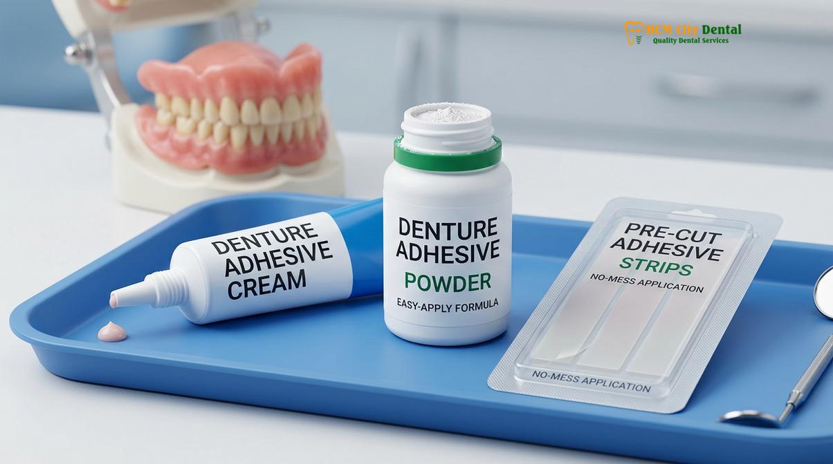
Dental Adhesive Strips: Also known as wafers or cushions, dental adhesive strips are pre-cut, dry polymer layers that are placed onto the denture base. They offer the ultimate in application control and cleanliness. Because the amount of adhesive is predetermined by the manufacturer, the risk of over-application and subsequent oozing is virtually eliminated. Strips are highly recommended for patients with compromised manual dexterity, such as those suffering from arthritis or neurological conditions, who may struggle to squeeze a tube or evenly distribute a powder. While they provide a reliable, mess-free hold, strips can sometimes be challenging to adapt to highly irregular or severely resorbed ridge anatomies, particularly on the mandibular (lower) arch.
| Adhesive Type | Retention Strength | Ease of Removal | Primary Clinical Indication | Risk of Overflow |
|---|---|---|---|---|
| Cream / Paste | High | Difficult | Moderate bone loss, high masticatory demand | High (if over-applied) |
| Powder | Moderate | Easy | Well-fitting dentures, minimal gap filling | Low |
| Pre-cut Strips | Moderate to High | Very Easy | Dexterity issues, travel convenience | None |
The ‘Less is More’ Rule: Application Step-by-Step
Applying adhesive correctly requires placing minimal, strategically located amounts on a thoroughly dried prosthetic to prevent material overflow, which can alter occlusion and irritate mucosal tissues.
The most common clinical error observed in patients utilizing removable prosthetics is the gross over-application of adhesive. Many patients operate under the misconception that more adhesive equates to a stronger, longer-lasting hold. In reality, the physics of surface tension dictate the exact opposite. An excessively thick layer of adhesive acts as a lubricant rather than a retentive matrix, causing the denture to slide over the mucosa. Furthermore, excess material inevitably oozes out from the peripheral borders, leading to ingestion, gagging, and a disruption of the carefully calibrated occlusal scheme. Adhering to a strict “less is more” protocol is paramount for both comfort and function.
The application process must always begin with meticulous preparation. The denture must be thoroughly cleaned using a soft-bristled brush and a non-abrasive cleanser to remove any residual adhesive, biofilm, or food particles from the previous day. Crucially, the intaglio surface of the denture must be completely dried before applying a cream or strip adhesive. If the acrylic is wet, the polymer will begin to hydrate prematurely, compromising its ability to bond effectively to the oral mucosa upon insertion. Conversely, if using a powder, the denture should be left slightly damp to facilitate the initial adherence of the powder particles.
“In our clinical practice, we constantly remind patients that adhesive is an aid, not a substitute for a well-fitting prosthetic. Applying more than three small dots of cream disrupts the precise adaptation we strive for in the laboratory. If you need a continuous ribbon of glue to keep your teeth in place, it is time for a comprehensive clinical reassessment.”
— Dr. Nguyen Van Cuong, Lead Prosthodontist
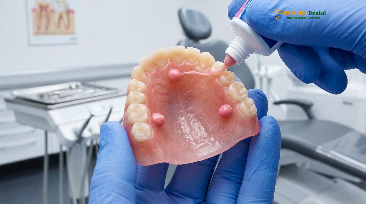
When applying a denture adhesive cream, the technique varies slightly between the maxillary (upper) and mandibular (lower) arches. For the upper denture, apply three or four small, pea-sized dots of cream: one in the anterior region behind the incisors, one on each side of the posterior ridge, and optionally, one small dot in the center of the palate. Never apply the adhesive directly to the peripheral borders, as the pressure of insertion will force the material outward. For the lower denture, which has a much smaller surface area, apply three small dots along the crest of the ridge: one in the anterior and one on each posterior side.
Once the adhesive is strategically placed, the patient should rinse their mouth with water to ensure the mucosa is adequately hydrated, as the polymers require this moisture to activate. The denture is then inserted and seated firmly into position. The patient must apply even, bilateral pressure—either by pressing with their fingers or biting down gently and evenly—for approximately 5 to 10 seconds. This sustained pressure allows the adhesive to spread into a microscopically thin, uniform layer, maximizing surface tension and initiating the cross-linking process. Following insertion, it is advisable to wait at least 15 to 30 minutes before consuming hot liquids or heavy meals to allow the cohesive matrix to fully mature and stabilize.
How to Safely Remove Sticky Adhesives from Gums and GBR Resin
Complete daily removal of adhesive residue from both the oral mucosa and the acrylic resin base is critical to prevent bacterial proliferation, tissue inflammation, and compromised subsequent adhesion.
The tenacious nature of modern adhesives, while beneficial for daytime retention, presents a significant challenge during nighttime removal. Leaving residual adhesive on the oral mucosa or the denture base overnight is a severe breach of oral hygiene protocols. The hydrated polymer matrix provides an ideal scaffolding for the colonization of pathogenic bacteria and fungi, particularly Candida albicans, leading to conditions such as denture stomatitis. Furthermore, applying fresh adhesive over old residue prevents the new material from bonding directly to the acrylic or the tissue, resulting in a weak, unreliable seal the following day[5].
Removing the denture itself should be done carefully to avoid traumatizing the tissues. The patient should swish vigorously with warm water or an alcohol-free mouthwash for 30 to 60 seconds. The warmth and the fluid dynamics help to break the surface tension and soften the adhesive seal. For the upper denture, placing the thumb against the palatal surface and gently pushing upward and outward breaks the suction. For the lower denture, a gentle rocking motion while pulling upward is usually sufficient. Never use sharp objects or excessive force to pry a denture loose.
Once the prosthetic is removed, the focus shifts to cleaning the oral mucosa. The gums, palate, and tongue must be meticulously debrided. The most effective and tissue-friendly method is to use a soft-bristled toothbrush or a clean, damp washcloth. Using warm water, gently massage the tissues in a circular motion. The mechanical friction, combined with the warm water, will roll the adhesive residue into small, easily expectorated balls. Avoid using regular toothpaste on the gums, as the detergents and abrasives can cause chemical irritation and micro-tears in the delicate mucosal lining. For stubborn residue, a gauze pad soaked in warm saline can provide slightly more texture without causing trauma.
Cleaning the denture base requires equal diligence, particularly if the prosthetic incorporates specialized materials like Guided Bone Regeneration (GBR) resin bases or soft liners, which can be easily damaged by aggressive scrubbing. The denture should be brushed under warm running water using a dedicated denture brush—which has softer bristles than a standard toothbrush—and a mild dish soap or specialized denture cleanser. Never use boiling water, as this can warp the PMMA acrylic. Ensure that all traces of the adhesive are removed from the deep recesses of the intaglio surface. After mechanical cleaning, the denture should be submerged in an effervescent soaking solution overnight to eliminate microscopic pathogens and keep the acrylic properly hydrated.
The Danger of Zinc Toxicity: What Neurological Symptoms to Watch For
Excessive, long-term use of zinc-containing adhesives can lead to severe copper deficiency, resulting in irreversible neurological damage, hematological abnormalities, and profound systemic health complications.
While denture adhesives are generally safe when used as directed, a significant and potentially life-altering risk arises from the chronic over-application of products containing zinc. Zinc is frequently added to adhesive formulations to enhance the initial tack and cohesive strength of the polymer matrix. In small, appropriate doses, the incidental ingestion of this zinc poses no threat, as the body requires trace amounts of zinc for normal metabolic function. However, when patients use excessive amounts of adhesive—often applying multiple thick ribbons several times a day to stabilize a poorly fitting denture—they can inadvertently ingest massive quantities of zinc over months or years[3].
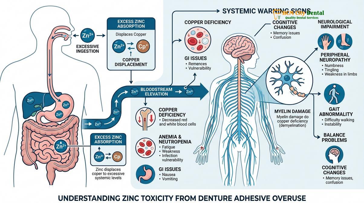
The pathophysiology of zinc toxicity is intrinsically linked to the body’s absorption of copper. In the gastrointestinal tract, zinc and copper compete for the same absorption pathways, specifically through the upregulation of a binding protein called metallothionein. When the body is flooded with excessive zinc from swallowed adhesive, metallothionein production spikes, binding tightly to copper and preventing its absorption into the bloodstream. This leads to a profound, systemic copper deficiency (hypocupremia). Copper is an essential trace element required for the maintenance of the central nervous system, the production of red and white blood cells, and the synthesis of myelin, the protective sheath surrounding nerve fibers.
Clinical Warning: Zinc Toxicity Symptoms
Patients utilizing large quantities of adhesive must be vigilant for signs of neurological impairment. Early zinc toxicity symptoms include unexplained numbness or tingling in the extremities (peripheral neuropathy), loss of balance or coordination (ataxia), and unexplained weakness. If these symptoms occur, discontinue the adhesive immediately, switch to a zinc-free denture glue, and seek urgent medical evaluation, as prolonged copper deficiency can lead to irreversible nerve damage.
The clinical manifestations of this zinc-induced copper deficiency are primarily neurological and hematological. Patients may initially present with subtle sensory changes, such as paresthesia (tingling or “pins and needles”) in the hands and feet. As the demyelination of the nerve fibers progresses, these symptoms can escalate to severe peripheral neuropathy, muscle weakness, spasticity, and a staggering, uncoordinated gait (ataxia) that mimics conditions like multiple sclerosis or severe spinal cord compression. Hematologically, the lack of copper impairs iron metabolism, leading to anemia that does not respond to iron supplementation, as well as neutropenia, which severely compromises the immune system’s ability to fight infections.
Because of these severe risks, the dental community strongly advocates for the use of zinc-free denture glue, especially for patients who require larger amounts of adhesive to achieve stability. Manufacturers have responded by reformulating many of their flagship products to eliminate zinc entirely, utilizing alternative cross-linking agents that provide comparable retention without the systemic risks. However, the ultimate solution to adhesive dependency is not merely switching brands, but addressing the underlying anatomical changes that make the adhesive necessary in the first place.
Why Adhesive Dependency is a Red Flag for a Professional Reline
Relying on increasing amounts of adhesive to stabilize a denture typically indicates underlying alveolar bone loss, requiring a clinical reline to restore proper fit and prevent accelerated tissue destruction.
A well-fabricated complete or partial denture is designed to fit intimately against the patient’s unique oral anatomy at the time of delivery. However, the human body is dynamic, and the extraction of natural teeth initiates an irreversible, lifelong process known as alveolar bone resorption. Without the mechanical stimulation previously provided by the tooth roots during chewing, the jawbone gradually shrinks in both height and width. As this jawbone shrinkage compensation occurs, the rigid acrylic base of the denture no longer matches the changing contours of the gums. The once-tight seal is lost, and the prosthetic begins to loosen, shift, and rock during function[4].
When patients experience this inevitable loosening, their first instinct is often to compensate by using more adhesive. While this may provide a temporary, superficial fix, it initiates a dangerous cycle of adhesive dependency. The thick layer of adhesive acts as a crude gasket, masking the poor fit but failing to distribute occlusal forces evenly. As the loose denture shifts and grinds against the shrinking ridge, it creates localized pressure points that accelerate the rate of bone resorption and cause chronic mucosal trauma, such as epulis fissuratum (hyperplastic tissue growth). The more the bone shrinks, the more adhesive the patient uses, further exacerbating the tissue destruction.
Clinical Case Review: Overcoming Adhesive Dependency
A 68-year-old patient presented to HCMC Dental Clinic in Ho Chi Minh City complaining of chronic gum soreness and the need to apply adhesive four times daily. Clinical examination by Dr. Cuong revealed severe mucosal erythema and significant alveolar resorption beneath a 5-year-old maxillary denture. Instead of recommending a different adhesive, the team at this leading dental clinic Saigon performed a comprehensive hard reline. By capturing the new tissue contours in a fresh layer of acrylic, the prosthetic’s natural suction was fully restored, completely eliminating the patient’s need for adhesive and halting the accelerated bone loss.
Therefore, a sudden or gradual increase in the need for adhesive is a critical clinical red flag. It is the body’s mechanical signal that the prosthetic requires professional intervention. The standard of care for a loose denture is a clinical reline. During a reline procedure, the dentist removes a thin layer of the intaglio surface of the denture and uses a specialized impression material to capture the current, exact anatomy of the patient’s gums. The dental laboratory then replaces this impression material with new, hard acrylic, effectively resurfacing the denture to fit the resorbed ridge perfectly.
In cases where bone resorption is severe and a conventional reline cannot provide adequate stability, the dentist may recommend transitioning to an implant-supported overdenture. By strategically placing dental implants into the jawbone, the prosthetic can be physically anchored in place, bypassing the need for mucosal surface tension and eliminating the need for adhesives entirely. Regular visits for comprehensive general dentistry check-ups are essential to monitor these changes and intervene before adhesive dependency compromises the patient’s oral health.
When to See a Doctor
Patients should seek immediate clinical evaluation if they experience chronic mucosal irritation, require multiple adhesive applications daily, or notice any neurological changes indicating potential systemic toxicity.
While denture adhesives are over-the-counter products, their use occurs within a complex medical and anatomical environment. Patients must recognize the threshold between routine maintenance and the need for professional clinical intervention. You should schedule an appointment with your prosthodontist or general dentist if you experience any of the following:
- Chronic Mucosal Trauma: If you develop persistent sore spots, ulcers, redness, or bleeding on your gums or palate that do not resolve within a few days, discontinue adhesive use and seek evaluation. These lesions may indicate a severely ill-fitting denture or an allergic reaction to the adhesive components.
- Excessive Application Requirements: If you find yourself needing to apply adhesive more than once a day, or if you must use large quantities (more than a few small dots) to achieve stability, your denture no longer fits properly and requires a professional reline or rebase.
- Neurological Symptoms: As discussed, any signs of numbness, tingling, weakness, or loss of coordination—especially if you have a history of heavy, long-term use of zinc-containing adhesives—warrant immediate medical and dental attention to rule out zinc toxicity and copper deficiency.
- Post-Surgical Healing: Never use adhesives immediately following tooth extractions, implant placement, or any oral surgery unless explicitly instructed by your surgeon. Adhesives can disrupt the healing process and introduce infection into open wounds.
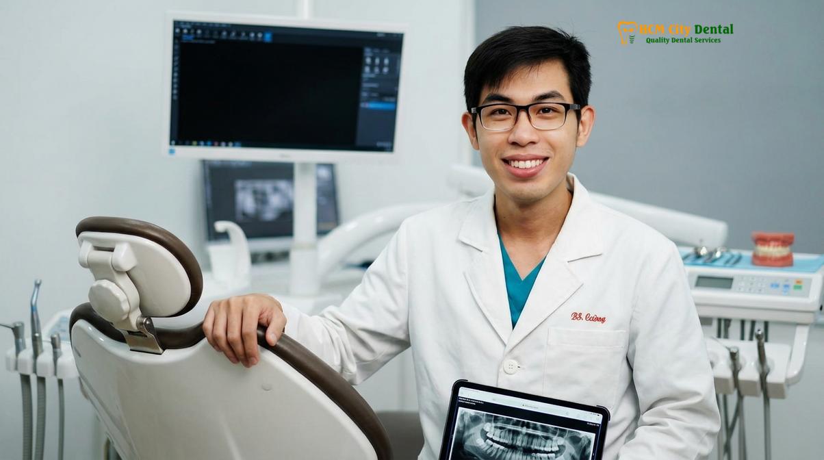
Maintaining a healthy, functional smile with removable prosthetics requires a partnership between the patient and the dental professional. Do not rely on adhesives to mask underlying structural problems. For a personalized assessment of your prosthetic fit and to explore advanced retention solutions, we encourage you to schedule a consultation with the specialists at HCMC Dental Clinic.
Frequently Asked Questions
Is it safe to use denture adhesive every day?
Yes, it is generally safe to use denture adhesive daily provided you apply the correct minimal amount and utilize a zinc-free formulation. However, daily reliance on large quantities of adhesive to keep a loose prosthetic in place indicates a need for a professional clinical evaluation and potential relining. Chronic use of excessive adhesive can mask severe bone loss and lead to tissue hyperplasia.
How do I get denture adhesive off the roof of my mouth?
The safest method to remove adhesive from the palate is to swish vigorously with warm water, then gently wipe the mucosa using a soft, damp washcloth or gauze. Avoid using stiff-bristled toothbrushes on the roof of your mouth, as this can cause micro-abrasions and tissue irritation. The warmth of the water helps break the polymer cross-links, making the residue easier to roll off the tissue.
Which is better: denture powder or cream adhesive?
Neither is universally better; the choice depends on clinical needs. Powders provide a thinner, more natural-feeling layer ideal for well-fitting dentures requiring slight stabilization, whereas creams offer a stronger, more viscous hold suitable for patients needing enhanced retention, though they are harder to clean. Patients with dry mouth often struggle with powders, as they require natural saliva to activate.
Can I use adhesive immediately after a tooth extraction?
No, you should never apply denture adhesive directly over fresh extraction sites or unhealed surgical wounds. Adhesives can introduce bacteria into the socket, disrupt blood clot formation, and severely delay the natural healing process; always wait for your dentist’s explicit clearance. If you have an immediate denture, your dentist will provide specific instructions on when and how to safely begin using adhesives.
How long does a single application of adhesive last?
A proper application of high-quality adhesive should provide a secure hold for 8 to 12 hours. If you find yourself needing to reapply the product multiple times throughout the day, your prosthetic likely suffers from poor adaptation to the underlying alveolar ridge. Factors such as excessive saliva production, consumption of very hot liquids, and heavy masticatory forces can slightly reduce this duration.
References
- Journal of Prosthetic Dentistry. Biomechanics of denture adhesives and mucosal surface tension. (2021).
- International Journal of Prosthodontics. Clinical efficacy of denture adhesive creams vs powders. (2020).
- Journal of the American Dental Association. Zinc toxicity associated with excessive denture adhesive use. (2019).
- Clinical Oral Implants Research. Alveolar bone resorption and the need for denture relining. (2022).
- Journal of Oral Rehabilitation. Protocols for the removal of denture adhesives from acrylic resin. (2018).