Effective yellow stains around braces brackets prevention requires aggressive plaque biofilm management, specialized orthodontic brushing techniques, and strict dietary control. These stains, often early signs of enamel demineralization, can generally be avoided through daily interdental cleaning and routine professional scaling to maintain pristine oral hygiene throughout your orthodontic journey.
Clinical Summary:
The phenomenon of yellow staining and white spot lesions during orthodontic therapy is a well-documented clinical challenge stemming from prolonged plaque retention. Orthodontic hardware, including brackets, archwires, and elastomeric ligatures, creates complex micro-environments that shield cariogenic bacteria from natural salivary clearance. As these bacteria metabolize fermentable carbohydrates, they excrete lactic acid, leading to localized drops in oral pH and subsequent enamel demineralization. Effective prevention necessitates a multifaceted approach combining rigorous mechanical plaque disruption—using V-trim brushes and interdental aids—with chemical remineralization via topical fluorides. Furthermore, dietary modification to reduce acidogenic challenges is paramount. Routine professional prophylaxis, utilizing ultrasonic scaling and air polishing, remains a critical adjunct to home care, ensuring the removal of calcified calculus and preserving the structural integrity and aesthetic appearance of the enamel upon appliance removal.
Key Takeaways:
- Yellow stains are primarily caused by plaque biofilm trapping chromogenic dietary pigments and bacterial byproducts.
- White spot lesions indicate early-stage enamel demineralization and require immediate remineralization protocols.
- Specialized tools, including interdental brushes and water flossers, are mandatory for effective plaque removal around brackets.
- Frequent consumption of acidic and sugary beverages exponentially increases the risk of permanent enamel scarring.
- Professional scaling every 3-4 months is highly recommended to manage calculus buildup during active orthodontic treatment.
Why Yellow Stains and White Spots Form Around Braces
Yellow stains and white spots develop when plaque biofilm accumulates around orthodontic hardware, leading to bacterial acid production that leaches essential minerals from the enamel surface.

The journey to a perfectly aligned smile through comprehensive orthodontic treatment is a transformative process, but it introduces unique challenges to maintaining optimal oral hygiene. One of the most prevalent clinical complications encountered by patients is the development of discoloration around the bonded hardware. Understanding the pathophysiology behind a yellow stains braces bracket scenario is the first critical step in prevention. The discoloration observed by patients is rarely a single entity; rather, it is a spectrum of enamel changes ranging from superficial extrinsic staining to deep intrinsic demineralization.
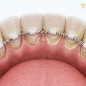
Extrinsic staining occurs when chromogenic substances from the diet—such as tannins in tea, coffee, and dark berries, or synthetic dyes in processed foods—become physically trapped in the porous matrix of dental plaque. Because orthodontic brackets create physical ledges on the facial surfaces of the teeth, they interrupt the natural self-cleansing mechanism of the lips, cheeks, and tongue. Consequently, plaque accumulates rapidly at the bracket periphery. If this biofilm is not meticulously removed, it absorbs these dietary pigments, resulting in a noticeable yellow or brownish halo around the bracket base.
More concerning from a clinical perspective is the formation of white spot lesions (WSLs). These chalky, opaque areas represent the earliest macroscopic signs of dental caries. The process begins when acidogenic bacteria within the plaque biofilm metabolize dietary sugars. The resulting acidic byproducts lower the local pH at the enamel surface below the critical threshold of 5.5. At this pH, the hydroxyapatite crystals that form the structural foundation of tooth enamel begin to dissolve, a process known as demineralization [1]. The loss of calcium and phosphate ions alters the refractive index of the enamel, causing it to appear chalky white. If the porous, demineralized enamel subsequently absorbs dietary stains, the white spots can transition into stubborn yellow or brown lesions.
The Enemy: Plaque Biofilm Trapped by Metal and Ceramic Brackets
Orthodontic brackets and archwires create complex micro-environments that shield plaque biofilm from natural salivary clearance, accelerating bacterial colonization and localized enamel erosion.
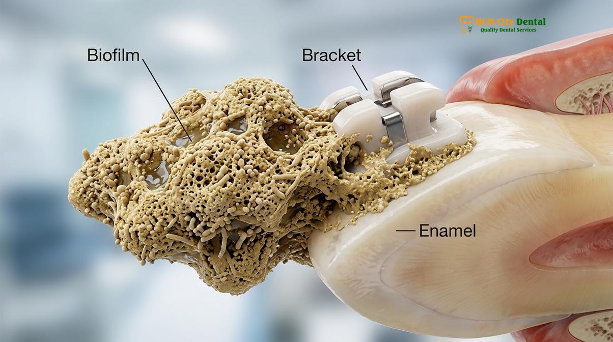
To effectively combat orthodontic staining, one must understand the biological adversary: dental plaque biofilm. Biofilm is not merely a loose collection of food debris; it is a highly organized, complex microbial ecosystem. When a patient undergoes orthodontic bonding, the smooth, easily cleanable surface of the natural tooth is suddenly interrupted by a complex topography of metal or ceramic brackets, archwires, elastomeric ties, and power chains. These components create numerous stagnation areas—microscopic safe havens where biofilm can mature undisturbed by the mechanical forces of chewing or the flushing action of saliva.
The maturation of plaque biofilm is a time-dependent process. Within minutes after brushing, a salivary pellicle forms on the enamel and bracket surfaces. This protein-rich layer acts as an adhesive base for early bacterial colonizers. If left undisturbed, the biofilm thickens and shifts in microbial composition, favoring anaerobic, acid-producing bacteria that are highly cariogenic and serve as the primary culprits behind enamel decalcification [2].
“The introduction of fixed orthodontic appliances fundamentally alters the oral microbiome, shifting the bacterial flora toward a more acidogenic and cariogenic profile. Meticulous mechanical disruption of this biofilm is the only definitive method to prevent structural enamel damage.”
The physical presence of the archwire further complicates hygiene efforts. It acts as a barrier, preventing the bristles of a standard toothbrush from reaching the gingival third of the tooth—the area between the bracket and the gumline. This specific zone is highly susceptible to plaque accumulation, leading not only to enamel demineralization but also to hyperplastic gingivitis. Swollen, inflamed gums can overgrow the brackets, creating even deeper pseudopockets that harbor aggressive bacteria and make effective plaque removal techniques exceedingly difficult.
Must-Have Tools: Orthodontic Brushes, Interdentals, and Mouthwashes
Effective orthodontic hygiene demands a multi-tool approach, utilizing V-trim toothbrushes, interdental brushes, and fluoridated mouthwashes to mechanically disrupt plaque in hard-to-reach bracket crevices.

Mastering oral hygiene with fixed appliances requires upgrading from a standard toothbrush to a specialized arsenal of dental tools. The complex geometry of braces means that a single cleaning instrument cannot adequately access all surfaces of the teeth and hardware. Implementing advanced orthodontic toothbrushing tips and utilizing the correct tools is non-negotiable for preventing yellow stains and white spot lesions.
The foundation of the orthodontic hygiene routine is the V-trim (or orthodontic) toothbrush. Unlike flat-bristled brushes, a V-trim brush features shorter bristles in the center and longer bristles on the edges. This specific design allows the longer outer bristles to clean the tooth surface above and below the bracket, while the shorter inner bristles clean the face of the bracket itself. Patients must employ the Modified Bass technique, angling the brush at 45 degrees toward the gumline to sweep plaque away from the gingival sulcus, followed by angling the brush 45 degrees toward the biting edge to clean under the archwire.
| Hygiene Tool | Primary Clinical Function | Recommended Frequency |
|---|---|---|
| V-Trim Orthodontic Toothbrush | Cleans facial enamel and bracket surfaces simultaneously. | After every meal (minimum 3x daily). |
| Interdental Brushes (Proxabrushes) | Disrupts biofilm beneath archwires and between brackets. | At least once daily, preferably before bedtime. |
| Water Flosser (Oral Irrigator) | Flushes out loose food debris and disrupts unattached biofilm. | Daily, as an adjunct to mechanical brushing and flossing. |
| Fluoride Mouthrinse (0.05% NaF) | Promotes chemical remineralization of early white spot lesions. | Nightly, after all mechanical cleaning is complete. |
Chemical plaque control is the final layer of defense. A daily fluoridated mouthwash is critical for orthodontic patients. Fluoride ions integrate into the enamel structure, converting hydroxyapatite into fluorapatite, which is significantly more resistant to acid dissolution [3]. Rinsing with a fluoride solution right before bed, after all mechanical cleaning is complete, allows the fluoride to remain on the teeth overnight, maximizing its remineralizing potential. For patients evaluating braces vs Invisalign dental tourist decisions, understanding this daily hygiene commitment is a crucial factor in selecting the right treatment modality.
Dietary Guidelines: Cutting Sugars and Acidic Drinks During Treatment
Minimizing fermentable carbohydrates and acidic beverages is critical during orthodontic treatment to prevent rapid pH drops that accelerate enamel demineralization and bracket staining.

Mechanical hygiene alone is often insufficient if the patient’s diet continuously fuels bacterial acid production. The relationship between diet and enamel health is governed by the Stephan Curve, a graphical representation of how oral pH drops immediately following the consumption of fermentable carbohydrates and slowly recovers over time. For a patient wearing braces, the recovery time is significantly prolonged because the hardware traps the carbohydrates and restricts the neutralizing flow of saliva.
Sucrose, fructose, and highly processed starches are the primary substrates for cariogenic bacteria. Sticky candies, caramels, and gummies are particularly detrimental; not only do they pose a mechanical risk of dislodging brackets or bending archwires, but they also adhere stubbornly to the hardware, providing a sustained sugar release that keeps the oral pH in the danger zone for extended periods. Continuous snacking or sipping on sugary beverages throughout the day prevents the saliva from ever neutralizing the acidic environment, leading to rapid and severe enamel decalcification [4].
“The frequency of carbohydrate intake is often more destructive than the total volume consumed. Orthodontic patients must consolidate their sugar intake to mealtimes to allow salivary buffering systems adequate time to restore a neutral oral pH.”
Acidic beverages pose a dual threat. Sodas, sports drinks, citrus juices, and even sparkling waters often have a very low pH. When consumed, these liquids bathe the teeth in acid, causing direct chemical erosion of the enamel that bypasses bacterial metabolism entirely. Patients are strongly advised to consume water as their primary beverage and to use a straw if acidic drinks are occasionally consumed, directing the fluid past the teeth to minimize contact time.
Clinic Scaling: The Importance of regular cleanings at HCMC Dental
Routine professional scaling is an indispensable component of orthodontic care, ensuring the removal of hardened calculus that home hygiene routines cannot eliminate.
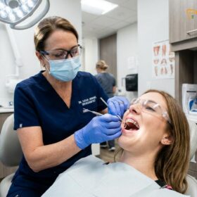
Even with the most meticulous home care routine, microscopic areas of plaque can calcify into tartar (calculus) around orthodontic brackets and beneath the gumline. Once calculus forms, it cannot be removed with a toothbrush or floss; it requires professional clinical intervention. This is why regular clinic scaling is absolutely vital during active orthodontic treatment. The presence of calculus not only contributes to severe yellow staining but also acts as a porous reservoir for further bacterial colonization, accelerating gingival inflammation and enamel breakdown.
Orthodontic patients are generally advised to undergo professional scaling and prophylaxis every three to four months, rather than the standard six-month interval. During these sessions, dental hygienists utilize specialized ultrasonic scalers and fine hand instruments designed to safely navigate around brackets and archwires without compromising the bond strength. Additionally, air polishing devices using fine powders like glycine or erythritol are highly effective at removing stubborn extrinsic stains from the complex topography of the braces. For patients who are moving abroad during Invisalign treatment or traditional braces therapy, establishing a reliable schedule for professional cleanings in their new location is critical to maintaining enamel health.
Clinical Case Study: Managing Demineralization in Ho Chi Minh City
A proactive approach combining professional scaling and enhanced home care can successfully reverse early signs of enamel demineralization and prevent permanent staining.
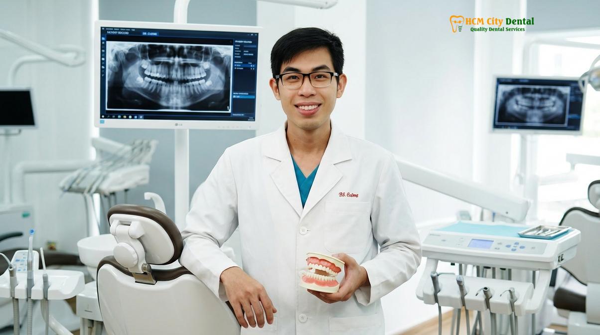
Dr. Nguyen Van Cuong, a leading specialist at HCMC Dental Clinic, emphasizes that early detection is paramount. He frequently advises patients that proactive plaque management is the only way to prevent irreversible enamel scarring. Under Dr. Cuong’s guidance, our clinical team implements rigorous hygiene protocols for every orthodontic case to ensure optimal aesthetic outcomes.
Clinical Case Study: A 22-year-old patient visited HCMC Dental Clinic in Ho Chi Minh City for routine orthodontic adjustments. During the fourth month of treatment, the clinical team noted early chalky white opacities forming along the gingival margins of the upper incisors, accompanied by mild yellow extrinsic staining around the ceramic brackets. The patient admitted to frequent consumption of sugary iced coffees and inconsistent use of interdental brushes. The intervention protocol was immediate and comprehensive. The clinical team performed a thorough ultrasonic scaling and air polishing procedure to remove all calcified deposits and extrinsic stains without damaging the orthodontic hardware. The patient was then re-educated on proper brushing techniques and prescribed a high-concentration fluoride toothpaste (5000 ppm) to use nightly. By the next adjustment appointment, the yellow staining was completely resolved, and the white spot lesions had begun to remineralize, losing their chalky appearance and blending back into the natural enamel shade. This highlights the importance of professional oversight, as untreated lesions can weaken the tooth structure and even contribute to complications like orthodontic relapse if the underlying dental health is compromised.
When to See an Orthodontist for Staining
Patients should seek immediate professional evaluation if they notice persistent white spots, bleeding gums, or dark yellow halos around brackets that do not improve with standard brushing.
Important Note: Do not attempt to use harsh abrasive whitening toothpastes or over-the-counter bleaching kits while wearing traditional braces. These products can scratch the enamel, damage the brackets, and result in uneven tooth coloration once the hardware is removed.
While minor dietary staining can often be managed at home, certain clinical signs warrant prompt professional intervention. According to preventative guidelines aligned with the Vietnam Ministry of Health (MOH) and general dental health authorities, patients should not wait for their next scheduled adjustment if they observe rapid changes in enamel color or gingival health. Persistent white, chalky lines framing the brackets are a definitive warning sign of active demineralization. If these spots begin to turn brown or yellow, it indicates that the porous enamel is absorbing stains and may be progressing toward cavitation.
Additionally, if the gums become swollen, red, and bleed easily during brushing, this gingival inflammation (gingivitis) can quickly escalate. Swollen gums create deeper pockets that trap more plaque, creating a vicious cycle of infection and enamel damage. In such cases, a professional cleaning is mandatory. Advanced treatments, such as resin infiltration or microabrasion, may be required post-treatment to address stubborn lesions and restore the natural gloss of the enamel [5]. Patients navigating these treatments should also review their dental insurance coverage for orthodontics to understand their benefits for preventative and restorative care.
Maintaining a pristine smile during orthodontic treatment requires dedication, but the results are well worth the effort. For comprehensive orthodontic care and personalized hygiene strategies, schedule a consultation with the experts at HCMC Dental Clinic, Ho Chi Minh City. Explore our full range of services, including custom nightguards and mouthguards, to protect your newly aligned smile for years to come.
References
- American Journal of Orthodontics and Dentofacial Orthopedics. Enamel demineralization during orthodontic treatment: etiology and prevention.
- Journal of Clinical Orthodontics. The efficacy of mechanical plaque control in fixed appliance patients.
- International Journal of Dentistry. Dietary influences on white spot lesion formation in orthodontics.
- Journal of Dental Hygiene. Ultrasonic scaling and air polishing protocols for orthodontic patients.
- European Journal of Orthodontics. Long-term aesthetic outcomes following white spot lesion microabrasion.
For pricing, booking, and a free clinical assessment, visit our Dental Braces & Orthodontics service page at HCMC Dental Clinic in Ho Chi Minh City.