Replacing a failing front tooth is widely considered one of the most complex challenges in modern oral implantology. When a natural tooth is extracted, the delicate facial bone—known as the bundle bone—inevitably loses its blood supply and begins to resorb. This biological reality often leads to a collapsed gum line, grey shadowing, and an artificial-looking dental implant. However, the advent of the socket shield technique dental implant in Vietnam protocol has revolutionized aesthetic dentistry. By intentionally retaining a micro-thin fragment of the natural tooth root, specialists can preserve the periodontal ligament, maintain the original blood supply, and completely prevent the collapse of the labial plate. This advanced procedure, also known as Partial Extraction Therapy (PET), ensures that the final implant crown emerges from the gums exactly like a natural tooth.
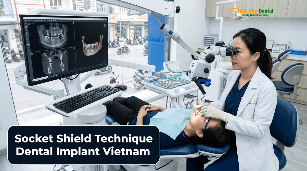
At HCMC Dental Clinic, located in District 1, Ho Chi Minh City, international patients and dental travelers have access to world-class biological implantology. Under the clinical direction of Dr. Nguyen Van Cuong, DDS (Clinical Director — MOH License 0003265/BL-CCHN), the clinic utilizes 3D CBCT imaging, Piezosurgery, and custom CAD/CAM prosthetics to execute the Socket Shield Technique with unparalleled precision. For expats and medical tourists seeking an immediate front tooth implant in the aesthetic zone in Vietnam, this protocol offers a minimally invasive alternative to extensive bone grafting, delivering superior long-term aesthetic stability at a highly accessible price point.
Clinical Summary:
The Socket Shield Technique (Partial Extraction Therapy) is an advanced surgical protocol used during immediate anterior implant placement. Instead of completely extracting a failing tooth, the surgeon uses microsurgical instruments to section the root, leaving a 1.5mm buccal fragment attached to the facial bone. This preserves the periodontal ligament (PDL) and its vital blood supply, effectively preventing the bundle bone resorption that typically follows tooth loss. At HCMC Dental Clinic, Dr. Nguyen Van Cuong, DDS combines this technique with 3D-guided surgery, Osstell ISQ resonance frequency analysis, and custom Zirconia abutments to deliver flawless, natural-looking front teeth for international patients, eliminating the need for invasive delayed bone grafting procedures.
Key Takeaways:
- Biological Preservation: Retaining a 1.5mm buccal root fragment maintains the PDL blood supply, acting as a natural barrier to prevent bundle bone resorption and dental implant failure.
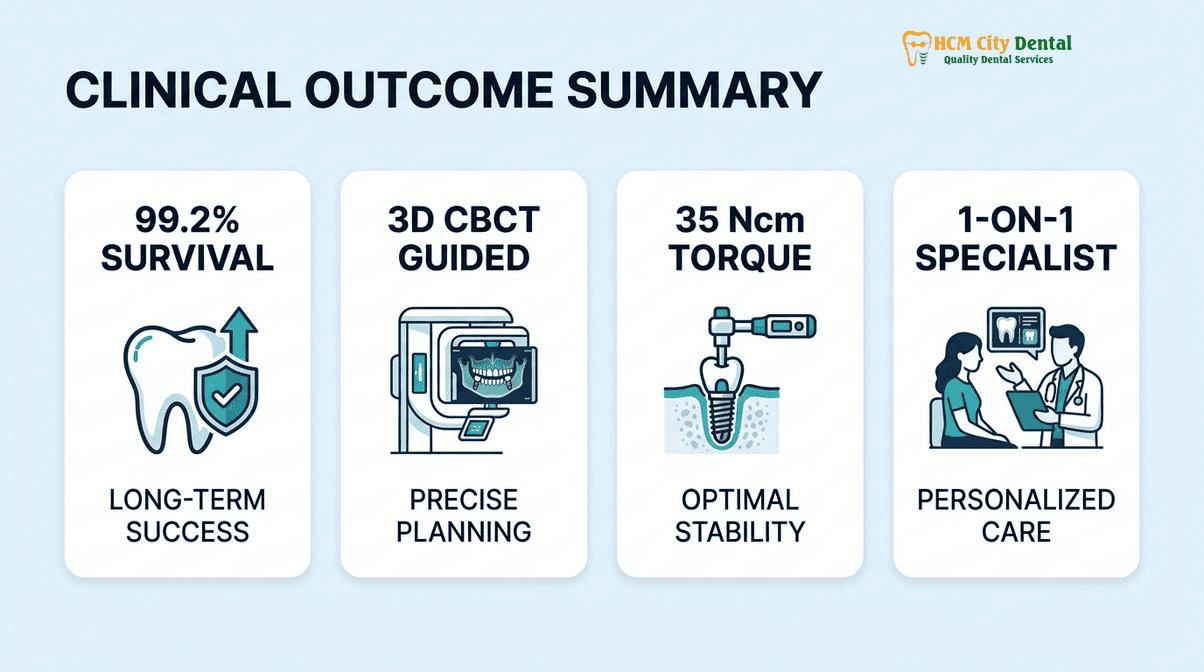
- Immediate Aesthetics: Patients can receive a custom CAD/CAM provisional crown on the same day, provided the implant achieves an insertion torque of ≥35 Ncm and an Osstell ISQ score of ≥70.
- Advanced Technology: The procedure utilizes Piezosurgery for atraumatic micro-sectioning and Conical Morse Taper Platform Switching implants to ensure <0.2mm crestal bone loss at 36 months.
- Cost Efficiency: A single European/Swiss implant ranges from $962 to $1,346 USD online, offering world-class treatment at a fraction of Western prices.
- Specialist Expertise: All procedures are performed by Dr. Nguyen Van Cuong, DDS, utilizing strict EN 13060 Class B infection control and biomaterial traceability protocols.
What Is the Socket Shield Technique (Partial Extraction Therapy) and Why Does the Labial Plate Collapse After Normal Extraction?
To understand the profound impact of the socket shield technique dental implant, one must first understand the biological consequences of a standard tooth extraction. The human tooth is anchored into the jawbone by the periodontal ligament (PDL). The bone immediately surrounding the tooth, known as the bundle bone, is entirely dependent on the blood vessels within the PDL for its nourishment and survival. When a tooth is fully extracted, the PDL is severed and removed. Deprived of its blood supply, the bundle bone rapidly undergoes ischemic necrosis and resorbs. Clinical studies show that up to 50% of the alveolar ridge width can be lost within the first six months following a conventional extraction.
In the anterior maxilla (the upper front teeth), this bundle bone is exceptionally thin—often measuring less than 1mm in thickness. When it resorbs, the overlying gum tissue collapses with it. This leads to a concave defect in the jaw, a higher gum line, and a loss of the natural scalloped architecture of the soft tissue. If a standard dental implant is placed into this collapsing socket, the result is often an aesthetic failure: the metal collar of the implant may show through the thinned gums, or the final crown may appear disproportionately long to compensate for the lost tissue.
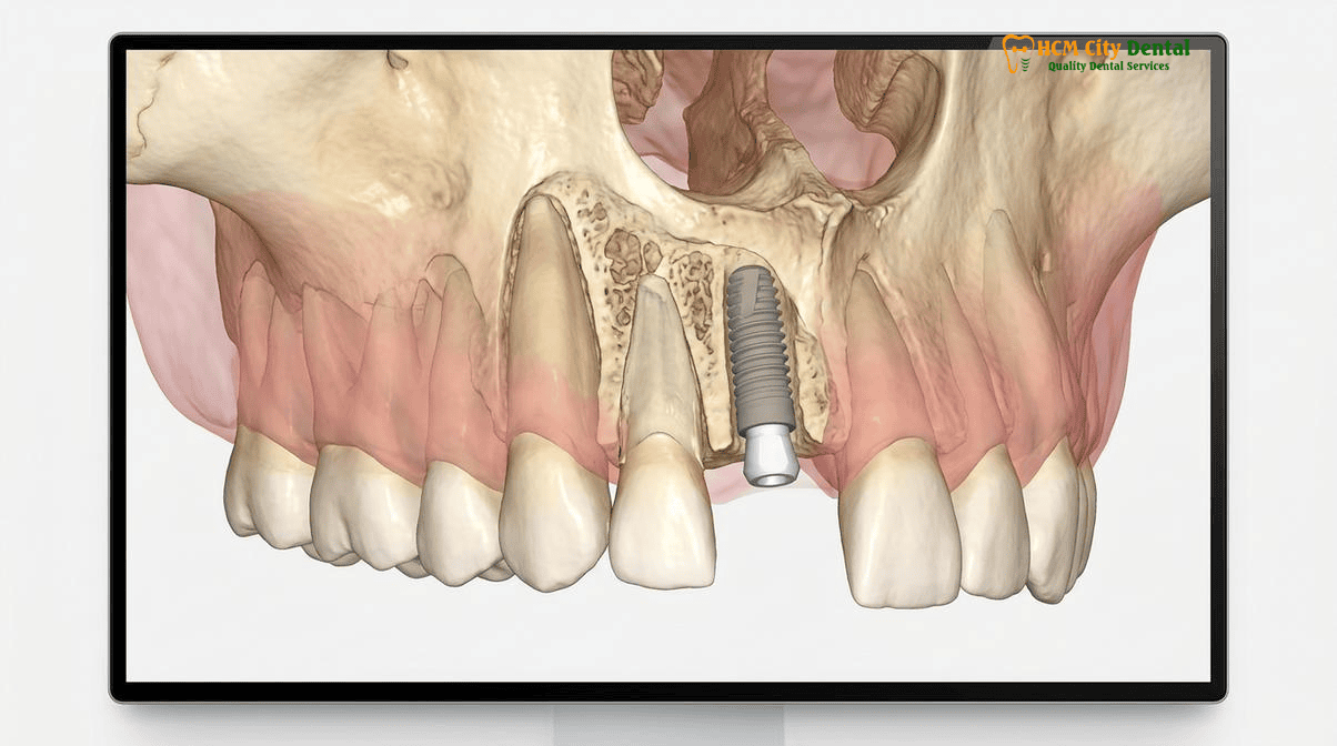
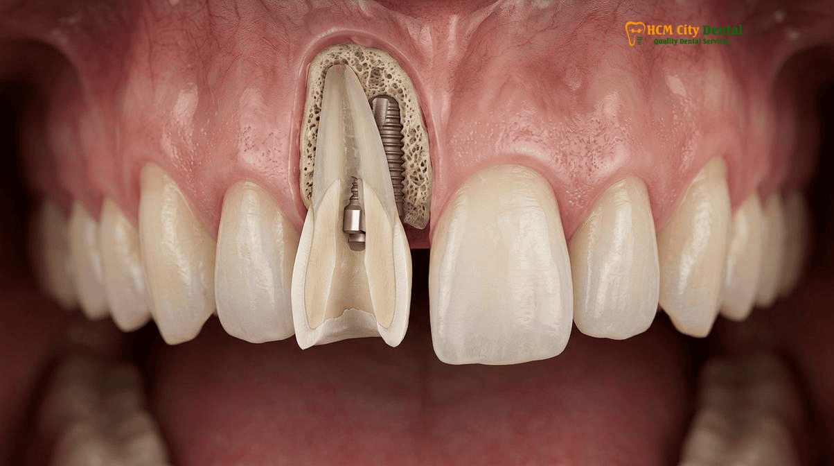
The Socket Shield Technique, a form of Partial Extraction Therapy (PET) anterior implant surgery, was developed to circumvent this biological inevitability. Instead of removing the entire root, the surgeon carefully decoronates the tooth and sections the root vertically. The palatal portion of the root is removed to make space for the implant, but the buccal (facial) portion of the root is intentionally left completely intact and attached to the bone. This “shield” typically measures about 1.5mm in thickness. Because the buccal root fragment remains untouched, its PDL remains vital. The blood supply to the bundle bone is uninterrupted, and the body does not recognize that the tooth has been “lost.” Consequently, the labial plate does not collapse, and the natural gum architecture is perfectly preserved.
“The Socket Shield Technique represents a paradigm shift in aesthetic implantology. By respecting the body’s natural biology and preserving the buccal root membrane, we can achieve soft tissue stability that is virtually impossible to replicate with artificial bone grafts alone. It is the ultimate method to prevent gum recession and front tooth implant complications.”
— Dr. Nguyen Van Cuong, DDS
This technique is particularly crucial for international patients seeking 3D-Guided Dental Implants & Specialist Oral Surgery in Vietnam, as it drastically reduces the total treatment time. By eliminating the need for complex, multi-stage Guided Bone Regeneration (GBR) and soft tissue grafting, patients can achieve a final aesthetic result much faster and with significantly less surgical trauma.
Biological Mechanism: How Preserving a 1.5mm Buccal Root Fragment Maintains the Periodontal Ligament (PDL) Blood Supply
The success of the buccal root membrane preservation implant protocol lies entirely in its biological foundation. The periodontium is a highly specialized complex comprising the cementum (the outer layer of the tooth root), the periodontal ligament, the bundle bone, and the gingiva. The PDL is a highly vascularized connective tissue network that acts as a shock absorber and a nutrient conduit. The blood vessels within the PDL anastomose (connect) with the supraperiosteal vessels of the gums and the intraosseous vessels of the alveolar bone.
When Dr. Nguyen Van Cuong, DDS performs the Socket Shield Technique, the goal is to preserve this delicate vascular network. The buccal root fragment is reduced to a thickness of approximately 1.5mm and reduced coronally to sit 1mm below the crest of the buccal bone. This specific dimension is critical. If the shield is too thick, there will not be enough room for the implant and the necessary “jumping gap.” If it is too thin, it may fracture during the procedure or resorb over time. [1]
Once the shield is prepared, the dental implant is placed palatal to the shield. It is a strict biomechanical requirement that the implant surface must never touch the root shield. A gap must be maintained between the titanium or zirconia implant threads and the dentin of the shield. This gap is typically filled with a xenograft or allograft bone substitute. As the implant osseointegrates, the bone graft consolidates, creating a solid matrix. Because the shield is completely decoupled from the implant, the occlusal forces (biting pressure) applied to the implant are not transferred to the fragile root fragment. [2]
Furthermore, the choice of implant design plays a massive role in the biological success of this procedure. At HCMC Dental Clinic, we utilize implants featuring Conical Morse Taper Platform Switching. This design moves the implant-abutment microgap away from the crestal bone and creates a tight, cold-weld seal that prevents bacterial microleakage. Clinical data demonstrates that platform-switched implants placed in conjunction with a socket shield exhibit less than 0.2mm of crestal bone loss at 36 months. This biomechanical stability, combined with the biological preservation of the PDL, ensures that the bundle bone resorption prevention strategy for dental implants is successful over the patient’s lifetime.
For patients with specific systemic concerns, such as those requiring Dental Implants for Patients on Corticosteroids or Biologics (Rheumatoid Arthritis, Lupus, Thyroid Conditions), the minimally invasive nature of the Socket Shield Technique is highly advantageous. Because it avoids large periosteal releasing incisions and extensive bone grafting, the inflammatory response is minimized, promoting faster and more predictable healing even in compromised immune states.
Strict Clinical Inclusion & Exclusion Criteria: When Socket Shield Succeeds vs. When Guided Bone Regeneration (GBR) Is Required
While the Socket Shield Technique offers unparalleled aesthetic benefits, it is not a universal solution for every failing tooth. It is a highly technique-sensitive procedure that requires strict adherence to clinical inclusion and exclusion criteria. Proper patient selection is the cornerstone of predictable oral implantology.
Inclusion Criteria (When Socket Shield Succeeds):
- Intact Buccal Bone and Root: The facial aspect of the tooth root and the overlying buccal bone must be completely intact, without any fenestrations (windows) or dehiscences (clefts).
- Adequate Apical Bone: There must be at least 3 to 4 millimeters of healthy native bone beyond the apex of the tooth root. This is essential for the implant to achieve primary stability. The implant must engage this apical bone to achieve an insertion torque of ≥35 Ncm, which is required for immediate provisionalization.
- Healthy Periodontium: The surrounding gum tissue should be relatively healthy, with a thick or medium gingival biotype preferred, as it provides better camouflage and resistance to recession.
- Absence of Acute Infection: While chronic, asymptomatic apical lesions can often be thoroughly curetted and managed during surgery, the presence of an acute, purulent apical abscess is generally a contraindication for immediate implant placement.
Clinical Precaution: If you are experiencing severe pain, swelling, or active pus discharge from a failing front tooth, immediate implant placement may carry a high risk of failure. Contact Dr. Nguyen Van Cuong, DDS immediately via WhatsApp at +84 853 020 003 for a comprehensive 3D CBCT evaluation and appropriate antibiotic management prior to surgical intervention.
Exclusion Criteria (When GBR Is Required):
- Vertical Root Fractures on the Buccal Aspect: If the tooth has fractured down the facial side, the PDL is already compromised, and bacteria have likely infiltrated the fracture line. The shield cannot be reliably maintained.
- Severe Periodontitis: Teeth with advanced periodontal disease, deep probing depths, and significant horizontal or vertical bone loss are not candidates. The bundle bone has already been destroyed.
- Internal or External Root Resorption: If the root structure is pathologically resorbing, it lacks the structural integrity to serve as a long-term shield.
- Inadequate Shield Thickness: If the root is exceptionally thin or curved, it may be impossible to prepare a 1.5mm shield without perforating it.
In cases where the Socket Shield Technique is contraindicated, Dr. Nguyen Van Cuong, DDS will perform a conventional atraumatic extraction followed by Guided Bone Regeneration (GBR). This involves placing a particulate bone graft into the socket and covering it with a resorbable collagen membrane to rebuild the lost labial plate. While highly effective, GBR requires a longer healing period (typically 4 to 6 months) before the final crown can be placed, and it may not perfectly replicate the original scalloped gum architecture as predictably as Partial Extraction Therapy. [3]
Step-by-Step 3D CBCT & Piezosurgery Micro-Sectioning Protocol Performed by Dr. Nguyen Van Cuong, DDS
The execution of the socket shield technique dental implant in Vietnam protocol requires extreme precision. A deviation of even a millimeter can result in the displacement of the shield or the failure of the implant. At HCMC Dental Clinic, Dr. Nguyen Van Cuong, DDS employs a rigorous, technology-driven workflow to ensure flawless results.
Step 1: 3D CBCT Diagnostics and Digital Planning
Every procedure begins with a high-resolution 3D Cone Beam Computed Tomography (CBCT) scan. This allows the specialist to evaluate the exact thickness of the buccal bone, the morphology of the root, and the volume of the apical bone. Using advanced CAD/CAM software, Dr. Cuong virtually plans the implant position, ensuring it is angled slightly palatally to avoid any contact with the future root shield. A digital surgical guide is then 3D-printed to transfer this virtual plan to the patient’s mouth with sub-millimeter accuracy.
Step 2: Atraumatic Decoronation
Under profound local anesthesia, the clinical crown of the failing tooth is amputated (decoronated) at the level of the gum line using a high-speed diamond bur under copious sterile saline irrigation. No incisions are made in the gums, and the periosteum is not elevated. This flapless approach is crucial for maintaining the blood supply to the soft tissues.
Step 3: Piezosurgery Micro-Sectioning
Instead of using aggressive rotary burs that can easily slip and damage the buccal bone, Dr. Cuong utilizes Piezosurgery. This advanced ultrasonic technology cuts hard tissue (tooth and bone) with micro-vibrations while leaving soft tissue (the PDL and gums) completely unharmed. The root is sectioned vertically in a mesio-distal direction. The palatal half of the root is carefully elevated and extracted using micro-forceps, leaving the buccal half firmly attached to the socket wall.
Step 4: Shaping the Shield
The remaining buccal root fragment is then thinned and shaped using long-shank diamond burs. It is reduced to a concave shape, approximately 1.5mm thick, and its top edge is reduced to sit 1mm below the crest of the bone. This creates a “submerged” shield that will eventually be covered by healthy gum tissue, preventing any exposure to the oral cavity.
Step 5: 3D-Guided Implant Placement and Osstell ISQ Verification
Using the 3D-printed surgical guide, the osteotomy is prepared in the palatal aspect of the socket. The implant—either a Grade 4 pure titanium fixture or a 100% metal-free Y-TZP Zirconia implant ($1,154 USD online) with MELISA lymphocyte testing for patients with metal sensitivities—is slowly inserted. Dr. Cuong then uses Osstell ISQ (Implant Stability Quotient) Resonance Frequency Analysis. If the ISQ value is ≥70 and the insertion torque is ≥35 Ncm, the implant is deemed stable enough for immediate loading.
Clinical Case Study: Immediate Aesthetic Restoration
A 42-year-old expat from Australia presented to HCMC Dental Clinic with a horizontally fractured central incisor (Tooth #11). Traditional extraction would have resulted in a severe aesthetic defect. Dr. Nguyen Van Cuong, DDS performed the Socket Shield Technique, placing a Straumann BLT-SLA implant palatal to the preserved root fragment. The jumping gap was grafted with particulate xenograft, and a custom CAD/CAM provisional crown was attached on the same day. The patient experienced zero gum recession, and the final Zirconia crown placed 4 months later was indistinguishable from the adjacent natural teeth.
Step 6: Gap Management and Immediate Provisionalization
The space between the implant and the shield is packed with a bone graft material. Finally, a custom CAD/CAM temporary crown is fabricated and screwed onto the implant. This provisional crown is carefully designed to be out of occlusion (not touching the opposing teeth when biting) but perfectly contoured to support the soft tissue and seal the socket.
Socket Shield + Immediate Custom Provisional Crown vs. Conventional Extraction and Delayed Grafting: 5-Year Aesthetic Comparison
When evaluating the long-term success of an immediate front tooth implant in the aesthetic zone in Vietnam, the Pink Esthetic Score (PES) is the gold standard metric. The PES evaluates seven variables: mesial papilla, distal papilla, soft-tissue level, soft-tissue contour, alveolar process deficiency, soft-tissue color, and soft-tissue texture. [4]
In conventional extraction and delayed grafting protocols, achieving a perfect PES is exceedingly difficult. Even with meticulous Guided Bone Regeneration and connective tissue grafts, the natural scalloping of the interdental papillae (the small triangles of gum between the teeth) is often blunted. The labial tissue may appear slightly scarred or discolored due to the lack of underlying bundle bone and the presence of foreign graft materials.
Conversely, 5-year clinical comparisons of the Socket Shield Technique demonstrate consistently superior PES outcomes. Because the buccal root membrane preservation implant protocol retains the native bundle bone and PDL, the soft tissue architecture remains entirely unchanged. The papillae do not recede, the gum margin remains stable, and the natural color and texture of the gingiva are perfectly preserved. The transition from the implant crown to the natural gum line is seamless.
To maximize these aesthetic results, HCMC Dental Clinic utilizes Custom CAD/CAM Hybrid Zirconia-TiBase abutments rather than stock titanium abutments. A stock abutment is round and generic, forcing the gum tissue to adapt to an unnatural shape. A custom Zirconia abutment is digitally milled to replicate the exact cross-sectional anatomy of the natural tooth that was lost. It provides optimal support for the gingival zenith and prevents the grey hue that can sometimes occur when titanium shines through thin biotype gums.
“The combination of Partial Extraction Therapy and custom Zirconia prosthetics allows us to achieve biomimetic perfection. We are not just replacing a tooth; we are regenerating the entire dentogingival complex. For patients traveling from abroad, this means fewer surgeries, less downtime, and a final smile that looks completely natural.”
— Dr. Nguyen Van Cuong, DDS
It is also worth noting that HCMC Dental Clinic is at the forefront of advanced biological surgery. In addition to PET, the clinic explores cutting-edge protocols such as 3D-printed donor tooth replica autotransplantation, where extra-oral PDL time is kept under 60 seconds to ensure maximum cell viability. This dedication to biological principles is why patients researching Dental Implants in Vietnam: Complete Cost Breakdown & How to Avoid Hidden Fees consistently choose Dr. Cuong’s expertise.
2026 Anterior Immediate Implant & Custom Zirconia Abutment Cost at HCMC Dental Clinic (District 1)
For international patients, the cost of complex aesthetic implantology in countries like the US, Australia, or the UK can be prohibitive, often exceeding $5,000 to $7,000 USD for a single front tooth. At HCMC Dental Clinic, we provide transparent, world-class care at a fraction of the cost, utilizing the exact same premium implant systems and digital technologies found in top-tier Western clinics.
By utilizing our Official 2026 Dental Price List & -40% Online Booking Fee Schedule, patients can access significant savings without compromising on clinical excellence or biomaterial safety.
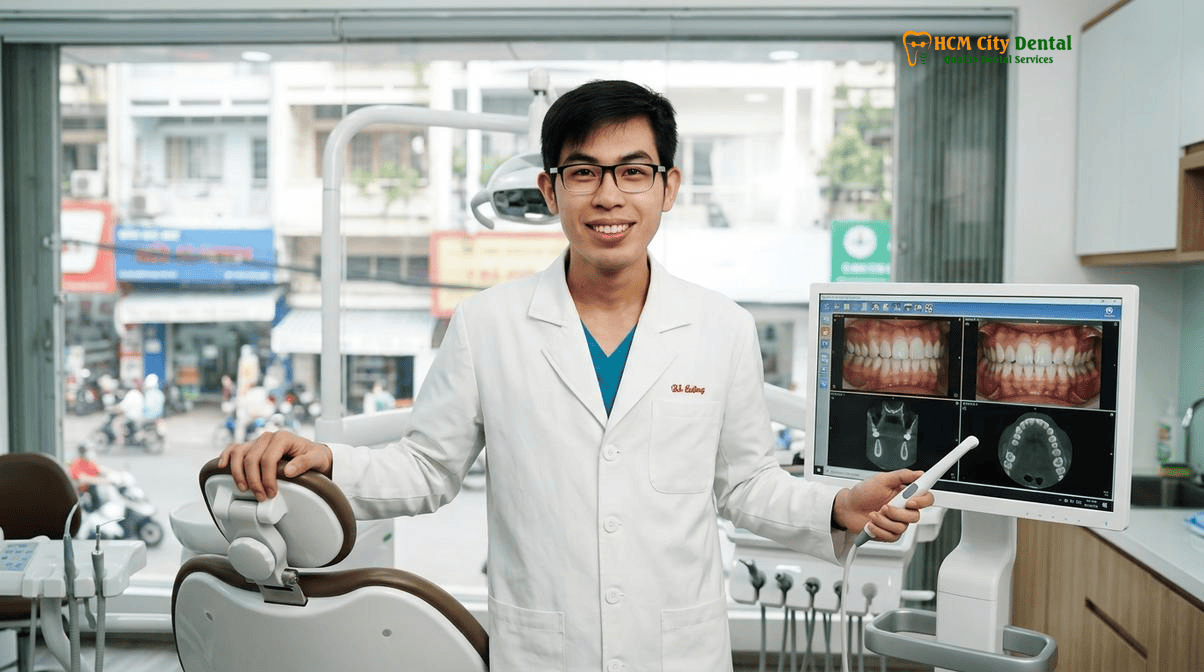
Below is the detailed 2026 fee schedule for single implant restorations at HCMC Dental Clinic. Please note that the Socket Shield micro-surgical preparation and any necessary gap grafting are highly specialized procedures; the specific clinical fees for these adjuncts will be transparently outlined during your comprehensive 3D CBCT consultation with Dr. Nguyen Van Cuong, DDS.
| Implant System / Procedure | 2026 Online Booking Price (-40%) | Walk-In / Standard Price |
|---|---|---|
| Single Korean Implant (Dentis / Neo Biotech / Dentium fixture + abutment) | $462 – $654 USD (12,000,000 – 17,000,000 VND) | $769 – $1,090 USD |
| Single European / Swiss Implant (Ritter / B&B / Straumann BLT-SLA / Straumann BLA-Active) | $962 – $1,346 USD (25,000,000 – 35,000,000 VND) | $1,603 – $2,244 USD |
| 100% Metal-Free Zirconia Ceramic Implant (Biocompatible / MELISA tested) | $1,154 USD | Consultation Required |
| Implant Zirconia HT Crown + Custom Abutment | $154 USD (4,000,000 VND) | Standard Pricing Applies |
| 3D CBCT Scan (Diagnostic & Surgical Planning) | $18 USD (480,000 VND) | Standard Pricing Applies |
For patients requiring more extensive rehabilitations, such as full-mouth restorations, HCMC Dental Clinic also offers highly competitive rates. For instance, you can review our comprehensive guide on the All-on-6 Dental Implants Vietnam Cost: Full-Arch Clinical Guide to understand the exceptional value provided for complex implantology. [5]
Frequently Asked Questions
Is leaving a thin buccal root shield inside the socket safe long-term during dental implant placement?
Yes, extensive clinical research and long-term histological studies have demonstrated that leaving a 1.5mm buccal root shield is highly safe and biologically sound. Because the root fragment remains attached to the periodontal ligament (PDL), it retains its natural blood supply and continues to nourish the surrounding bundle bone. This prevents the foreign body response and avoids root resorption, provided the shield is properly reduced, completely decoupled from the implant surface, and free of any active endodontic infection.
Who is NOT a candidate for the Socket Shield Technique?
Patients are not candidates for the Socket Shield Technique if the tooth in question has a vertical root fracture on the buccal aspect, severe active periodontal disease with significant bone loss, or an acute apical abscess that cannot be thoroughly debrided. Additionally, if the buccal root is internally resorbed or too thin to maintain a 1.5mm thickness after preparation, conventional extraction followed by Guided Bone Regeneration (GBR) is the required clinical protocol.
Can I leave HCMC Dental Clinic with a fixed temporary front tooth on the exact same day as Socket Shield implant surgery?
Yes. If the implant achieves an insertion torque of at least 35 Ncm and an Osstell ISQ Resonance Frequency Analysis score of 70 or higher, Dr. Nguyen Van Cuong, DDS can immediately load the implant. You will leave the clinic on the same day with a custom CAD/CAM provisional crown fixed to the implant, which supports the soft tissue architecture and provides immediate aesthetic restoration while the implant osseointegrates.
How does 3D CBCT surgical guide planning prevent the implant threads from displacing the buccal root shield?
3D CBCT imaging combined with digital surgical guides allows the specialist to plan the exact trajectory, depth, and angulation of the implant osteotomy. The protocol dictates that the implant must be placed slightly palatal to the root shield, leaving a small ‘jumping gap’ (often filled with bone graft material). The surgical guide ensures the drill never deviates buccally, preventing the implant threads from physically contacting, displacing, or fracturing the delicate root shield during insertion.
How much does an anterior immediate dental implant with Socket Shield cost in Ho Chi Minh City?
At HCMC Dental Clinic, the cost is highly transparent. A single European or Swiss implant (such as Straumann BLT-SLA) ranges from $962 to $1,346 USD online. A 100% metal-free Zirconia ceramic implant is $1,154 USD online. The final Implant Zirconia HT Crown with a Custom Abutment is an additional $154 USD online. The Socket Shield micro-surgical preparation and any necessary gap grafting are discussed during your consultation, ensuring you receive world-class aesthetic outcomes at a fraction of Western prices.
References
- Hürzeler, M. B., Zuhr, O., Schupbach, P., Rebele, S. F., Emmanouilidis, N., & Fickl, S. (2010). The socket-shield technique: a proof-of-principle report. Journal of Clinical Periodontology, 37(9), 855-862.
- Gluckman, H., Salama, M., & Du Toit, J. (2016). Partial Extraction Therapies (PET) Part 1: Maintaining Alveolar Ridge Contour at Pontic and Immediate Implant Sites. The International Journal of Periodontics & Restorative Dentistry, 36(5), 681-687.
- Bramanti, E., Norcia, A., Cicciù, M., Matacena, G., Cervino, G., Troiano, G., Zhurakivska, K., & Laino, L. (2018). Postextraction Dental Implant in the Aesthetic Zone, Socket Shield Technique Versus Conventional Protocol. Journal of Craniofacial Surgery, 29(4), 1037-1041.
- Abd-Elrahman, A., Shaheen, M., Askar, N., & Atef, M. (2020). Socket shield technique vs conventional immediate implant placement with immediate temporization. Randomized clinical trial. Clinical Implant Dentistry and Related Research, 22(5), 602-611.
- Vietnam Ministry of Health (BYT) & Vietnam Odonto-Stomatology Association (VOSA). National Clinical Guidelines on Specialist Dental Care, Biomaterial Traceability & EN 13060 Class B Infection Control. (2024).