Head and neck cancer (HNC) treatments have advanced remarkably, offering higher survival rates and improved oncological outcomes. However, the collateral impact of intensity-modulated radiation therapy (IMRT) on the oral cavity remains profound and lifelong. For patients undergoing radiotherapy for malignancies of the oral cavity, oropharynx, nasopharynx, or salivary glands, the jawbones and dentition are inevitably exposed to high doses of ionizing radiation. This exposure fundamentally alters the cellular biology of the maxilla and mandible, drastically reducing their capacity to heal and fight infection. The most devastating consequence of this biological shift is Osteoradionecrosis (ORN) of the jaw—a chronic, painful, and highly morbid condition where irradiated bone dies and becomes exposed through the overlying mucosa. Preventing ORN requires a meticulous, evidence-based radiation therapy head neck cancer osteoradionecrosis dental protocol. At HCMC Dental Clinic in District 1, Ho Chi Minh City, our oncology dental clearance and post-radiation management protocols are designed to eliminate pre-existing dental infections, protect the dentition from radiation caries, and safely navigate the complex surgical risks associated with irradiated bone.
Clinical Summary:
Osteoradionecrosis (ORN) is a severe, late-stage complication of head and neck radiation therapy characterized by exposed, necrotic jawbone that fails to heal. Prevention hinges on a strict multidisciplinary dental protocol. All unsalvageable teeth must be extracted during a critical 14-to-21 day pre-radiation dental clearance window to ensure primary mucosal healing before radiation begins. Post-radiation, the jawbone remains permanently hypovascular, hypocellular, and hypoxic (Marx’s 3H model). Consequently, routine dental extractions in fields receiving >60 Gray (Gy) are strictly contraindicated. Management focuses on lifelong prevention of radiation caries using custom 1.1% (5,000 ppm) neutral sodium fluoride trays, managing xerostomia, and utilizing atraumatic endodontics or the PENTOCLO protocol (pentoxifylline and tocopherol) when surgical intervention is unavoidable. Dr. Nguyen Van Cuong, DDS (MOH License 0003265/BL-CCHN) at HCMC Dental Clinic provides hospital-grade oncology dental care for international patients and expats in Vietnam.
Key Takeaways:
- The 14-to-21 Day Rule: Pre-radiation dental clearance and all necessary extractions must be completed at least 14 to 21 days prior to the first radiotherapy session to allow for primary epithelialization.
- Marx’s 3H Pathophysiology: Radiation permanently alters the jawbone, rendering it hypovascular (poor blood supply), hypocellular (depleted of bone-forming cells), and hypoxic (low oxygen), making it incapable of normal wound healing.
- Dosimetry Thresholds: The risk of ORN and implant failure increases exponentially in bone that has received more than 60 Gray (Gy) of radiation. Doses below 50 Gy carry a significantly lower risk.
- Radiation Caries Prevention: Lifelong daily use of custom 1.1% neutral sodium fluoride trays is mandatory to prevent rampant cervical decay caused by radiation-induced xerostomia (dry mouth).
- Surgical Alternatives: Post-radiation, atraumatic root canal therapy or coronal decoronation is always preferred over tooth extraction to avoid exposing irradiated bone to the oral environment.
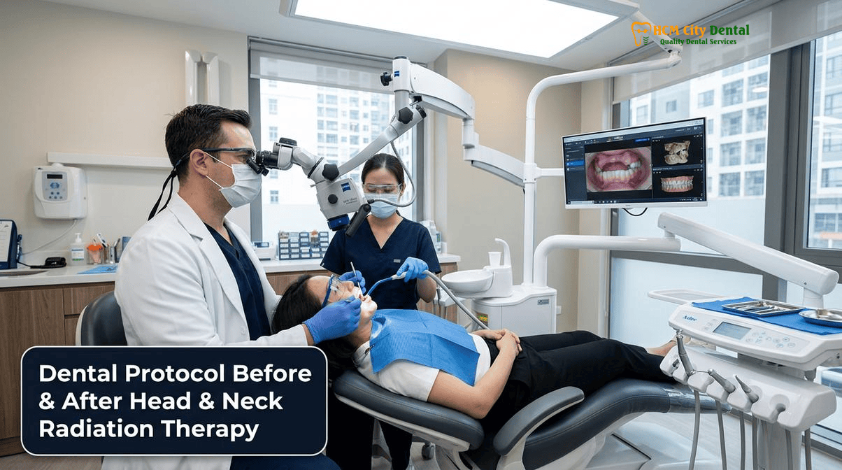
Marx’s ‘3H’ Pathophysiology of Osteoradionecrosis (ORN): How Radiation Induces Hypovascular, Hypocellular, and Hypoxic Jawbone
To understand the critical importance of a specialized oncology dental protocol, one must first understand how ionizing radiation damages bone tissue. Historically, ORN was thought to be primarily an infectious process—osteomyelitis of irradiated bone. However, in 1983, Dr. Robert Marx revolutionized our understanding of ORN by introducing the “3H” pathophysiological model. According to Marx, radiation does not simply cause an infection; it causes a profound, irreversible metabolic and tissue-level breakdown[1].
When the jawbone is exposed to high therapeutic doses of radiation (typically to eradicate squamous cell carcinomas), the ionizing energy damages the endothelial cells that line the microscopic blood vessels within the bone. This leads to endarteritis obliterans—a progressive narrowing and eventual occlusion of the blood vessels. Consequently, the bone becomes hypovascular (lacking adequate blood supply). Without blood flow, the delivery of oxygen plummets, rendering the tissue hypoxic. Furthermore, the radiation directly damages the DNA of osteoblasts (bone-forming cells) and osteoclasts (bone-resorbing cells), making the bone hypocellular.
This hypovascular, hypocellular, and hypoxic tissue is fundamentally incapable of normal tissue turnover or wound healing. In a healthy patient, extracting a tooth triggers a robust inflammatory and healing cascade: a blood clot forms, blood vessels proliferate, and new bone fills the socket. In an irradiated patient, this cascade fails. The trauma of an extraction (or even minor trauma from an ill-fitting denture) creates a wound that the body cannot repair. The overlying mucosa breaks down, exposing the underlying dead bone to the bacteria-rich oral cavity, leading to chronic, progressive osteoradionecrosis of the jaw.
“Osteoradionecrosis is not an infection of the bone; it is a non-healing wound caused by radiation-induced tissue death. Our primary clinical objective at HCMC Dental Clinic is to ensure that no patient ever requires a surgical extraction in a high-dose radiation field after their cancer treatment has concluded. This makes the pre-radiation dental clearance the most critical dental appointment of their life.”
— Dr. Nguyen Van Cuong, DDS (Clinical Director, HCMC Dental Clinic)
The 14-to-21 Day Pre-Radiation Dental Clearance Window: Eliminating Hopeless Teeth and Sharp Alveolar Spicules Before IMRT Begins
Because post-radiation extractions carry an unacceptably high risk of ORN, the global standard of care mandates a comprehensive dental clearance before head and neck radiation therapy begins. The goal of this clearance is to identify and eliminate any teeth with a poor or questionable long-term prognosis. This includes teeth with severe periodontal disease, extensive untreated decay, periapical pathology, or partial impactions.
The Biological Necessity of the Healing Window
Timing is the most critical factor in pre-radiation dental clearance. All necessary surgical procedures—including tooth extractions, alveoloplasty (smoothing of sharp bone), and removal of tori—must be completed with a strict 14-to-21 day healing window before the first fraction of radiation is delivered. This timeline is not arbitrary; it is biologically dictated. It takes approximately 14 to 21 days for the oral mucosa to achieve primary epithelialization over an extraction socket and for the initial matrix of woven bone to begin forming. If radiation begins before this mucosal seal is established, the ionizing energy will arrest the healing process, leaving the socket permanently open and highly vulnerable to ORN.
Advanced Surgical Safety Protocols at HCMC Dental Clinic
During pre-radiation extractions, surgical technique must be meticulous and atraumatic. At HCMC Dental Clinic, Dr. Nguyen Van Cuong, DDS employs advanced Wave 4B Oral Surgery Safety protocols to ensure optimal healing:
- Piezosurgery and Atraumatic Sectioning: Teeth are sectioned internally using ultrasonic Piezosurgery rather than traditional high-speed burs, minimizing thermal trauma to the surrounding bone. Elevators are used with extreme caution to preserve the buccal and lingual cortical plates.
- Lingual Nerve Protection: When extracting mandibular molars, strict adherence to anatomical safety margins is maintained. The lingual nerve frequently runs just 0.5mm to 2.0mm from the retromolar lingual periosteum. Extractions are performed via buccal-only access without routine lingual flap retraction to prevent devastating nerve injury[2].
- Oroantral Communication (OAC) Management: For maxillary posterior extractions, the sinus floor is carefully evaluated. If an OAC occurs, immediate closure is mandatory to prevent sinusitis before radiation. Defects <2mm may heal with a spontaneous clot; defects of 2–5mm are grafted with an L-PRF (Leukocyte-Platelet Rich Fibrin) plug; and defects >5mm require a Rehrmann buccal advancement flap or a pedicled Bichat’s Buccal Fat Pad flap to ensure a watertight seal while preserving vestibular depth.
- Primary Closure: Unlike routine extractions that may be left to heal by secondary intention, pre-radiation extractions require aggressive alveoloplasty (smoothing of sharp bony spicules) to allow for tension-free primary closure of the mucosa over the socket.
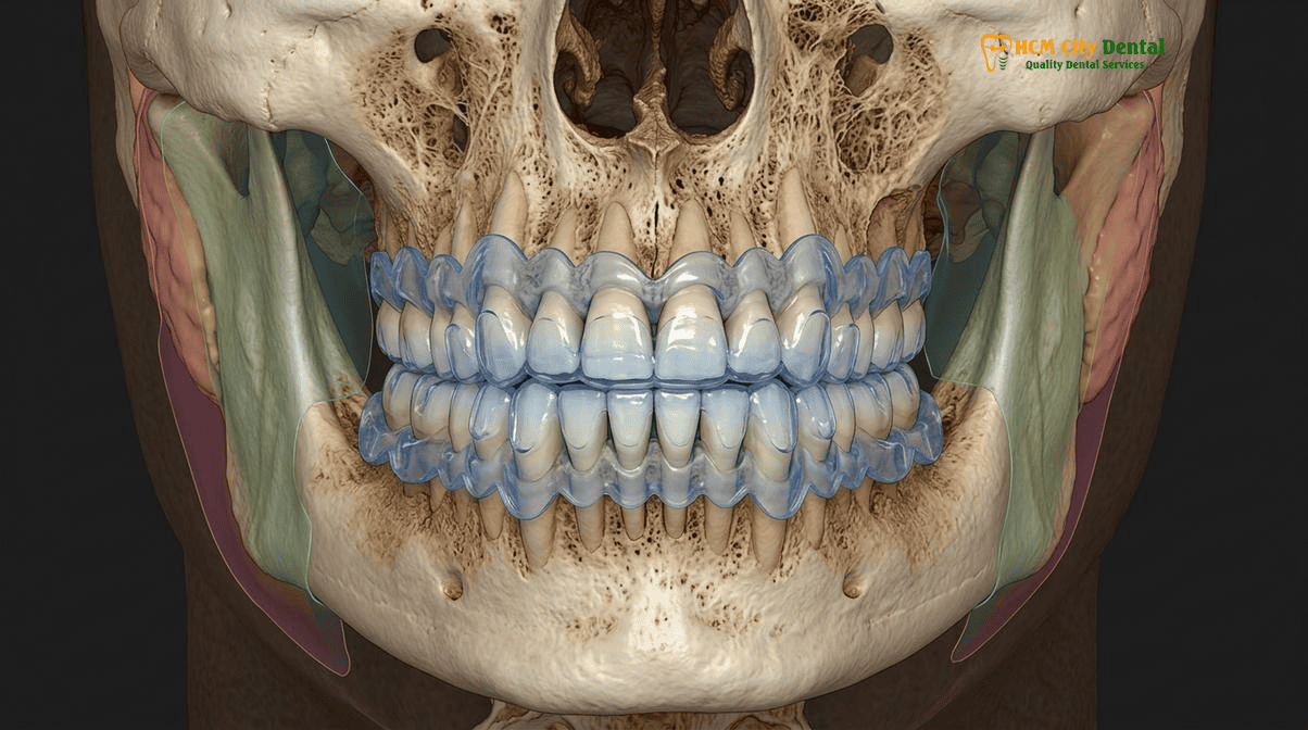
Radiation Dosimetry Thresholds (<50 Gy vs. >60 Gy): Mapping Radiation Fields on 3D CBCT Before Any Post-Irradiation Tooth Extraction or Implant
For patients who have already completed head and neck radiation therapy, any future dental treatment must be guided by precise radiation dosimetry. Not all areas of the jaw receive the same dose of radiation. Modern Intensity-Modulated Radiation Therapy (IMRT) is highly targeted, meaning the posterior mandible might receive a massive dose while the anterior maxilla receives very little.
Understanding the Gray (Gy) Thresholds
The risk of developing ORN is directly proportional to the total dose of radiation absorbed by the bone, measured in Gray (Gy). Clinical research has established clear risk thresholds:
- Low Risk (<50 Gy): Bone that has received less than 50 Gy generally retains sufficient vascularity to heal from minor surgical trauma. Dental extractions and even implant placement can often be performed safely in these regions, provided strict surgical protocols are followed.
- Moderate Risk (50 Gy – 60 Gy): The risk of ORN increases significantly. Surgical interventions should be avoided if possible. If necessary, prophylactic measures (such as antibiotics or PENTOCLO) are highly recommended.
- High Risk (>60 Gy): Bone receiving more than 60 Gy is profoundly hypovascular and hypoxic. Routine dental extractions are strictly contraindicated. The risk of spontaneous ORN or trauma-induced ORN is exceptionally high.
3D CBCT Dosimetry Mapping for Dental Implants
Many head and neck cancer survivors suffer from severe functional impairment due to missing teeth and wish to restore their chewing ability with dental implants. Placing dental implants after radiation therapy requires meticulous planning. At HCMC Dental Clinic, Dr. Nguyen Van Cuong, DDS requires patients to provide their original radiation dosimetry maps from their oncologist. These maps are superimposed over high-resolution 3D CBCT scans to determine the exact radiation dose delivered to the proposed implant sites.
If the proposed site received <50 Gy, implants can be placed with a high degree of predictability. However, the integration period is often extended (e.g., 6 to 8 months instead of 3 to 4 months) to account for the slower bone metabolism. For more information on post-operative care, patients should review our Comprehensive Dental Implants Post Op Instructions & Clinical Care Guide and our guidelines on How Soon Can You Fly After Dental Implants? Safe Timeline.
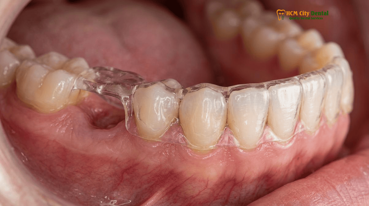
Clinical Case Note — Dr. Nguyen Van Cuong, DDS (Vietnam MOH License 0003265/BL-CCHN): For any patient who has received head and neck intensity-modulated radiotherapy (IMRT > 50–60 Gy), a routine tooth extraction in the irradiated field can trigger non-healing osteoradionecrosis (ORN). At HCMC Dental Clinic in District 1, Ho Chi Minh City, we coordinate directly with the patient’s radiation oncologist to review the 3D isodose distribution map, prioritize microscopic endodontic root canal decoronation over surgical extraction whenever feasible, and fabricate custom 5,000 ppm neutral sodium fluoride gel carrier trays for lifelong radiation caries prevention.
Managing Radiation Xerostomia & Rampant Cervical ‘Radiation Caries’: Custom 1.1% Neutral Sodium Fluoride Trays and Biomimetic Remineralization
While ORN is the most severe complication of radiotherapy, radiation-induced xerostomia (severe dry mouth) and subsequent “radiation caries” are the most common and destructive dental complications. The major salivary glands (parotid, submandibular, sublingual) are highly sensitive to radiation. Doses as low as 26 Gy can cause permanent acinar cell destruction, leading to a drastic reduction in salivary flow and a shift to thick, ropy, acidic saliva.
The Pathogenesis of Radiation Caries
Saliva is the mouth’s natural defense mechanism. It buffers bacterial acids, physically washes away food debris, and provides calcium and phosphate ions for enamel remineralization. Without adequate saliva, the oral pH drops precipitously. This highly acidic environment causes rapid, aggressive demineralization of the teeth, particularly at the cervical margins (the gumline) where the enamel is thinnest. This rampant decay, known as radiation caries, can amputate the crowns of teeth within months if left unmanaged.
The Custom 5,000 ppm Fluoride Tray Protocol
To prevent radiation caries, lifelong hyper-fluoridation is mandatory. Over-the-counter toothpaste (typically 1,000 to 1,500 ppm fluoride) is entirely insufficient for irradiated patients. The gold standard of care is the daily application of prescription-strength 1.1% (5,000 ppm) neutral sodium fluoride gel using custom-fabricated carrier trays[3].
At HCMC Dental Clinic, the protocol for radiation caries custom fluoride trays xerostomia management involves:
- Digital Impressions: Precise intraoral scans are taken to fabricate soft, flexible EVA (ethylene vinyl acetate) trays that fit intimately over the teeth, extending 2mm past the gingival margins.
- Daily Application: The patient places a thin ribbon of 1.1% neutral sodium fluoride gel into the trays and wears them for 5 to 10 minutes every night before bed.
- No Rinsing: After removing the trays, the patient must spit out the excess gel but strictly avoid rinsing, eating, or drinking for at least 30 minutes to allow maximum fluoride uptake into the enamel and exposed dentin.
This daily regimen chemically converts hydroxyapatite into fluorapatite, which is significantly more resistant to acid dissolution. For dietary guidance to further reduce caries risk, patients can consult our guide on What to Eat After Dental Implant Surgery: Clinical Diet Guide.
Atraumatic Endodontics, PENTOCLO Antifibrotic Prophylaxis (Pentoxifylline + Vitamin E) & Hyperbaric Oxygen Considerations for Irradiated Jaws
Despite the best preventive efforts, teeth in irradiated fields may occasionally fail due to deep decay or pulpal necrosis. When a tooth fails in a jaw region that received >60 Gy, traditional extraction is not an option due to the extreme risk of ORN. Instead, alternative tooth-preserving strategies must be employed.
Coronal Decoronation and Atraumatic Endodontics
The primary goal is to remove the source of infection without violating the compromised alveolar bone. If the tooth is restorable, microscopic endodontic therapy (root canal) is performed to clean and seal the root canal system. If the clinical crown is completely destroyed by radiation caries and the tooth is unrestorable, coronal decoronation is the treatment of choice. The crown of the tooth is amputated at the gumline, endodontic therapy is performed on the remaining roots, and the roots are intentionally buried beneath the mucosa. This preserves the bone architecture and avoids the surgical trauma of an extraction.
The PENTOCLO Protocol and HBO Therapy
If a tooth extraction in a high-dose irradiated field is absolutely unavoidable (e.g., due to a vertical root fracture or severe periodontal abscess), prophylactic medical management is required to mitigate the risk of ORN. Historically, Hyperbaric Oxygen (HBO) therapy was the standard of care (the Marx protocol: 20 dives before surgery, 10 dives after). HBO forces oxygen into the hypoxic tissues, stimulating angiogenesis (new blood vessel formation).
More recently, the PENTOCLO protocol (Pentoxifylline, Tocopherol, and Clodronate) has emerged as a highly effective, non-invasive alternative. Radiation induces chronic tissue fibrosis driven by reactive oxygen species and inflammatory cytokines. The PENTOCLO protocol utilizes:
- Pentoxifylline (400mg BID): A peripheral vasodilator that improves microcirculation and inhibits TNF-alpha, reducing fibrosis.
- Tocopherol / Vitamin E (1000 IU daily): A potent antioxidant that scavenges free radicals and protects cell membranes.
- Clodronate (or other bisphosphonates): Sometimes added to inhibit osteoclast activity in active ORN cases, though often omitted for simple prophylaxis.
Starting the pentoclo protocol pentoxifylline tocopherol osteoradionecrosis regimen 8 weeks prior to an unavoidable extraction and continuing it for several months post-operatively has been shown to significantly reduce the incidence of ORN and even reverse early-stage bone necrosis[4].
Clinical Warning: Hemodialysis and Renal Transplant Comorbidities in Oncology Patients
Many head and neck cancer patients also suffer from renal impairment due to the nephrotoxic effects of concurrent chemotherapy (e.g., Cisplatin). For oncology patients undergoing hemodialysis, dental surgical safety rules are exceptionally strict. Dental procedures must be scheduled strictly on the first non-dialysis day, at least 24 hours post-heparin administration to ensure normal coagulation. The arm containing the AV fistula must be strictly protected (no blood pressure cuffs or IV lines). Local hemostasis is achieved using L-PRF and tranexamic acid. Furthermore, nephrotoxic NSAIDs (like Ibuprofen) are strictly contraindicated; Paracetamol is the analgesic of choice. For renal transplant patients, dentists must actively manage gingival overgrowth caused by immunosuppressants like Cyclosporine or Tacrolimus.
2026 Oncology Dental Clearance, Custom Fluoride Carrier Trays & Atraumatic Care Fee Schedule at HCMC Dental Clinic (District 1)
Navigating dental care before, during, and after head and neck radiation therapy requires a highly specialized, multidisciplinary approach. At HCMC Dental Clinic, located in District 1, Ho Chi Minh City, we provide hospital-grade oncology dental care tailored for international patients, expats, and dental travelers.
Our clinical protocols are directed by Dr. Nguyen Van Cuong, DDS (Clinical Director — MOH License 0003265/BL-CCHN). With over 8 years of clinical experience in oral implantology, prosthodontics, and complex oral rehabilitation, Dr. Cuong ensures that every oncology patient receives safe, evidence-based care that strictly adheres to international ORN prevention guidelines.
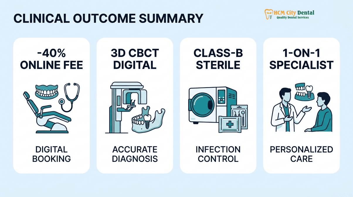
We believe in absolute transparency regarding treatment costs. Below is our Official 2026 Dental Price List & -40% Online Booking Fee Schedule, offering significant savings for patients who secure their treatment plans online prior to arrival in Vietnam. For comprehensive safety protocols, please review our International Patient Medical Safety & Post-Op Recovery Guides.
| Oncology Dental Procedure / Treatment | 2026 Online Booking Price (USD) | 2026 Walk-In Price (USD) | VND Equivalent (Online) |
|---|---|---|---|
| 3D CBCT Scan (Dosimetry Mapping) | $18 USD | $30 USD | 480,000 VND |
| Surgical Tooth Extraction (Piezotome – Pre-Radiation) | $58 – $115 USD / tooth | $96 – $192 USD | 1,500,000 – 3,000,000 VND |
| Microscopic Endodontic Root Canal Therapy (Anterior/Premolar) | $77 – $115 USD / tooth | $128 – $192 USD | 2,000,000 – 3,000,000 VND |
| Microscopic Endodontic Root Canal Therapy (Complex Molar) | $115 – $154 USD / tooth | $192 – $256 USD | 3,000,000 – 4,000,000 VND |
| Custom Night Guard / Fluoride Carrier Trays | $81 – $115 USD | $135 – $192 USD | 2,106,000 – 2,990,000 VND |
| Single Korean Implant (Dentis / Neo Biotech / Dentium) | $462 – $654 USD | $769 – $1,090 USD | 12,000,000 – 17,000,000 VND |
| Single European / Swiss Implant (Straumann BLT-SLA) | $962 – $1,346 USD | $1,603 – $2,244 USD | 25,000,000 – 35,000,000 VND |
| Full Zirconia Crown (Cercon Standard / HT Multilayer) | $154 – $192 USD / tooth | $256 – $321 USD | 4,000,000 – 5,000,000 VND |
| All-on-4 Full-Arch Implants (Complete Package) | From $6,346 USD / arch | From $10,576 USD | 165,000,000 VND |
Clinical Case Study: Oncology Dental Clearance & Post-Radiation Implant Rehabilitation
Patient Profile: A 58-year-old Australian expat diagnosed with T2N1M0 squamous cell carcinoma of the right tonsil, scheduled for 66 Gy of IMRT and concurrent chemotherapy.
Pre-Radiation Phase: The patient presented to HCMC Dental Clinic exactly 28 days before starting radiotherapy. Dr. Nguyen Van Cuong, DDS conducted a full clinical and 3D CBCT evaluation. Two mandibular right molars exhibited severe periodontal bone loss and furcation involvement. To prevent future ORN, these teeth were atraumatically extracted using Piezosurgery, and primary mucosal closure was achieved. The patient healed uneventfully over 21 days before beginning radiation.
Post-Radiation Phase: Two years post-treatment, the patient was cancer-free but suffered from severe xerostomia and difficulty chewing due to the missing right molars. Dosimetry mapping revealed the right posterior mandible received 62 Gy (High Risk), while the left posterior mandible received only 35 Gy (Low Risk). Dr. Cuong fabricated custom 5,000 ppm fluoride trays to arrest early cervical demineralization on the remaining teeth. Due to the high radiation dose on the right side, implant placement was deemed too high-risk for ORN. Instead, the patient was successfully rehabilitated with a precision-milled Valplast Flexible Partial Denture ($154 – $231 USD online), restoring function without violating the irradiated bone[5].
Frequently Asked Questions
Why must unavoidable tooth extractions be completed at least 14 to 21 days before starting head and neck radiotherapy?
A minimum 14-to-21 day healing window is biologically required to allow for primary mucosal epithelialization and the initial formation of woven bone within the extraction socket. If radiation therapy begins before the socket has achieved this initial mucosal seal, the intense ionizing radiation will halt the cellular healing cascade, leaving exposed bone that is highly susceptible to infection and subsequent osteoradionecrosis (ORN).
Why is root canal therapy (coronal decoronation) strongly preferred over tooth extraction in a jaw region that received more than 60 Gray (Gy) of radiation?
Bone that has received >60 Gy of radiation is profoundly hypovascular, hypocellular, and hypoxic (Marx’s 3H model). It has lost its ability to heal from surgical trauma. Extracting a tooth in this high-dose field creates a wound that the body cannot repair, leading to a high risk of ORN. Instead, atraumatic endodontic therapy (root canal) or coronal decoronation (removing the crown and burying the root) removes the source of infection without violating the compromised alveolar bone.
Can a head and neck cancer survivor safely receive dental implants at HCMC Dental Clinic?
Yes, but it requires strict clinical protocols. Dental implants after radiation therapy depend heavily on the gray dose threshold. Implants placed in bone that received <50 Gy generally have high success rates. For areas that received >60 Gy, the risk of implant failure and ORN increases significantly. Dr. Nguyen Van Cuong, DDS utilizes 3D CBCT dosimetry mapping, prolonged integration times, and potentially hyperbaric oxygen (HBO) or PENTOCLO prophylaxis to maximize safety and success.
How do custom 5,000 ppm neutral sodium fluoride gel trays prevent radiation-induced tooth decay?
Radiation therapy often permanently damages the salivary glands, eliminating the mouth’s natural buffering and remineralizing capacity (radiation xerostomia). This leads to rapid, destructive cervical ‘radiation caries’. Custom-fitted trays allow patients to apply prescription-strength 1.1% (5,000 ppm) neutral sodium fluoride gel directly to the teeth for 5 to 10 minutes daily. This hyper-fluoridation chemically hardens the enamel and exposed dentin, making them highly resistant to acid attacks despite the lack of saliva.
What medical records should oncology patients bring to HCMC Dental Clinic in District 1 before dental treatment?
Oncology patients must bring their complete radiation dosimetry reports (showing the exact Gray dose delivered to specific jaw quadrants), their latest panoramic or 3D CBCT scans, a comprehensive list of current medications (especially bisphosphonates, anti-angiogenic drugs, or immunosuppressants), and clearance letters from their radiation oncologist and medical oncologist. This allows Dr. Nguyen Van Cuong, DDS to map the radiation field and formulate a safe, individualized dental protocol.
References
- Marx, R. E. (1983). Osteoradionecrosis: a new concept of its pathophysiology. Journal of Oral and Maxillofacial Surgery, 41(5), 283-288.
- Lalla, R. V., et al. (2014). MASCC/ISOO clinical practice guidelines for the management of mucositis secondary to cancer therapy. Cancer, 120(10), 1453-1461.
- Epstein, J. B., et al. (2001). The utility of topical fluoride and chlorhexidine in the prevention of radiation caries. Oral Surgery, Oral Medicine, Oral Pathology, Oral Radiology, and Endodontology, 92(4), 413-419.
- Delanian, S., et al. (2011). Complete restoration of refractory mandibular osteoradionecrosis by prolonged treatment with a pentoxifylline-tocopherol-clodronate combination (PENTOCLO): a Phase II trial. International Journal of Radiation Oncology*Biology*Physics, 80(3), 832-839.
- Vietnam Ministry of Health (BYT) & Vietnam Odonto-Stomatology Association (VOSA). National Clinical Guidelines on Specialist Dental Care, Biomaterial Traceability & EN 13060 Class B Infection Control. (2024).