Is a crown after root canal necessary? Yes, for most posterior teeth (molars and premolars), a full-coverage crown is clinically mandatory to prevent catastrophic fractures. While some front teeth may only require a composite filling, back teeth endure immense chewing forces that demand robust structural protection for long-term survival.
Clinical Summary:
Following endodontic therapy, a tooth loses its internal blood supply and significant structural dentin, leading to increased brittleness. Clinical evidence strongly supports placing a permanent restoration—typically a full-coverage crown—shortly after treatment to seal the root canal system and distribute occlusal (biting) forces. Advanced digital workflows utilizing iTero and Medit i700 intraoral scanners allow for the precise fabrication of Zirconia HT or E.max crowns. For posterior teeth, the risk of a vertical root fracture without a crown is exceptionally high, often resulting in tooth loss and the subsequent need for a dental implant. Proper tooth preparation, including achieving an adequate ferrule and biologic width, ensures the longevity of the restoration.
Key Takeaways:
- Molars and premolars almost universally require a crown after a root canal to withstand heavy chewing forces.
- Endodontically treated teeth lose moisture and structural integrity, making them highly susceptible to fracturing.
- Delaying crown placement increases the risk of bacterial microleakage and irreversible tooth splitting.
- Modern CAD/CAM milling and digital intraoral scanners eliminate messy impressions and ensure a flawless marginal seal.
- Premium materials like Zirconia HT and E.max offer exceptional durability and lifelike aesthetics for restored teeth.
Why Root Canal Treated Teeth Become Brittle
Root canal therapy removes the living pulp, eliminating the internal blood supply and hydration, which gradually alters the dentin structure and increases its susceptibility to cracking under stress.
To understand why a restoration is required, one must first examine the biological changes that occur within the tooth during endodontic therapy. The core objective of a root canal is to remove infected or necrotic pulp tissue from the inner chambers of the tooth. This pulp contains the nerves, blood vessels, and connective tissues that nourish the tooth during its development and maintain its internal hydration throughout life.
Once this vital tissue is removed and the canals are shaped, disinfected, and sealed with gutta-percha, the tooth is technically considered “dead” or non-vital. Without a continuous blood supply, the moisture content within the dentinal tubules gradually decreases. This dehydration process alters the collagen cross-linking within the dentin matrix, leading to what is commonly referred to as a brittle teeth root canal scenario[1]. The tooth loses its natural elasticity and shock-absorbing capabilities, making it significantly less resilient to the repetitive mechanical stresses of mastication (chewing).
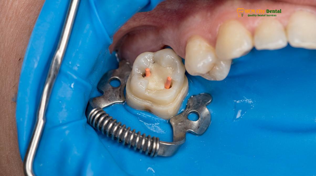
Furthermore, the loss of vitality means the tooth no longer has a defensive mechanism against deep decay, nor can it transmit temperature sensations. A patient will not feel hot or cold, but the periodontal ligament surrounding the root remains intact, meaning pressure and bite discrepancies can still be felt. Because the tooth cannot signal early warning signs of structural fatigue, a sudden fracture can occur without any prior symptoms if the tooth is left unprotected.
Structural Loss and Fracture Risks
The primary cause of post-endodontic tooth failure is the significant loss of structural dentin required to access and clean the infected root canals, severely weakening the tooth’s architecture.
While the loss of moisture contributes to brittleness, the most critical factor dictating the need for a crown is the sheer volume of missing tooth structure. By the time a tooth requires a root canal, it has typically already suffered extensive damage from deep dental caries (cavities), repeated large fillings, or physical trauma.
During the endodontic procedure, the dentist must create an access cavity through the biting surface of the tooth to locate and clean the root canals. This access preparation often removes the “roof” of the pulp chamber and compromises the marginal ridges—the reinforced outer edges of the tooth that hold its cusps together. Without these intact marginal ridges, the tooth is structurally compromised, much like a barrel missing its metal hoops.
“The survival of an endodontically treated tooth is directly proportional to the amount of remaining healthy coronal tooth structure. Full-coverage restorations are critical for distributing occlusal forces and preventing cuspal deflection.”
When a patient bites down, the forces act like a wedge inside the hollowed-out tooth, pushing the remaining walls outward. Over time, this wedging effect leads to a tooth fracture post endodontic treatment[2]. If the crack propagates above the gum line, the tooth might still be salvageable with a crown lengthening procedure and a new restoration. However, if the fracture extends vertically down the root (a vertical root fracture), the tooth is deemed non-restorable and must be extracted, often necessitating a dental implant or a dental bridge to replace the missing unit.
Clinical Warning: Delaying the placement of a permanent crown after a root canal significantly increases the risk of bacterial microleakage. Temporary fillings are porous and designed to last only a few weeks. If bacteria seep past the temporary seal, the root canal system can become reinfected, requiring costly retreatment or leading to tooth loss.
Anterior vs. Posterior Teeth Requirements
Molars and premolars absorb heavy vertical chewing forces, making full-coverage crowns mandatory, whereas incisors and canines experience lighter shearing forces and may sometimes be restored with bonded fillings.
The necessity of a crown is heavily dependent on the anatomical location of the tooth and its functional role in the dental arch. The human bite generates varying degrees of force, and different teeth are designed to handle specific types of stress.
Posterior Teeth (Molars and Premolars): These teeth are the workhorses of the mouth. They are located at the back of the jaw, closer to the temporomandibular joint (TMJ), where the bite force is the strongest—often exceeding 150 to 200 pounds of pressure per square inch. Molars have multiple cusps designed for grinding and crushing food. Because of this heavy vertical loading, a molar crown after root canal is almost universally required[3]. Without a crown, the weakened cusps will inevitably fracture under the immense pressure.
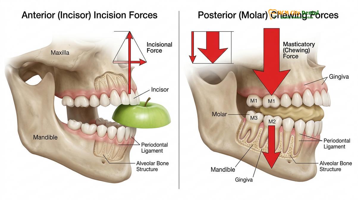
Anterior Teeth (Incisors and Canines): The front teeth are primarily used for tearing and shearing food, rather than heavy grinding. They endure lateral (side-to-side) forces rather than direct vertical compression. If an anterior tooth requires a root canal due to trauma but remains largely structurally intact (e.g., a small access hole on the back of the tooth with no large cavities), a crown may not be strictly necessary. In such cases, the access hole can be sealed with a high-quality composite resin.
| Tooth Type | Primary Function | Bite Force Exposure | Standard Post-Root Canal Restoration |
|---|---|---|---|
| Molars | Crushing and heavy grinding | Very High (Vertical compression) | Full-coverage Crown (Zirconia or PFM) |
| Premolars | Tearing and initial grinding | High (Mixed forces) | Full-coverage Crown or Onlay |
| Canines | Tearing and lateral guidance | Moderate (Shearing forces) | Crown or Composite Filling (case dependent) |
| Incisors | Cutting and aesthetics | Low to Moderate | Composite Filling or E.max Crown/Veneer |
Alternatives to Full Crowns: Direct Fillings and Onlays
When significant healthy tooth structure remains, conservative alternatives like direct composite fillings or custom-milled porcelain onlays can provide adequate functional restoration without requiring full crown preparation.
While full-coverage custom dental crowns and bridges are the gold standard for posterior teeth, modern adhesive dentistry offers conservative alternatives when clinically appropriate. The goal of any prosthodontic treatment is to preserve as much natural, healthy tooth structure as possible while ensuring long-term stability.
A simple dental filling after root canal is generally reserved for anterior teeth with minimal structural loss. High-strength composite resins are bonded directly to the dentin and enamel, sealing the access cavity. However, composites have limitations; they can shrink slightly upon curing and may wear down faster than ceramic materials under heavy occlusal loads.
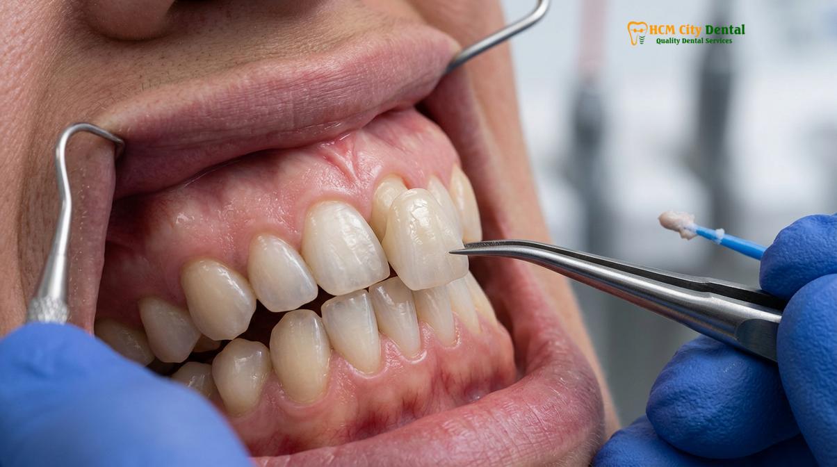
For premolars or molars that have thick, intact buccal (cheek-side) and lingual (tongue-side) walls, an Inlay or Onlay (Sứ Lab) may be recommended. An onlay is an indirect restoration, meaning it is fabricated in a dental laboratory or milled via CAD/CAM technology, just like a crown. However, instead of capping the entire tooth down to the gum line, an onlay only covers the chewing surface and the specific cusps that are weakened. This approach preserves the healthy lower portions of the tooth while still providing the necessary cuspal coverage to prevent the wedging effect that causes fractures.
Clinical Guidelines for Longevity
Maximizing the lifespan of a root-canal-treated tooth requires precise tooth preparation margins, high-quality core buildups, and the use of advanced CAD/CAM digital scanning for a flawless marginal seal.
The success of a crown after a root canal relies heavily on meticulous clinical execution. It is not merely about placing a “cap” on a tooth; it involves a complex interplay of biomechanics, material science, and periodontal health.
The Ferrule Effect and Core Buildup
Before a crown can be placed, the tooth must have a solid foundation. Because the root canal removes the internal core of the tooth, the dentist must perform a “core buildup” using specialized composite materials to replace the missing dentin. In cases of severe structural loss, a fiber or titanium post may be cemented into one of the treated root canals to anchor the core material securely.
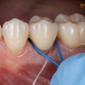
Crucially, the tooth preparation must incorporate a “ferrule.” A ferrule is a band of healthy, solid tooth structure (usually 1.5 to 2 millimeters in height) that extends above the gum line, which the crown will encircle. This ferrule acts like the metal band around a barrel, hugging the natural tooth and preventing the root from fracturing under lateral forces. If decay extends below the gum line, a minor surgical procedure called crown lengthening may be required to expose enough healthy tooth structure to establish a proper ferrule and respect the biologic width (the natural attachment of the gums to the tooth)[4].
Digital Scanning and Precision Fit
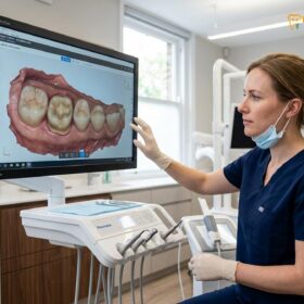
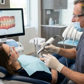
At advanced practices, traditional messy silicone impressions have been entirely replaced by state-of-the-art digital scanning technology. Utilizing the iTero and Medit i700 Intraoral Scanners, the dentist captures highly accurate 3D topographical maps of the prepared tooth, the adjacent teeth, and the opposing bite.
“Digital intraoral scanning significantly reduces marginal discrepancies in CAD/CAM restorations, ensuring a micro-precise fit that prevents bacterial leakage and secondary caries beneath the crown.”
This digital file is transmitted instantly to the laboratory, where the crown is designed using CAD (Computer-Aided Design) software. This ensures perfect shade matching, precise interproximal contacts (so food doesn’t get stuck between teeth), and accurate bite adjustment, minimizing the need for extensive grinding during the final cementation appointment.
Clinical Case Review: A dental tourism patient visited HCMC Dental Clinic in Ho Chi Minh City presenting with a fractured temporary filling on a lower molar, three months post-root canal. Dr. Nguyen Van Cuong evaluated the tooth using a 3D CBCT scan, confirming the root was intact. Utilizing the Medit i700 scanner, Dr. Cuong designed a custom Zirconia HT crown. The precise digital workflow allowed the patient to receive a perfectly fitted, permanent restoration within just 4 days, saving the tooth from imminent extraction.
Advanced Prosthodontic Workflows and Pricing
Modern restorative dentistry combines rapid digital fabrication with transparent pricing, allowing patients to receive premium, custom-milled crowns in just a few days without compromising clinical precision.
For patients seeking high-quality restorations, understanding material options and laboratory workflows is essential. A Direct Lab Partnership enables complete quality control and direct collaboration between the prosthodontist and the master ceramist. This synergy is particularly beneficial for complex cases, such as matching a single central incisor or fabricating a multi-unit dental bridge.
Material Certification and Selection
The choice of material dictates both the aesthetic outcome and the functional durability of the crown. Reputable clinics use only authentic, certified materials from industry-leading manufacturers in Germany and Liechtenstein:
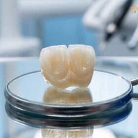
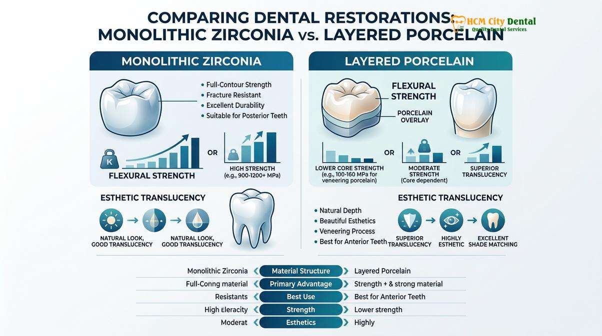
- Zirconia HT (Cercon HT, Germany): Known for its ultimate flexural strength and high translucency, making it the premier choice for molars and multi-unit bridges. It is milled from a single block of monolithic zirconia, making it virtually indestructible under normal chewing forces.
- E.max (Ivoclar, Liechtenstein): A lithium disilicate glass-ceramic that offers outstanding, lifelike aesthetics. Its light-transmitting properties mimic natural enamel perfectly, making it the gold standard for front teeth[5].
- PFM Crown (Porcelain-Fused-to-Metal): A traditional option that combines a strong metal substructure with a porcelain overlay. While durable, it lacks the natural translucency of all-ceramic options and may eventually show a dark line at the gum margins.
Express Turnaround and Transparent Pricing
Tailored specifically for dental tourists and expats, an Express 3-5 Day Turnaround allows complete smile makeovers or multiple crowns to be fabricated and fitted within a single week. Dr. Nguyen Van Cuong emphasizes that this rapid timeline does not compromise quality, as the CAD/CAM milling process operates with micron-level precision.
Understanding the financial investment is crucial. According to the latest clinic fee schedule at HCMC Dental Clinic in Ho Chi Minh City, patients can access premium restorations at highly competitive rates, especially when utilizing the 40% pre-arrival discount for WhatsApp bookings:
- PFM Crown: Walk-in: ~$140 (3.5M VND) | WhatsApp pre-booking discount: From $84 (2.1M VND) (-40%).
- Zirconia Crown (Cercon): Walk-in: ~$200 (5.0M VND) | WhatsApp pre-booking discount: From $120 (3.0M VND) (-40%).
- Zirconia HT Crown (Cercon HT): Walk-in: ~$260 (6.5M VND) | WhatsApp pre-booking discount: From $156 (3.9M VND) (-40%).
- E.max Crown (Ivoclar): Walk-in: ~$280 (7.0M VND) | WhatsApp pre-booking discount: From $168 (4.2M VND) (-40%).
- 3-Unit Bridge (Zirconia HT): Walk-in: ~$480 (12.0M VND) | WhatsApp pre-booking discount: From $288 (7.2M VND) (-40%).
- 4-Unit Bridge (Zirconia HT): Walk-in: ~$640 (16.0M VND) | WhatsApp pre-booking discount: From $384 (9.6M VND) (-40%).
- Inlay / Onlay (Sứ Lab): Walk-in: ~$180 (4.5M VND) | WhatsApp pre-booking discount: From $108 (2.7M VND) (-40%).
To further assist patients, comprehensive care includes a global warranty policy with remote follow-up support via WhatsApp (utilizing photo and video assessment protocols), as well as 0% interest monthly credit card installment plans for eligible treatments.
When to See a Doctor
If you have recently undergone root canal therapy and are currently wearing a temporary filling or temporary crown, it is imperative to monitor the tooth closely. You should schedule an immediate clinical evaluation if you experience any of the following:
- The temporary filling or crown becomes loose, dislodged, or falls out completely.
- You feel a sharp edge or notice a visible crack in the remaining tooth structure.
- You experience pain when biting down, which could indicate a developing fracture or an issue with the bite adjustment.
- There is swelling, redness, or a pimple-like bump (fistula) on the gums near the treated tooth, suggesting a potential reinfection.
- More than four weeks have passed since your root canal, and you have not yet scheduled the placement of your permanent restoration.
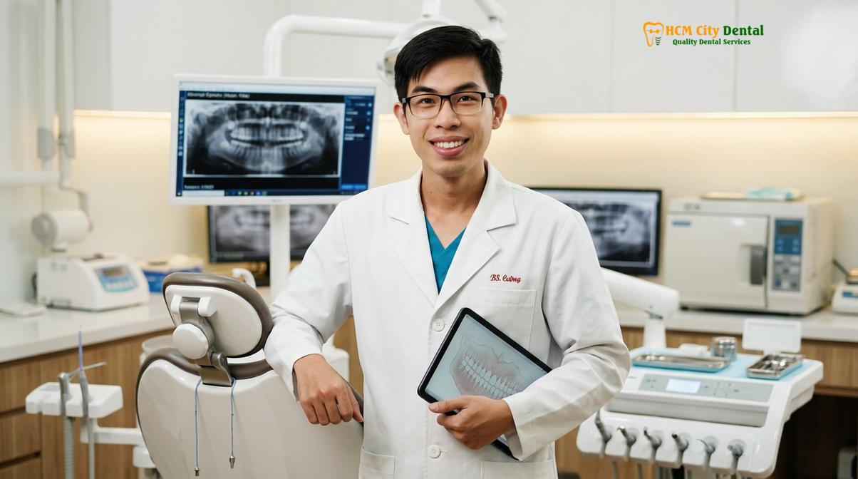
Protecting your investment in endodontic therapy requires timely prosthodontic care. If you are unsure whether your tooth requires a full crown, an onlay, or a simple filling, a comprehensive evaluation utilizing digital diagnostics will provide clarity. Based on clinical diagnostics, a personalized treatment plan will ensure your tooth remains functional and aesthetically pleasing for years to come.
Frequently Asked Questions
Why does a tooth need a crown after a root canal?
A crown is necessary to restore structural integrity, prevent catastrophic fractures, and seal the tooth against bacterial reinfection. Because endodontic therapy removes the internal blood supply and significant dentin, the remaining tooth structure becomes highly susceptible to breaking under normal chewing forces without full-coverage protection.
Can I just get a filling instead of a crown after a root canal?
Yes, but typically only for front teeth (incisors and canines) that have minimal structural damage and endure lower chewing forces. For molars and premolars, a simple dental filling after root canal is rarely sufficient, as these teeth bear heavy vertical loads that easily fracture unprotected enamel.
How long can a root canal tooth survive without a crown?
An unprotected root-canal-treated molar may fracture within a few weeks to a few months under normal chewing stress. While front teeth might survive longer, clinical guidelines strongly recommend placing a permanent crown or onlay within two to four weeks to ensure long-term survival and prevent irreversible splitting.
Does getting a crown on a root-canal-treated tooth hurt?
No, the crown preparation and placement process is generally painless because the tooth’s nerve has already been removed during the root canal. You may experience mild, temporary gum sensitivity around the margins after the procedure, which typically resolves within a few days of cementation.
What is the best material for a crown after a root canal?
The ideal material depends on the tooth’s location; Zirconia HT is excellent for molars due to its immense fracture resistance, while E.max (lithium disilicate) provides superior lifelike aesthetics for front teeth. Your prosthodontist will evaluate your bite forces and aesthetic needs to recommend the optimal ceramic.
References
- Journal of Endodontics. Biomechanical principles of post-endodontic restorations. (2021).
- International Journal of Prosthodontics. Survival rates of crowned versus uncrowned endodontically treated teeth. (2020).
- Journal of Prosthetic Dentistry. The ferrule effect and structural integrity of root-filled teeth. (2019).
- American Dental Association. Clinical guidelines for indirect restorations and dental crowns. (2022).
- Journal of Esthetic and Restorative Dentistry. Translucency and flexural strength of CAD/CAM ceramics. (2023).