Dentists pull teeth out using specialized instruments to gently loosen the tooth from its periodontal ligament before carefully lifting it from the socket. Depending on the tooth’s condition, the procedure is classified as either a simple manual extraction or a surgical extraction requiring tissue incision and bone modification. Understanding the biomechanics of this process helps demystify the procedure and highlights the precision required for safe removal [1].
Clinical Summary:
Tooth extraction, clinically known as exodontia, is a highly controlled dental procedure performed when a tooth cannot be salvaged through restorative means. The process begins with comprehensive radiographic imaging to assess root anatomy and proximity to vital structures like nerves and sinuses. Dentists utilize profound local anesthesia to eliminate pain, though patients will still perceive pressure. The mechanical process involves using dental elevators to expand the alveolar bone socket and sever the periodontal ligament, followed by extraction forceps to deliver the tooth. While simple extractions involve visible teeth that can be grasped easily, surgical extractions are required for impacted, broken, or multi-rooted teeth, often necessitating gingival incisions and tooth sectioning. Post-operative healing relies heavily on the formation of a stable blood clot, making strict adherence to aftercare instructions critical for preventing complications such as alveolar osteitis (dry socket). Modern dentistry prioritizes atraumatic extraction techniques to preserve surrounding bone, facilitating future replacement with dental implants or bridges.
Key Takeaways:
- Extractions are categorized into simple (non-surgical) and surgical procedures based on tooth visibility and root complexity.
- Profound local anesthesia ensures the procedure is virtually painless, though mechanical pressure is a normal sensation.
- Specialized instruments called elevators and forceps are used to expand the bone and sever the ligament holding the tooth.
- Surgical extractions often require sectioning the tooth into smaller pieces to protect surrounding bone and nerves.
- Proper blood clot formation is the most critical factor in post-extraction healing and preventing dry socket.
Clinical Reasons for Tooth Extraction
Teeth are extracted when they are damaged beyond repair by severe decay or trauma, or to resolve overcrowding and impacted wisdom teeth.
While the primary goal of modern dentistry is the preservation of the natural dentition, there are specific clinical scenarios where removing a tooth is the most biologically sound and necessary treatment. The decision to extract a tooth is never taken lightly and is always preceded by a thorough diagnostic evaluation. One of the most common reasons for extraction is advanced dental caries (tooth decay) that has destroyed a significant portion of the tooth structure, rendering it impossible to restore with a filling or a dental crown. When decay penetrates deep into the pulp chamber and causes irreversible pulpitis or apical periodontitis, and root canal therapy is either not viable or has previously failed, extraction becomes necessary to prevent the spread of infection to the surrounding alveolar bone and systemic bloodstream.
Periodontal disease is another leading cause of tooth loss in adults. Chronic periodontitis involves the progressive destruction of the supporting structures of the teeth, including the gingiva, periodontal ligament, and alveolar bone. As the bone recedes, teeth become increasingly mobile. When the attachment loss is severe and the tooth exhibits class III mobility (severe looseness), extraction is often the only definitive way to eliminate the periodontal pocket and halt the localized inflammatory process.
Orthodontic considerations also frequently necessitate tooth extraction. In cases of severe dental crowding or significant skeletal discrepancies, there simply may not be enough space in the dental arch to align all the teeth properly. In such scenarios, an orthodontist may recommend the strategic removal of specific teeth—most commonly the first or second premolars. This creates the necessary space to retract anterior teeth, correct overjets, and achieve a stable, functional occlusion. Patients undergoing treatments detailed in our Buck Teeth Braces: Clinical Overjet Correction Guide often require careful extraction planning to ensure optimal facial aesthetics and bite alignment.
Pre-Extraction Clinical Assessment and Preparation
Before an extraction, dentists conduct a thorough clinical evaluation using 3D imaging and review medical histories to determine the safest anesthetic and surgical approach.
The foundation of a safe and successful tooth extraction lies in meticulous pre-operative planning. The clinical assessment begins with a comprehensive review of the patient’s medical history. The dentist must identify any systemic conditions that could complicate the surgery or the healing process. Conditions such as uncontrolled diabetes, hypertension, cardiovascular disease, or bleeding disorders require specific management protocols. Furthermore, a detailed pharmacological history is crucial. Patients taking anticoagulants (blood thinners) may require medical consultation to adjust their dosage.
Dr. Nguyen Van Cuong, a leading specialist in oral surgery, emphasizes that comprehensive pre-operative planning is the cornerstone of safe exodontia. Under Dr. Cuong’s clinical direction, the team utilizes advanced 3D CBCT imaging to map out nerve pathways before any procedure. Patients seeking comprehensive care can explore our General Dentistry services to understand how extractions fit into a broader oral health strategy.
“A successful extraction is heavily reliant on diagnosis and planning. By utilizing advanced 3D imaging, we can anticipate anatomical challenges, select the appropriate surgical instruments, and ensure the procedure is performed as atraumatically as possible, preserving vital bone for future restoration.”
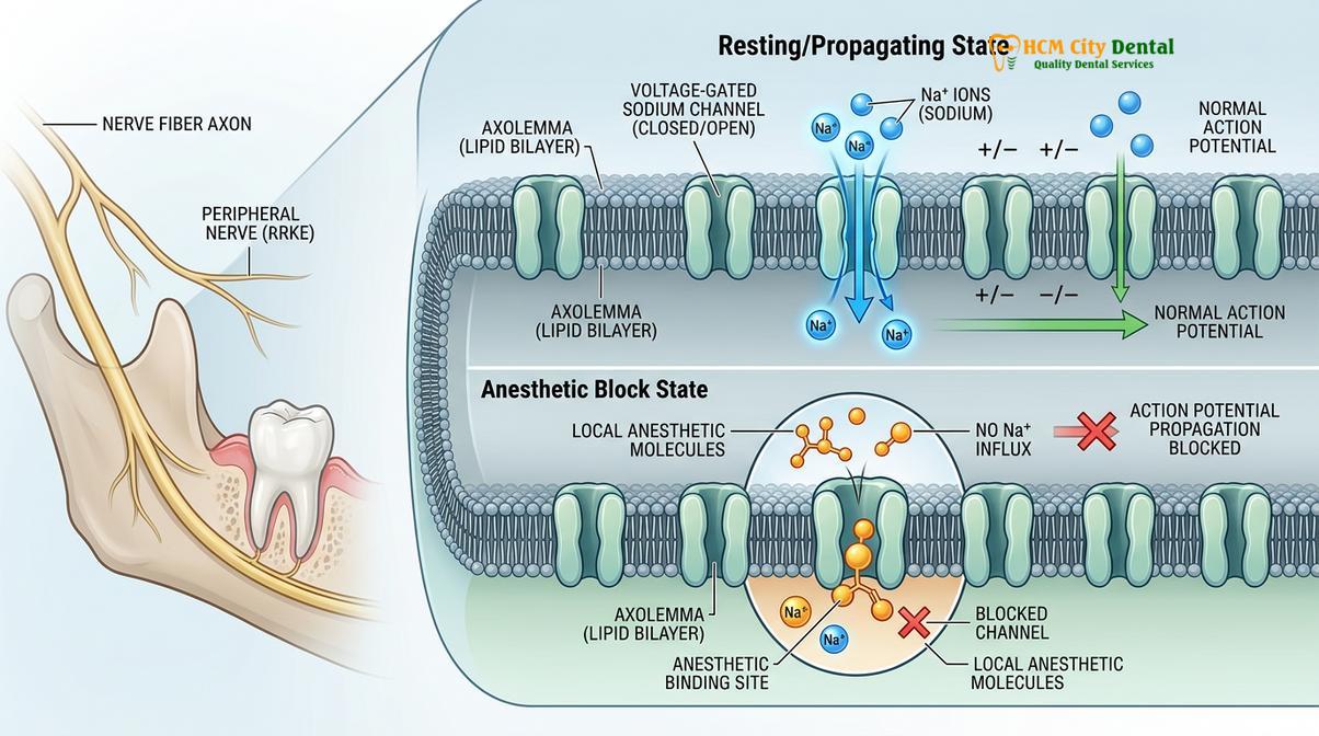
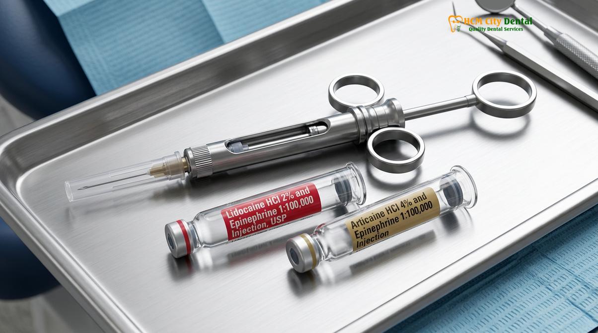
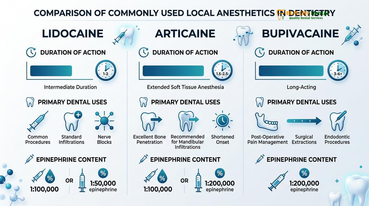
Radiographic imaging is an indispensable diagnostic tool in exodontia. Cone Beam Computed Tomography (CBCT) provides highly detailed, cross-sectional images of the maxillofacial region. This allows the dentist to precisely visualize the relationship between the tooth roots and critical structures such as the inferior alveolar nerve in the mandible or the maxillary sinus floor in the upper jaw. Once the assessment is complete, the appropriate anesthesia protocol is selected. Local anesthetics are typically combined with a vasoconstrictor like epinephrine to prolong the duration of the anesthetic effect and significantly reduce bleeding at the surgical site [3].
The Step-by-Step Procedure: How Dentists Pull Teeth Out
The extraction process involves numbing the area, severing the gingival attachment, expanding the bony socket using elevators, and delivering the tooth with extraction forceps.
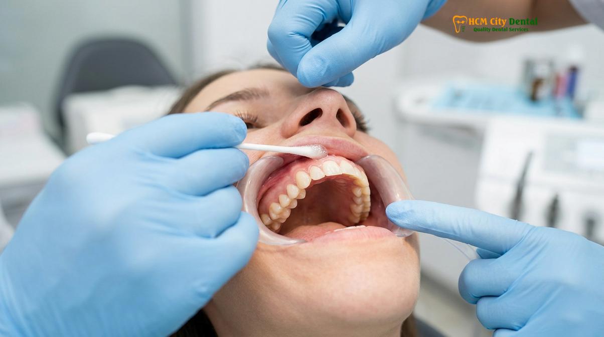
The actual mechanical process of extracting a tooth is a precise application of physics and biomechanics, rather than simply an exercise in brute force. The procedure begins only after the dentist has confirmed that the local anesthesia has achieved profound numbness in the target tooth and surrounding tissues. The patient will be informed that while they will not feel sharp pain, they will experience sensations of significant pressure and pushing.
The first step in the extraction sequence is the severing of the soft tissue attachment. The dentist uses a sharp instrument, typically a periosteal elevator or a scalpel, to carefully detach the gingival tissue (gums) from the neck of the tooth. This step, known as syndesmotomy, is crucial because it prevents the gums from tearing when the tooth is eventually removed. Once the soft tissue is freed, the dentist begins the process of luxation—loosening the tooth within its bony socket.
Luxation is achieved using instruments called dental elevators. An elevator acts as a wedge and a lever. The dentist carefully inserts the blade of the elevator into the periodontal ligament space. By applying controlled, rotational force, the elevator stretches and tears the periodontal ligament fibers that anchor the tooth. Simultaneously, this wedging action gently expands the surrounding alveolar bone.
| Extraction Type | Clinical Indication | Procedure Characteristics |
|---|---|---|
| Simple Extraction | Visible, fully erupted teeth with straight roots | Uses elevators and forceps; no incisions required |
| Surgical Extraction | Impacted, broken, or severely decayed teeth | Requires gingival incision, bone removal, or tooth sectioning |
Once the tooth is sufficiently mobile, the dentist utilizes extraction forceps to deliver the tooth from the mouth. Forceps are designed with specific beak shapes to adapt to the unique anatomical contours of different teeth. The forceps are seated as far down the root as possible to establish a secure mechanical advantage. The dentist then applies steady, controlled forces to further expand the bone and finally lift the tooth out of the socket.
Advanced Surgical Extractions and Wisdom Teeth
Surgical extractions require making a gingival incision, removing obstructing bone, and often sectioning the tooth into smaller pieces for safe removal without damaging surrounding nerves.
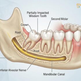
While simple extractions involve teeth that are fully visible and easily accessible, many clinical situations demand a surgical approach. A surgical extraction is defined as a procedure that requires the reflection of a mucoperiosteal flap (cutting and lifting the gums) and the removal of bone to access the tooth. This approach is most commonly associated with the removal of impacted wisdom teeth, but it is also necessary for teeth that have fractured at the gum line or teeth with severely curved roots [2].
Clinical Case Study: Complex Surgical Extraction
A 24-year-old patient presented to HCMC Dental Clinic in Ho Chi Minh City with severe pain in the lower right jaw. Clinical and CBCT radiographic examination revealed a horizontally impacted mandibular third molar (wisdom tooth) whose roots were in close proximity to the inferior alveolar nerve. The clinical team performed a surgical extraction utilizing an ultrasonic bone surgery device to safely remove the overlying bone without damaging soft tissues. The tooth was sectioned into three distinct pieces and removed atraumatically. The patient experienced minimal post-operative swelling and no nerve paresthesia, demonstrating the efficacy of advanced 3D planning.
One of the most critical techniques in surgical exodontia is tooth sectioning, clinically known as odontotomy. Instead of attempting to force a large, multi-rooted, or awkwardly positioned tooth out of a small opening, the dentist uses a surgical bur to cut the tooth into smaller, manageable fragments. For more information on the logistics of this procedure, patients often review our guide on Wisdom Teeth Removal: Hospital or Dentist? to understand the appropriate clinical setting for their specific case.
After the tooth fragments are completely removed, the surgical site is thoroughly debrided. The dentist uses a curette to scrape the walls of the socket, removing any infected granulation tissue, cyst remnants, or bone debris. The area is flushed with sterile saline. Finally, the mucoperiosteal flap is repositioned, and the site is closed using dental sutures. Understanding the financial aspects of these complex procedures is also important, which is why we provide resources like the Dentist Wisdom Teeth Removal Cost guide.
Post-Extraction Healing and Aftercare Workflows
Successful healing relies on the formation of a stable blood clot within the socket, requiring patients to avoid suction, smoking, and vigorous rinsing.
The biological process of healing following a tooth extraction is a complex, highly orchestrated cascade of cellular events. The immediate and most critical phase is hemostasis—the cessation of bleeding. Immediately after the tooth is removed, blood fills the empty alveolar socket. Within minutes, platelets aggregate and a fibrin mesh forms, creating a stable blood clot. This blood clot is the biological scaffold upon which all subsequent healing depends.
Important Clinical Note: To prevent alveolar osteitis (dry socket), patients must strictly avoid smoking, drinking through a straw, and vigorous rinsing for at least 72 hours post-extraction. Dislodging the blood clot exposes underlying bone and nerves, leading to severe pain and delayed healing.
If this blood clot fails to form, or if it is prematurely dislodged, the patient may develop alveolar osteitis, commonly known as a dry socket. This condition exposes the underlying alveolar bone and nerve endings to the oral environment, resulting in severe, radiating pain and delayed healing. Preventing dry socket requires strict adherence to post-operative instructions, including avoiding smoking, spitting, and using straws, which can create negative pressure in the mouth [4].
Over the following weeks, the blood clot is gradually replaced by granulation tissue, which is rich in blood vessels and fibroblasts. The surface of the socket undergoes epithelialization as gum tissue grows over the wound. Beneath the surface, osteoblasts begin the slow process of bone remodeling, filling the socket with new woven bone. In cases where a dental implant is planned, the dentist may perform a ridge preservation procedure at the time of extraction, placing a bone graft material into the socket to minimize natural bone resorption and maintain the structural integrity of the jaw [5].
“The first 48 hours post-extraction are critical. Patients must prioritize rest, maintain a soft diet, and avoid any actions that could disturb the fragile blood clot. Proper aftercare is just as important as the surgical technique itself in ensuring a smooth recovery.”
When to Contact Your Dentist Post-Extraction
While mild swelling and discomfort are normal, severe pain, prolonged bleeding, or signs of infection require immediate clinical evaluation.
Following an extraction, it is normal to experience some degree of localized swelling, mild bruising, and discomfort as the anesthesia wears off. These symptoms typically peak within 48 to 72 hours and can usually be managed effectively with over-the-counter analgesics and cold compresses applied to the outside of the face. However, patients must remain vigilant for signs of potential complications that warrant immediate professional attention.
You should contact your dental provider if you experience bleeding that cannot be controlled by applying firm pressure with a sterile gauze pad for 45 minutes. Additionally, if you develop a fever, notice pus or a foul-tasting discharge from the extraction site, or experience swelling that worsens after the third day or begins to restrict your breathing or swallowing, you must seek immediate care. These are clinical signs of a potential secondary infection. Dental clinics adhere strictly to the Vietnam Ministry of Health guidelines regarding infection control and surgical protocols to minimize these risks, but patient monitoring at home remains a vital component of the recovery phase [6].

If you are experiencing severe dental pain, have an impacted wisdom tooth, or require a professional evaluation for a potential tooth extraction, contact HCMC Dental Clinic in Ho Chi Minh City today. Our clinical team is ready to provide safe, painless care utilizing the latest in dental technology. Learn more about our comprehensive General Dentistry services and schedule your consultation.
References
- Journal of Oral and Maxillofacial Surgery. Biomechanics of tooth extraction and socket healing protocols. (2021).
- International Journal of Oral and Maxillofacial Surgery. Management of impacted third molars and surgical exodontia. (2020).
- British Dental Journal. Local anesthesia techniques and pharmacology in exodontia. (2019).
- Journal of the American Dental Association. Prevention, diagnosis, and treatment of alveolar osteitis. (2022).
- Clinical Oral Investigations. Ridge preservation and biological tissue response following tooth extraction. (2018).
- Vietnam Ministry of Health. National Guidelines on Infection Control and Surgical Protocols in Odonto-Stomatology. (2020).