Gneuromuscular dentistry is an advanced clinical discipline that evaluates the complex relationship between teeth, temporomandibular joints, and masticatory muscles to establish an optimal bite. By prioritizing muscle relaxation and joint alignment, this methodology provides comprehensive, evidence-based relief for chronic jaw pain and severe occlusal dysfunction, moving beyond traditional static bite analysis. For patients suffering from unresolved craniofacial pain, understanding the principles of neuromuscular dentistry can be the first step toward achieving lasting comfort and functional harmony[1].
Clinical Summary:
Gneuromuscular dentistry represents a paradigm shift in the treatment of temporomandibular joint disorders (TMD) and complex malocclusions. Unlike traditional approaches that focus primarily on static tooth contacts, this advanced methodology utilizes objective electronic instrumentation—including ultra-low frequency TENS, surface electromyography (sEMG), and computerized mandibular scanning (CMS)—to measure and optimize the dynamic function of the entire stomatognathic system. By erasing pathological muscle memory and establishing a true physiological rest position, clinicians can design custom orthotics and restorative treatments that harmonize the muscles, joints, and teeth. This comprehensive approach aims to alleviate chronic facial pain, tension headaches, and joint degradation while providing a stable foundation for long-term oral rehabilitation.
Key Takeaways:
- Neuromuscular dentistry evaluates the dynamic interplay between muscles, nerves, and the temporomandibular joints, rather than just static tooth alignment.
- Ultra-low frequency TENS is utilized to involuntarily relax hyperactive jaw muscles and erase pathological bite memory.
- Surface electromyography (sEMG) provides objective, quantifiable data on muscle resting states and functional activity.
- Treatment typically begins with a reversible Phase 1 orthotic appliance to stabilize the jaw and relieve chronic symptoms.
- Long-term stabilization (Phase 2) may involve orthodontics, prosthodontics, or targeted occlusal adjustments once the optimal bite is confirmed.
Neuromuscular vs. Traditional Dentistry: The Key Differences
Traditional dentistry primarily focuses on static tooth contacts and structural alignment, whereas neuromuscular dentistry evaluates the dynamic interplay of muscles, nerves, and joints to achieve a harmonious, symptom-free bite.
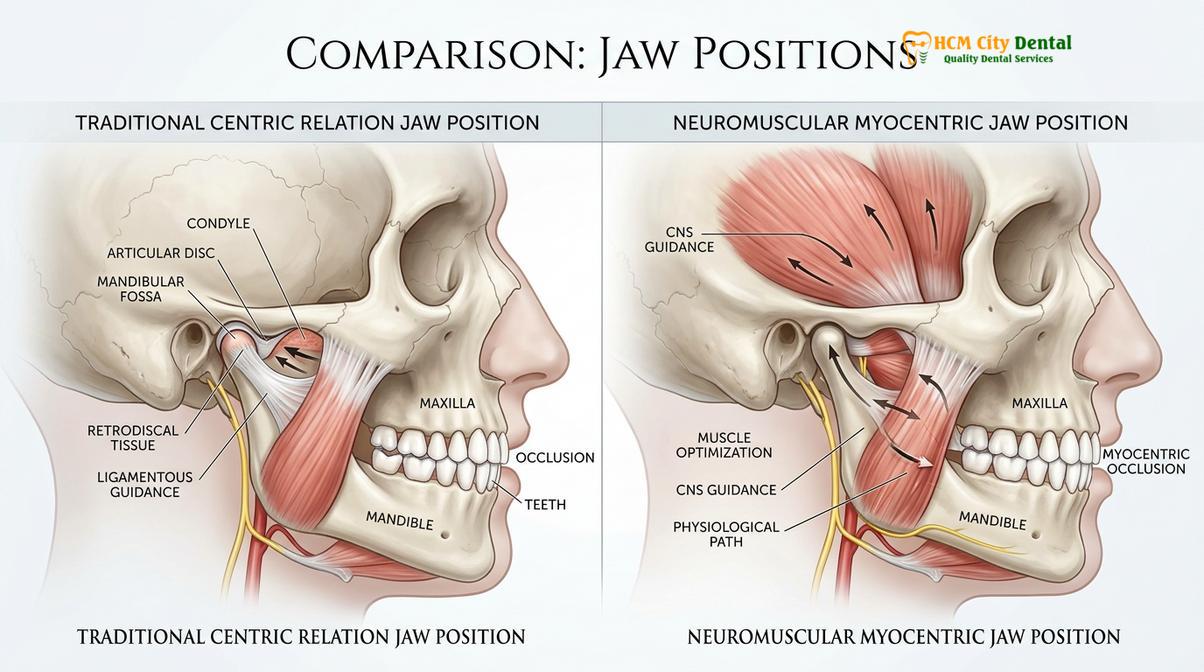
The evolution of dental science has seen a profound shift in how clinicians understand and treat the human bite (occlusion). For decades, traditional dentistry—often referred to as gnathology—relied heavily on the mechanical relationship of the teeth and the manipulation of the jaw by the dentist into a position known as “centric relation.” While this approach is highly effective for routine restorative work, it often falls short when addressing complex cases of chronic facial pain, severe attrition, and temporomandibular joint disorders (TMD)[2].
Traditional gnathological principles dictate that the temporomandibular joints should be seated in their most superior and posterior position within the glenoid fossa. However, this static, bone-to-bone philosophy frequently overlooks the most critical component of the stomatognathic system: the musculature. If the muscles are in a state of chronic spasm or hypertonicity, forcing the jaw into a predetermined mechanical position can exacerbate pain and lead to treatment failure. The neuromuscular approach recognizes that the teeth and joints are often victims of hyperactive or imbalanced musculature.
In contrast, gneuromuscular dentistry operates on the principle that the muscles dictate the position of the jaw. When a patient has a malocclusion (a bad bite), the brain constantly sends signals to the masticatory muscles to accommodate the misalignment, ensuring the teeth can still chew food. This constant accommodation leads to a phenomenon known as a “muscle engram”—a programmed, habitual trajectory that avoids premature tooth contacts but forces the muscles to work overtime, leading to lactic acid buildup, fatigue, and chronic pain.
Anatomy of the Stomatognathic System and Muscle Engrams
The stomatognathic system is a complex network of bones, joints, and muscles that must work in perfect synchrony; when misalignment occurs, the brain creates compensatory muscle engrams that lead to chronic fatigue.
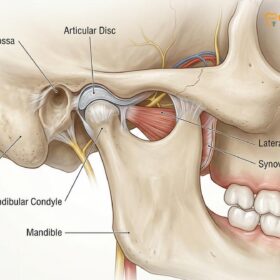
To fully appreciate the value of neuromuscular dentistry, one must understand the intricate anatomy of the stomatognathic system. The temporomandibular joint (TMJ) is one of the most complex joints in the human body. Unlike a simple hinge joint, the TMJ must both hinge and glide to allow for the complex movements required for speaking, chewing, and swallowing. Between the mandibular condyle (the ball of the joint) and the glenoid fossa (the socket in the skull) lies a fibrous articular disc that acts as a shock absorber.
The movement of this joint is controlled by a powerful group of masticatory muscles, primarily the masseter, the temporalis, and the medial and lateral pterygoids. The masseter and medial pterygoid are responsible for the heavy lifting of closing the jaw, while the temporalis helps position the mandible. The lateral pterygoid is crucial for opening the jaw and moving it side to side. In a healthy system, these muscles work in perfect, effortless coordination.
However, the central nervous system is highly protective of the teeth. The periodontal ligaments surrounding each tooth are embedded with sensitive mechanoreceptors. If a single tooth is slightly out of alignment, these receptors send an immediate warning signal to the brain upon contact. To prevent damage to the tooth, the brain instantly reprograms the masticatory muscles to alter the jaw’s trajectory, avoiding the premature contact. This reprogrammed pathway is the muscle engram, which forces muscles to operate outside their optimal physiological length.
Restoring the Physiological Rest Position of the Mandible
The physiological rest position is the mandibular posture where masticatory muscles exhibit their lowest electrical activity, serving as the foundational starting point for neuromuscular bite reconstruction.
Understanding the concept of the physiological rest position is paramount to grasping the efficacy of neuromuscular dentistry. In a healthy stomatognathic system, the teeth should only make contact during swallowing and active mastication—amounting to roughly 15 to 20 minutes per day. For the remaining hours, the jaw should be suspended in a relaxed state, with a small gap between the upper and lower teeth known as the “freeway space” or interocclusal rest space. This space typically measures between 1 to 3 millimeters, allowing the muscles to rest and recover[3].
“The foundation of all successful neuromuscular rehabilitation lies in our ability to bypass the patient’s conscious bite memory. Until we achieve profound muscular relaxation, any bite registration we take is merely capturing a state of pathology. Our goal is to find the exact spatial coordinates where the muscles are at peace.”
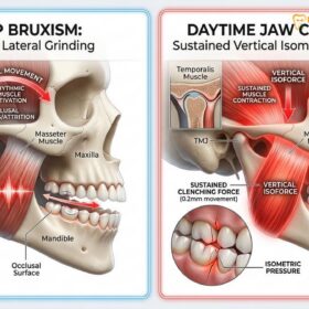
However, in patients suffering from TMD, bruxism, or severe malocclusion, this resting state is rarely achieved. The constant neurological feedback loop created by a misaligned bite prevents the jaw from ever reaching its true physiological resting posture. The muscles are kept in a state of chronic hypertonicity, constantly bracing the jaw to protect the teeth or grinding in a subconscious attempt to find a stable, comfortable position.
When the muscles are denied their natural rest, they become ischemic, meaning they are deprived of adequate blood flow. This lack of oxygen forces the muscle cells to switch to anaerobic metabolism, which produces lactic acid as a byproduct. The accumulation of lactic acid and other metabolic waste products triggers pain receptors, leading to the classic symptoms of TMD: tension headaches, facial pain, neck stiffness, and referred pain behind the eyes.
TENS (Transcutaneous Electrical Nerve Stimulation) in TMJ Diagnostics
Ultra-low frequency TENS is utilized to involuntarily stimulate the facial nerves, effectively erasing pathological muscle engrams and allowing the mandible to settle into its true relaxed trajectory.
The cornerstone of neuromuscular diagnosis and treatment is the application of Transcutaneous Electrical Nerve Stimulation (TENS). However, it is crucial to distinguish the specific type of TENS used in neuromuscular dentistry from the standard TENS units commonly found in physical therapy clinics or purchased over the counter for general pain relief.
Standard medical TENS units operate at high frequencies and are designed to block pain signals from reaching the brain. While effective for temporary analgesia, high-frequency TENS does not alter the resting length of the muscle or erase muscle memory. In contrast, neuromuscular dentistry employs an ultra-low frequency TENS (ULF-TENS) device, which operates at a highly specific frequency of approximately 0.5 to 4 Hz. This specific frequency is designed not to block pain, but to induce a rhythmic, involuntary contraction of the muscles[4].
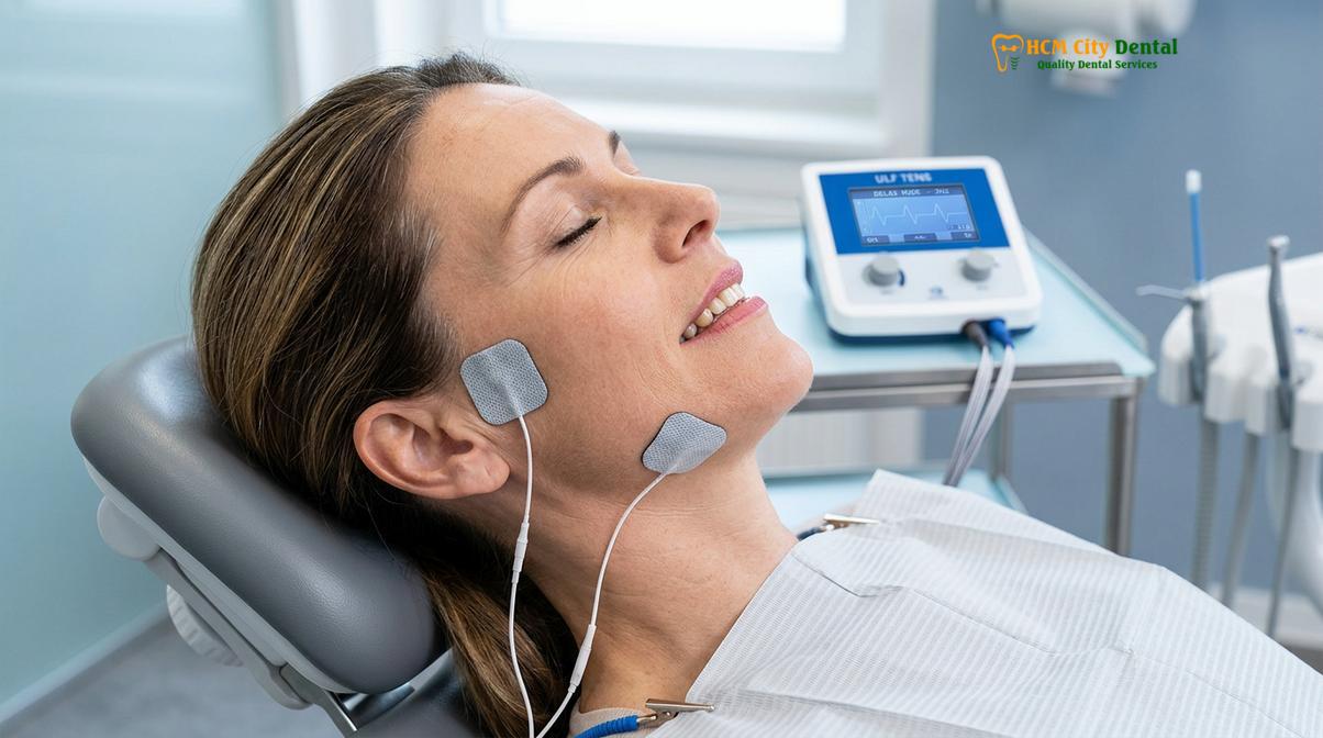
During a clinical session, specialized electrode pads are placed over the preauricular area to target the mandibular division of the trigeminal nerve, and often over the facial nerve to target the facial muscles. When the ULF-TENS device is activated, it delivers a mild, rhythmic electrical impulse that travels along the nerve pathways. This impulse triggers a synchronous, involuntary contraction of the masticatory and facial muscles every one to two seconds, acting as a vascular massage that increases local blood flow and flushes out accumulated lactic acid.
Electromyography (EMG) and Jaw Tracking Protocols
Surface electromyography (sEMG) and computerized jaw tracking provide objective, quantifiable data on muscle resting states and mandibular movement patterns before and after TENS application.
A defining characteristic of neuromuscular dentistry is its reliance on objective, measurable data rather than subjective clinical guesswork. To accurately assess the state of the stomatognathic system and verify the efficacy of the TENS therapy, clinicians utilize highly sophisticated computerized instrumentation: surface electromyography (sEMG) and computerized mandibular scanning (CMS), also known as jaw tracking. According to guidelines from the Vietnam Odonto-Stomatology Association (VOSA), integrating objective measurement tools is highly recommended for managing complex temporomandibular joint disorders in Vietnamese populations[5].
Similar to how an electrocardiogram measures the electrical activity of the heart muscle, sEMG measures the electrical action potentials of the masticatory and cervical muscles. Small, non-invasive sensor pads are placed over key muscle groups, typically the anterior temporalis, the superficial masseter, and the anterior digastric muscles. The sEMG software records the microvolt output of these muscles in real-time. A healthy, resting muscle should display very low electrical activity, whereas an elevated baseline indicates chronic spasm.
While sEMG measures muscle activity, CMS measures the precise kinematics of the jaw in three-dimensional space. A lightweight sensor array is placed on the patient’s head, and a small, temporary magnet is adhered to the lower incisors. As the patient opens, closes, and swallows, the sensor array tracks the movement of the magnet with sub-millimeter accuracy. This allows the dentist to visualize the exact trajectory of the jaw, identifying any deviations, restrictions, or irregular movements that indicate joint dysfunction.
Phase 1 and Phase 2 Treatment Protocols
Neuromuscular treatment is typically divided into two distinct phases: initial stabilization with a reversible orthotic, followed by long-term restorative or orthodontic solutions once symptoms have resolved.
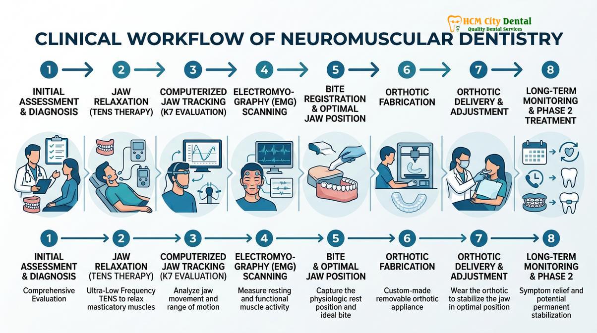
The transition from diagnosis to treatment in neuromuscular dentistry is carefully staged to ensure patient comfort and verify the efficacy of the new bite position before any permanent changes are made to the teeth. This staged approach is generally divided into Phase 1 (Stabilization) and Phase 2 (Long-Term Restoration).
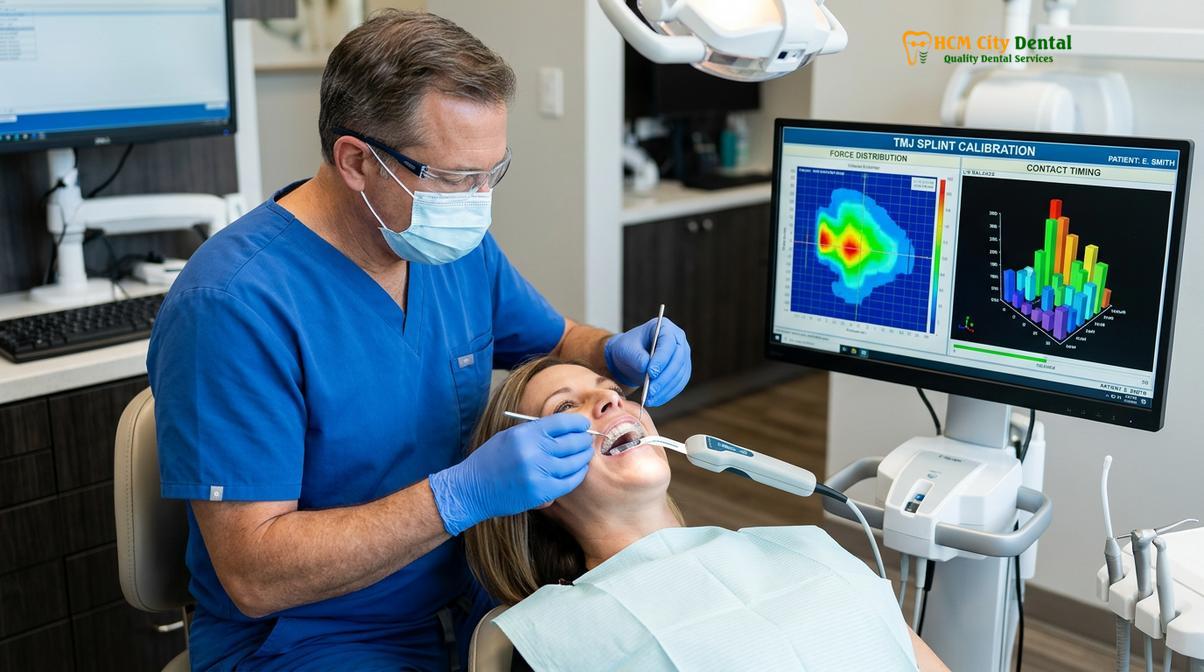
During Phase 1, the initial treatment is strictly reversible. Using the precise myocentric bite registration captured during the diagnostic phase, the dental laboratory fabricates a custom neuromuscular orthotic. This appliance is fundamentally different from standard Nightguards tại HCMC Dental Clinic, as it actively repositions the jaw rather than just protecting teeth from friction. The patient wears this orthotic over their lower teeth, allowing the jaw to close into the newly established, relaxed position.
“Phase 1 therapy is our diagnostic proving ground. By utilizing a removable orthotic, we can confirm that the new mandibular position resolves the patient’s pain and dysfunction before we ever touch a dental drill. It is a conservative, patient-first approach to complex rehabilitation.”
Once the patient has been symptom-free for a sustained period and the muscles have adapted to their new resting length, they may choose to proceed to Phase 2. Phase 2 involves making the new bite permanent so the patient no longer needs to wear the orthotic. This can be achieved through various methods, including orthodontic tooth movement, prosthodontic reconstruction (crowns and veneers), or targeted occlusal adjustments.
Comparison of TMJ Treatment Modalities
To better understand the advantages of the neuromuscular approach, it is helpful to compare it directly with traditional methods of bite management.
| Feature | Traditional Dentistry (Gnathology) | Gneuromuscular Dentistry |
|---|---|---|
| Primary Focus | Static tooth contacts and structural joint alignment | Dynamic muscle function and physiological rest position |
| Diagnostic Tools | Articulators, bite wax, manual manipulation | ULF-TENS, surface electromyography (sEMG), jaw tracking |
| Jaw Positioning | Centric relation (forced superior-posterior position) | Myocentric position (muscle-dictated relaxed trajectory) |
| Appliance Therapy | Flat-plane splints to protect enamel from grinding | Anatomical orthotics to actively reposition the mandible |
| Ideal Candidates | Routine restorative cases without chronic pain | Complex TMD, chronic facial pain, severe malocclusion |
Case Study: TMJ Rehabilitation at HCMC Dental Clinic
Clinical application of neuromuscular principles often yields profound improvements in patients suffering from long-standing craniofacial pain and occlusal instability.
Patient Profile: A 45-year-old patient visiting HCMC Dental Clinic in Ho Chi Minh City presented with severe, chronic tension headaches, limited jaw opening, and significant wear on their posterior teeth. Traditional bite guards had failed to provide lasting relief, as they only protected the enamel without addressing the underlying muscular hypertonicity.
Clinical Intervention: After a comprehensive evaluation utilizing ULF-TENS and sEMG, it was determined that the patient’s mandible was forced into a retruded position, causing chronic spasm of the masseter and temporalis muscles. Following a 60-minute TENS session, a new myocentric bite registration was captured, and a custom lower orthotic was fabricated.
Outcome: Within weeks of wearing the Phase 1 orthotic, the patient reported a complete cessation of tension headaches and a significant increase in their comfortable range of motion, demonstrating the profound impact of addressing the muscular component of the bite.
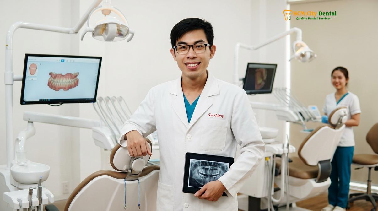
Dr. Nguyen Van Cuong, a leading specialist in complex bite rehabilitation, emphasizes that true occlusal harmony begins with relaxed musculature. Dr. Cuong utilizes advanced diagnostic protocols to ensure that every patient receives a customized treatment plan tailored to their unique physiological needs, focusing on long-term stability rather than temporary symptom management.
When to See a Doctor for TMJ and Bite Issues
Early intervention is crucial for preventing permanent joint damage; recognizing the signs of neuromuscular dysfunction can help you seek appropriate care before symptoms escalate.
Important Clinical Note: Delaying treatment for severe temporomandibular joint dysfunction can lead to irreversible cartilage degradation, osteoarthritis of the jaw, and chronic neuropathic pain. If you experience persistent jaw locking or severe facial pain, seek professional evaluation promptly.
Many patients endure jaw pain and headaches for years, mistakenly believing these symptoms are normal or unrelated to their dental health. You should consider scheduling a comprehensive neuromuscular evaluation if you experience any of the following persistent issues:
- Chronic tension headaches, particularly upon waking in the morning.
- Pain, tenderness, or a feeling of fatigue in the jaw muscles after chewing.
- Clicking, popping, or grating sounds in the temporomandibular joints when opening or closing your mouth.
- Limited range of motion, or instances where the jaw feels “locked” in an open or closed position.
- Unexplained neck and shoulder stiffness that accompanies facial pain.
For detailed information on diagnostics and custom splint therapy options, visit our comprehensive TMJ & Jaw Pain Treatment Ho Chi Minh City guidelines.
References
- Journal of the American Dental Association. Occlusal splint therapy for temporomandibular disorders: a clinical review.
- Journal of Oral Rehabilitation. Efficacy of Michigan repositioning splints in TMD patients.
- Journal of Prosthetic Dentistry. Computerized occlusal analysis and bite mapping in TMJ therapy.
- Clinical Oral Investigations. Masseter Botox injections for chronic jaw pain and bruxism.
- Vietnam Odonto-Stomatology Association (VOSA). Clinical guidelines on the management of temporomandibular joint disorders in Vietnamese populations.
If you are experiencing chronic jaw pain or suspect you have a complex bite misalignment, contact HCMC Dental Clinic in Ho Chi Minh City today. Our team is dedicated to providing advanced neuromuscular evaluations to help you achieve lasting comfort and optimal oral function. Learn more about our comprehensive solutions, including custom Nightguards tại HCMC Dental Clinic.