Root canal aftercare requires immediate protection of the temporary filling, meticulous oral hygiene, and timely placement of a permanent crown. Proper post-endodontic maintenance prevents bacterial reinfection, protects the structurally compromised tooth from fracture, and ensures the underlying bone heals completely for long-term clinical success.
Clinical Summary:
Effective root canal aftercare is a multi-phase clinical protocol designed to preserve the endodontically treated tooth and prevent secondary infections. Immediately following the procedure, patients must manage localized inflammation and protect the temporary restoration from occlusal stress. Long-term success relies heavily on preventing coronal leakage through a hermetic permanent crown seal, maintaining optimal periodontal health around the restoration margins, and mitigating excessive biomechanical forces such as nocturnal bruxism. Regular radiographic monitoring and professional prophylaxis are essential to verify periapical healing and ensure the structural integrity of the restored tooth.
Key Takeaways:
- Avoid chewing on the treated tooth until the permanent crown is securely cemented to prevent structural fractures.
- Coronal leakage is a primary cause of late-stage root canal failure and requires immediate restorative attention.
- Custom nightguards are highly recommended to prevent vertical root fractures in patients who clench or grind their teeth.
- Routine radiographic evaluations are necessary to confirm the resolution of periapical bone lesions over time.
- Meticulous interdental cleaning prevents gingival inflammation around the margins of the final restoration.
The Threat of Coronal Leakage: How Saliva Bacteria Can Re-Infect Roots
Coronal leakage occurs when saliva and bacteria bypass the dental restoration, infiltrating the disinfected root canal system. Preventing this microleakage is the most critical factor in avoiding secondary endodontic infections.
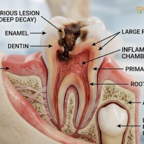
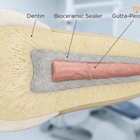
The success of endodontic therapy relies not only on the thorough debridement and disinfection of the root canal system but also on the establishment of an impenetrable seal at the coronal (top) portion of the tooth. When a tooth undergoes a root canal, the internal pulp chamber is hollowed out, sterilized, and filled with a biocompatible material, typically gutta-percha, combined with an endodontic sealer. However, gutta-percha alone does not provide a hermetic seal against moisture and bacteria. If the coronal restoration—whether a temporary filling or a permanent crown—fails, the entire root canal system becomes vulnerable to a phenomenon known as coronal leakage.
Coronal leakage is a microscopic process where saliva, which is heavily laden with complex bacterial biofilms, seeps through compromised margins between the restorative material and the natural tooth structure. The oral cavity hosts hundreds of bacterial species, and certain resilient pathogens, such as Enterococcus faecalis, are particularly adept at surviving in the nutrient-deprived environment of a treated root canal[1]. Once these bacteria breach the coronal seal, they can migrate down the length of the root canal alongside the gutta-percha, eventually reaching the apical foramen (the tip of the root) and re-infecting the surrounding periapical bone.
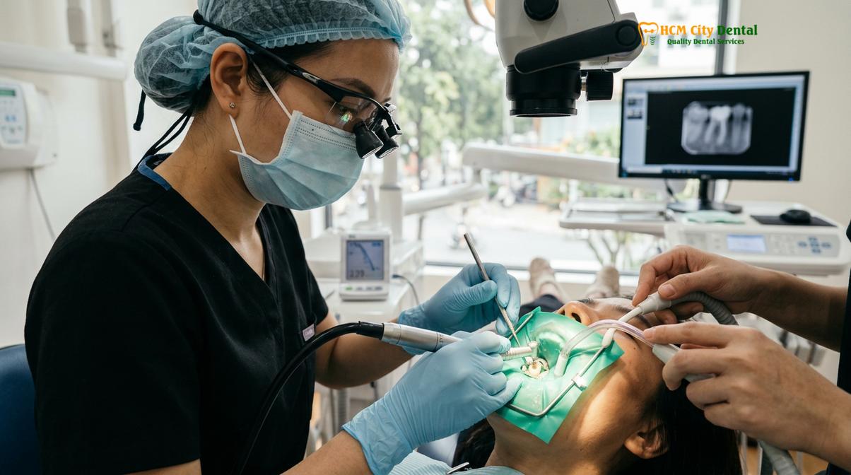
The timeline for coronal leakage can be alarmingly rapid. Clinical studies have demonstrated that if a temporary filling is lost or if a root canal is left exposed to the oral environment without a permanent restoration, salivary bacteria can contaminate the entire length of the root canal system within a matter of days to weeks[2]. This rapid ingress of pathogens underscores the absolute necessity of timely restorative care. A temporary filling is designed to last only a few weeks; it is highly susceptible to occlusal wear, thermal expansion, and marginal degradation. Therefore, transitioning from a temporary seal to a permanent, custom-fabricated crown is a non-negotiable step in coronal leakage prevention.
Furthermore, the chemical degradation of endodontic sealers plays a role in late-stage failures. When exposed to the enzymatic action of saliva and bacterial byproducts, the sealer that binds the gutta-percha to the dentinal walls can begin to dissolve. This dissolution creates microscopic voids, acting as highways for bacterial colonization. To combat this, modern endodontics often employs advanced bioceramic sealers that offer superior dimensional stability and biocompatibility, though they still require a robust coronal shield to function optimally.
At HCMC Dental Clinic, our clinical protocol mandates that the permanent restorative phase be initiated as soon as the tooth is asymptomatic and the initial healing phase is confirmed. By minimizing the time the tooth relies on a temporary filling, we drastically reduce the window of opportunity for bacterial microleakage, thereby safeguarding the long-term prognosis of the treated tooth.
Daily Hygiene Protocols: Brushing, Flossing, and Interdental Cleaning
Meticulous daily oral hygiene removes cariogenic biofilms and prevents localized gingival inflammation around the treated tooth. Utilizing proper brushing techniques and interdental tools is essential for maintaining the integrity of the restoration.
A common misconception among patients is that a tooth treated with a root canal and covered with a dental crown is impervious to future dental disease. While the inorganic materials of the crown (such as zirconia or porcelain) cannot decay, the underlying natural tooth structure at the margin—where the crown meets the root—remains highly susceptible to bacterial acid attacks. Therefore, rigorous daily hygiene protocols are paramount in root canal aftercare to protect this vulnerable junction and the surrounding periodontal tissues.
The foundation of post-endodontic home care is the mechanical disruption of dental plaque. Plaque is a sticky, colorless biofilm composed of bacteria that constantly forms on the surfaces of teeth and restorations. If not removed every 12 to 24 hours, this biofilm matures, calcifies into tartar (calculus), and shifts its microbial composition toward more aggressive, acid-producing, and tissue-destroying strains. For a tooth that has undergone root canal treatment, the accumulation of plaque at the crown margin can lead to recurrent decay (secondary caries) that silently undermines the restoration, as the tooth no longer has a nerve to signal pain.
“The success of endodontic therapy does not end in the operatory; it is sustained in the patient’s daily routine. A perfectly executed root canal will ultimately fail if the restorative margins are left to drown in cariogenic plaque.” — Dr. Nguyen Van Cuong
To effectively manage plaque, patients must adopt the Modified Bass brushing technique. This method involves placing a soft-bristled toothbrush at a 45-degree angle to the gumline, allowing the bristles to gently penetrate the gingival sulcus (the shallow crevice between the tooth and the gum). By using small, vibratory, circular motions rather than aggressive horizontal scrubbing, patients can dislodge plaque from the critical crown margin without causing gingival recession or abrading the root surface. Brushing should be performed at least twice daily using a fluoridated toothpaste, which helps remineralize any microscopic areas of demineralization on the exposed root structure.
However, brushing alone is insufficient, as toothbrush bristles cannot access the interproximal spaces (the areas between the teeth). This is where interdental cleaning becomes critical. Using high-quality dental floss root canal HCMC patients can effectively clear the contact points and the interproximal gumline. When flossing around a newly crowned root-canaled tooth, it is important to adapt the floss in a C-shape around the base of the tooth, sliding it gently beneath the gumline. If the contact between the teeth is particularly tight, pulling the floss through horizontally rather than snapping it upward can prevent accidental dislodgement of the restoration.
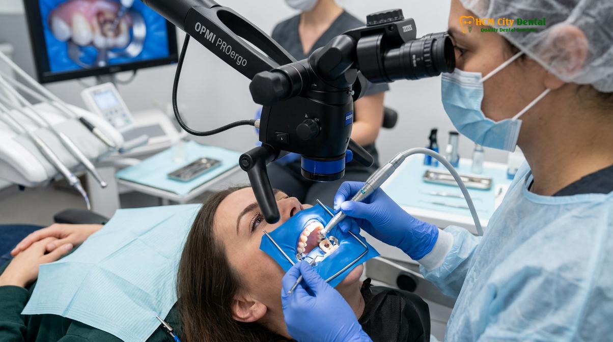
For patients with wider interdental spaces, mild gingival recession, or complex restorative work, interdental brushes or water flossers (oral irrigators) serve as excellent adjuncts. Water flossers utilize a pulsating stream of water to flush out loosely attached plaque, food debris, and inflammatory cytokines from the periodontal pockets. While they do not replace the mechanical wiping action of string floss, they significantly reduce gingival bleeding and inflammation, creating a healthier environment for the restored tooth to function.
Periodontal Health: Keeping Gums Inflame-Free Around the Crown Margin
Maintaining healthy, inflammation-free gums around the dental crown margin prevents periodontal pocketing and subsequent bone loss. A precise marginal fit combined with excellent tissue care ensures the biological width remains uncompromised.
The relationship between endodontic success and periodontal health is deeply intertwined. A tooth does not exist in isolation; it is anchored into the jawbone by the periodontium, which includes the gingiva (gums), periodontal ligament, cementum, and alveolar bone. Following a root canal and the placement of a permanent crown, the health of the gingival tissues immediately surrounding the restoration becomes a critical determinant of the tooth’s long-term survival. If the gums become chronically inflamed, the resulting periodontal disease can lead to bone loss that destabilizes the tooth, regardless of how perfectly the root canal was performed.
Central to this concept is the preservation of the “biological width.” The biological width is the natural dimension of the soft tissue attached to the portion of the tooth above the alveolar bone. It acts as a protective seal, preventing oral bacteria from accessing the underlying bone. When a dental crown is fabricated, the margin of the crown must be carefully placed so that it does not impinge upon this biological width. If the crown margin is placed too deeply beneath the gumline, the body perceives it as a foreign object, triggering a chronic inflammatory response. This localized inflammation manifests as red, swollen, and bleeding gums, eventually leading to tissue recession and localized bone destruction.
Even with a perfectly placed crown, the margin represents a microscopic transition zone between the restorative material and the natural tooth. This junction is inherently more plaque-retentive than smooth, intact enamel. Therefore, a routine crown margin seal check by a dental professional is necessary to ensure that the cement line remains intact and that no micro-gaps have developed over time. If plaque is allowed to stagnate at this margin, the bacteria release toxins that provoke an immune response. The host’s immune system, in its attempt to fight off the bacterial invasion, releases inflammatory mediators (such as cytokines and prostaglandins) that inadvertently destroy the collagen fibers attaching the gum to the tooth.
As these collagen fibers are destroyed, a periodontal pocket forms. This pocket provides an anaerobic (oxygen-free) environment where highly pathogenic bacteria can thrive, accelerating the destruction of the supporting alveolar bone. In severe cases, a combined endodontic-periodontal lesion can occur, where an infection from the deep periodontal pocket communicates with the root apex, or vice versa, severely complicating the prognosis of the tooth.
To keep the gums inflame-free, patients must be vigilant for early signs of gingivitis, such as bleeding during brushing or flossing, localized swelling, or a change in gum color from stippled pink to glossy red. At HCMC Dental Clinic, we emphasize that healthy gums should never bleed during normal oral hygiene routines. If bleeding occurs around a root-canaled tooth, it is an immediate signal that the biofilm management in that specific area is inadequate and requires professional intervention to prevent irreversible periodontal damage.
Preventive Maintenance: Why Professional Cleanings and Checkups Are Critical
Professional dental cleanings and routine radiographic examinations allow clinicians to monitor periapical healing and detect early signs of restoration failure. This proactive maintenance schedule is vital for the long-term retention of the tooth.
While diligent home care is the first line of defense, it cannot replace the diagnostic and therapeutic benefits of professional preventive maintenance. Root canal aftercare is an ongoing process that extends years beyond the initial procedure. The biological healing of the bone surrounding the root apex is a slow process, and the structural integrity of the coronal restoration is constantly challenged by the harsh environment of the oral cavity. Therefore, a structured schedule of professional cleanings (prophylaxis) and clinical examinations is essential to validate the success of the endodontic treatment.
Professional cleanings are necessary to remove calculus (tartar) that inevitably forms in areas missed by daily brushing and flossing. Calculus is highly porous and acts as a reservoir for bacterial biofilms, constantly irritating the gingival tissues. Dental hygienists and dentists use specialized ultrasonic scalers and hand instruments to meticulously debride the tooth surfaces and crown margins. Special care is taken when scaling around porcelain or zirconia crowns to avoid scratching the highly polished surfaces, which could otherwise increase future plaque retention.
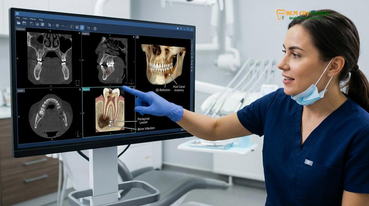
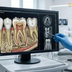
Equally important to the physical cleaning is the radiographic monitoring of the treated tooth. Following a root canal, the periapical bone (the bone at the tip of the root) requires time to heal, especially if a pre-existing infection or abscess had caused a localized area of bone destruction (radiolucency). Radiographic evidence of complete bone regeneration can take anywhere from six months to over a year[3]. Routine X-rays allow the clinician to compare the current bone density against baseline images taken immediately after the procedure.
| Timeline | Clinical Action | Diagnostic Purpose |
|---|---|---|
| 1-2 Weeks Post-Op | Initial Follow-up & Crown Prep | Assess acute symptom resolution; prepare tooth for permanent coronal seal. |
| 6 Months Post-Op | Radiographic Evaluation | Monitor early signs of periapical bone healing and verify crown margin integrity. |
| 12 Months Post-Op | Comprehensive Exam & X-ray | Confirm complete resolution of apical radiolucency; check for occlusal wear. |
| Every 6 Months | Professional Prophylaxis | Remove calculus, manage periodontal health, and perform routine oral cancer screening. |
During these checkups, the dentist will also perform a thorough clinical evaluation of the restoration. This includes checking the occlusal contacts to ensure the crown is not bearing excessive biting forces, inspecting the margins with a dental explorer to detect any microleakage or recurrent decay, and assessing the mobility of the tooth. Advanced imaging techniques, such as Cone Beam Computed Tomography (CBCT), may be utilized if a patient reports vague symptoms or if standard two-dimensional X-rays are inconclusive. CBCT provides a highly detailed, three-dimensional view of the root canal system and surrounding bone, allowing for the early detection of complications such as missed accessory canals or subtle root fractures.
Nighttime Clenching Protection: Using Custom Nightguards to Protect Root Canals
Nocturnal bruxism exerts excessive lateral and vertical forces on endodontically treated teeth, significantly increasing the risk of catastrophic root fractures. Custom-fabricated occlusal guards distribute these forces to protect the compromised tooth structure.
One of the most significant, yet frequently overlooked, threats to the longevity of a root-canaled tooth is bruxism—the unconscious clenching and grinding of teeth, typically occurring during sleep. To understand why bruxism is so dangerous to endodontically treated teeth, one must consider the biomechanical changes that occur following a root canal. When the dental pulp (the nerve and blood supply) is removed, the tooth loses its internal hydration mechanism. Over time, the dentin becomes slightly more brittle compared to a vital tooth. Furthermore, the access cavity created to perform the root canal, combined with any pre-existing decay, significantly reduces the overall structural volume of the tooth.
Additionally, the removal of the pulp eliminates the tooth’s internal proprioceptive feedback. In a healthy, vital tooth, the nerve provides sensory information about biting pressure. If you bite down too hard on a seed or a piece of ice, the nerve sends a sharp pain signal, causing an immediate reflex to release the pressure. A root-canaled tooth lacks this internal warning system. While the periodontal ligament surrounding the root still provides some pressure sensation, the threshold for detecting excessive force is altered. Consequently, patients with bruxism can exert immense, unregulated forces on a root-canaled tooth without realizing it.
Clinical Warning: Unprotected nocturnal bruxism is a leading cause of vertical root fractures in endodontically treated teeth. A vertical root fracture splits the tooth down the middle, rendering it completely unrestorable and necessitating immediate extraction.
During a bruxism episode, the masseter and temporalis muscles can generate forces far exceeding normal chewing pressures. When these forces are applied laterally (side-to-side) or heavily on the cusps of a structurally compromised tooth, the stress concentrates along the root axis. If the stress exceeds the tensile strength of the dentin, a micro-crack can initiate and propagate down the root, resulting in a vertical root fracture. Unlike a fractured crown, which can often be replaced, a fractured root is a catastrophic failure.
To mitigate this risk, the fabrication of a custom occlusal guard (nightguard) is highly recommended for patients who exhibit signs of bruxism, such as worn enamel, scalloped tongue margins, or frequent morning headaches. A custom nightguard, typically made from a durable hard acrylic or a dual-laminate material, acts as a protective shock absorber. It provides a smooth, even surface for the opposing teeth to glide against, preventing the interlocking of cusps that generates harmful lateral forces. By evenly distributing the occlusal load across the entire dental arch, the nightguard shields the vulnerable root-canaled tooth from concentrated stress, significantly extending its clinical lifespan.
Clinical Lifespan: What Contributes to a Root Canal Lasting a Lifetime
The lifetime survivability of a root-canaled tooth depends on the amount of remaining healthy dentin, the presence of a ferrule effect, and the quality of the final coronal restoration.
Patients frequently ask how long a tooth will last after a root canal. With optimal clinical execution and rigorous aftercare, an endodontically treated tooth has the potential to last a lifetime. However, this longevity is not guaranteed by the root canal procedure alone; it is heavily dependent on a combination of biomechanical factors, restorative excellence, and the patient’s biological response[4].
The most critical biomechanical factor determining the survival of a root-canaled tooth is the amount of remaining sound tooth structure, specifically the presence of a “ferrule effect.” The ferrule is a band of healthy dentin that extends above the gumline, completely encircling the tooth. For a dental crown to be structurally sound, it must grasp onto at least 1.5 to 2 millimeters of this solid dentin. The ferrule acts much like the metal band around a wooden barrel; it binds the tooth structure together, preventing the root from splitting under the wedging forces of chewing. If a tooth is severely broken down and lacks an adequate ferrule, the long-term prognosis drops significantly, even if the root canal itself was performed flawlessly.
Clinical Case Review: A 45-year-old patient presented to HCMC Dental Clinic in Ho Chi Minh City with a fractured temporary filling on a lower molar, treated endodontically three months prior at a different facility. Due to the delay in placing a permanent crown, coronal leakage had occurred, and the tooth lacked sufficient ferrule. Dr. Nguyen Van Cuong performed a crown lengthening procedure to expose more healthy dentin, followed by a meticulous core buildup and a precision-milled zirconia crown, successfully saving the tooth from extraction.
Following the establishment of a solid foundation, the core buildup material plays a vital role. The core replaces the missing dentin in the center of the tooth, providing a stable base for the crown. In cases where the tooth structure is severely compromised, a dental post (often made of flexible fiberglass or cast metal) may be cemented into one of the root canals to help retain the core material. However, it is important to note that a post does not strengthen the root; it merely anchors the core. The ultimate strength of the tooth relies on the ferrule and the final restoration.
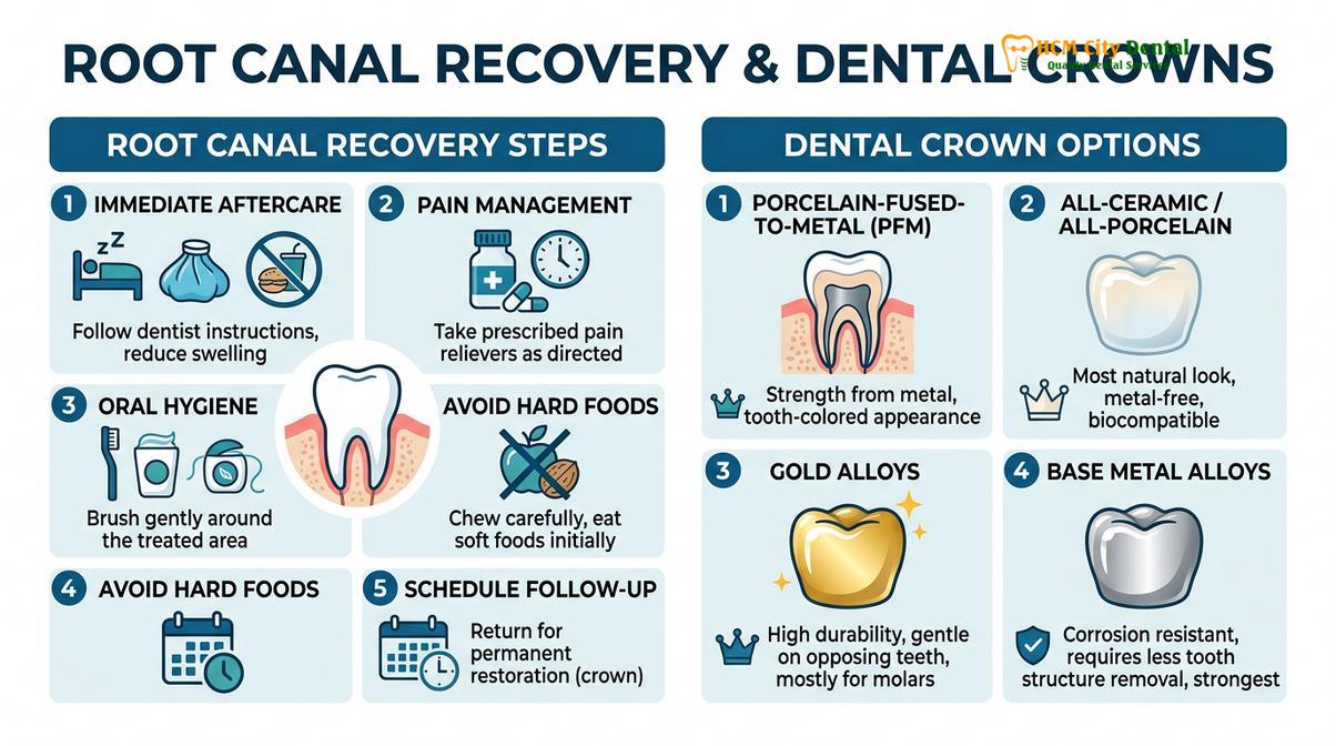
The material and fit of the final coronal restoration are the last lines of defense. Modern dentistry utilizes highly durable materials such as monolithic zirconia or lithium disilicate (E-max), which offer exceptional fracture resistance and aesthetic integration. The precision of the crown margins, achieved through digital scanning and CAD/CAM milling, ensures a tight seal against bacterial microleakage. When a robust ferrule, a stable core, and a precisely fitting crown are combined with excellent oral hygiene and routine professional monitoring, the endodontically treated tooth can function seamlessly within the dental arch for decades.
“A root canal saves the biological foundation, but the restorative phase dictates the mechanical longevity. We must engineer the final restoration to withstand the immense dynamic forces of the human bite.” — Dr. Nguyen Van Cuong
When to See a Doctor (Important Clinical Considerations)
While mild tenderness and slight tissue inflammation are normal for a few days following a root canal, certain symptoms indicate a deviation from the normal healing process and require immediate clinical evaluation. Patients must be vigilant in monitoring their recovery and should not hesitate to contact their dental provider if red flags arise.
You should seek immediate clinical attention if you experience progressive, severe pain that is not alleviated by over-the-counter analgesics, especially if the pain is sharp and throbbing when biting down. This could indicate an acute flare-up, a high bite on the temporary restoration, or a missed accessory canal. Additionally, the sudden onset of localized swelling in the gums, face, or jaw is a clear sign of an active infection that may require antibiotic therapy or further endodontic intervention[5].

Another critical warning sign is the appearance of a sinus tract, often described by patients as a small “pimple” or blister on the gums near the treated tooth. This tract is a drainage pathway for pus originating from a periapical infection. Even if the sinus tract is painless, its presence confirms that bacteria are actively destroying the surrounding bone. Furthermore, if the temporary filling falls out or feels loose, you must return to the clinic promptly. Leaving the root canal system exposed to the oral environment rapidly accelerates coronal leakage, jeopardizing the entire treatment. Early intervention based on clinical diagnostics is essential to manage these complications and preserve the tooth.
If you are experiencing any abnormal symptoms following your procedure, or if you are ready to proceed with your permanent coronal restoration, we encourage you to schedule a follow-up consultation. Contact our team at HCMC Dental Clinic to ensure your recovery is progressing optimally.
Frequently Asked Questions
Can a root-canaled tooth get a cavity?
Yes, a root-canaled tooth can still develop a cavity. Because the tooth no longer has a nerve, you will not feel pain if decay occurs, making regular dental checkups essential to detect and treat new cavities before they compromise the remaining tooth structure. The natural dentin at the base of the crown remains vulnerable to bacterial acid, emphasizing the need for meticulous daily brushing and flossing.
Why is gum health around a dental crown so important?
Healthy gums seal the margin where the crown meets the tooth, preventing bacteria from infiltrating the root. If gums become inflamed or recede, the vulnerable root surface and crown margin are exposed to plaque, significantly increasing the risk of secondary infection and structural failure. Periodontal disease can destroy the bone supporting the tooth, leading to tooth loss regardless of the root canal’s success.
Does teeth clenching damage root-canaled teeth?
Yes, chronic teeth clenching exerts severe biomechanical stress on root-canaled teeth, which are structurally weaker than natural teeth. This excessive pressure can lead to catastrophic vertical root fractures, often rendering the tooth unrestorable and requiring extraction. Wearing a custom-fitted nightguard is highly recommended to distribute these forces and protect the integrity of the restored tooth.
How long does it take for the bone to heal after a root canal?
Radiographic evidence of periapical bone healing typically takes between six months to over a year to become visible. While the acute symptoms usually resolve within a few days, the cellular regeneration of the bone surrounding the root apex is a gradual biological process. Your dentist will monitor this healing progress through routine X-rays during your follow-up appointments.
When can I resume eating normally after root canal therapy?
You should wait until the local anesthesia has completely worn off to avoid accidentally biting your cheek or tongue. Furthermore, you must avoid chewing hard or sticky foods on the treated side until the permanent crown is securely cemented to prevent fracturing the temporary filling. Once the final restoration is placed and adjusted, you can gradually return to your normal diet.
References
- Journal of Endodontics. Outcomes of root canal treatment and restoration longevity. (2020).
- International Endodontic Journal. Biofilms in endodontics and disinfection strategies. (2021).
- Journal of the American Dental Association. Microscope-assisted endodontics and missed canals. (2019).
- Clinical Oral Investigations. Biocompatibility of bioceramic root canal sealers. (2022).
- British Dental Journal. Pain management and dental anxiety in endodontics. (2018).