TMJ arthrocentesis in Vietnam is a minimally invasive, highly effective clinical procedure designed to treat severe jaw locking and chronic joint pain. By flushing the temporomandibular joint space with sterile fluids, specialists can remove inflammatory byproducts and restore normal jaw mobility without the need for open joint surgery.
Clinical Summary:
Temporomandibular joint (TMJ) arthrocentesis bridges the critical gap between conservative therapies and invasive open-joint surgeries. This minimally invasive lavage technique utilizes hydraulic pressure to expand the upper joint space, effectively washing away inflammatory mediators such as cytokines and prostaglandins that contribute to pain and cartilage degradation. In modern clinical practice, this joint fluid wash is frequently combined with intra-articular injections of hyaluronic acid or corticosteroids to enhance condylar lubrication and facilitate smooth articular disc movement. Indicated primarily for patients experiencing acute closed lock, severe arthralgia, or disc displacement without reduction, the procedure offers a rapid reduction in pain scores and a significant increase in maximal incisal opening. Performed under local anesthesia or conscious sedation, it presents a low-morbidity alternative for patients who have exhausted conservative options like occlusal splints or physical therapy.
Key Takeaways:
- Minimally invasive lavage technique targeting the upper compartment of the temporomandibular joint to restore function.
- Effectively removes pain-causing inflammatory cytokines and breaks down intra-articular fibrous adhesions.
- Often supplemented with a hyaluronic acid injection jaw protocol to restore synovial fluid viscosity and boundary lubrication.
- Serves as a critical intermediate surgical step when conservative treatments fail to resolve severe TMD locking HCMC.
- Performed as an outpatient procedure with a rapid recovery timeline, minimal surgical morbidity, and high clinical success rates.
What is TMJ Arthrocentesis? Clinical Overview
TMJ arthrocentesis is a minimally invasive surgical procedure that uses sterile fluid to flush the temporomandibular joint capsule, removing inflammatory debris and releasing adhesions to restore normal jaw function.
The temporomandibular joint is one of the most complex and frequently used joints in the human body. Classified as a ginglymoarthrodial joint, it allows for both hinging (rotation) and sliding (translation) movements. The joint is divided into an upper and lower compartment by a fibrocartilaginous articular disc. When the delicate biomechanical harmony of this system is disrupted—whether through macro-trauma, chronic micro-trauma from bruxism, or degenerative joint disease—patients often experience debilitating pain, restricted mouth opening, and joint noises. While initial management typically involves conservative measures such as physical therapy, pharmacological intervention, and the use of custom nightguards, a subset of patients will remain refractory to these non-surgical treatments.[1]
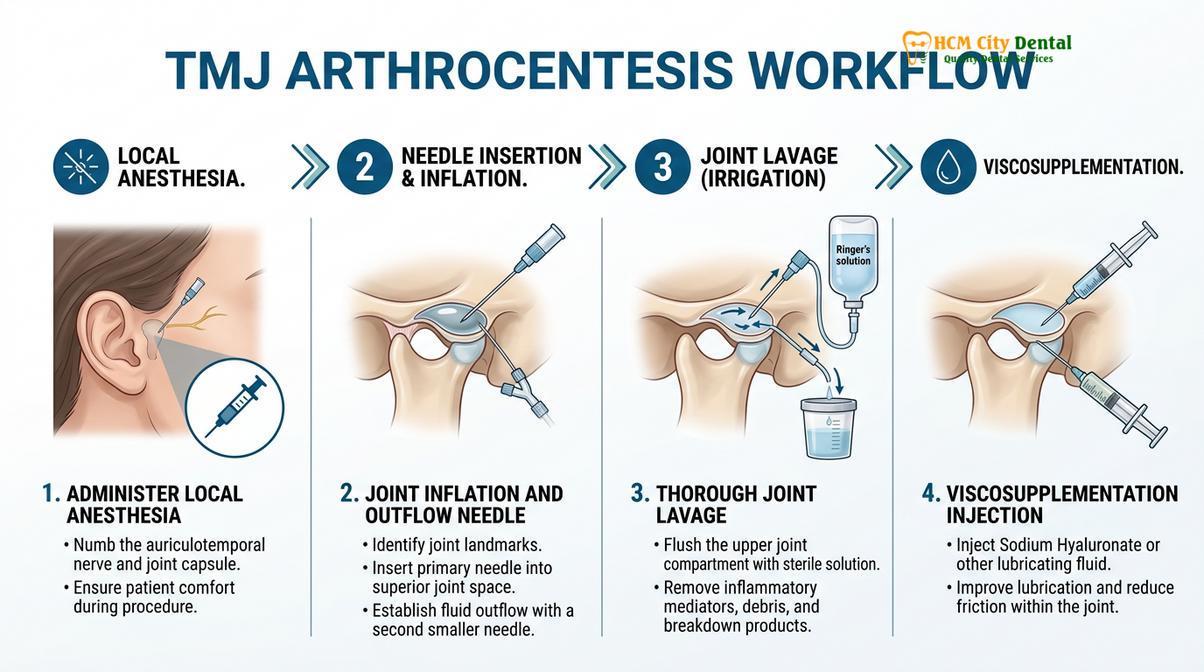
For these refractory cases, TMJ arthrocentesis serves as a highly effective, minimally invasive surgical intervention. Introduced in the early 1990s, the procedure fundamentally shifted the paradigm of temporomandibular disorder (TMD) management. Instead of immediately resorting to open joint surgery (arthrotomy), which carries higher risks of facial nerve injury and significant morbidity, maxillofacial surgeons can utilize arthrocentesis to therapeutic effect. The procedure involves the insertion of two small needles into the superior joint space. A sterile irrigant, typically Ringer’s lactate or normal saline, is then pumped through the joint under pressure. This continuous flow of fluid serves a dual purpose: it physically washes out the joint space and utilizes hydraulic pressure to expand the joint capsule, thereby releasing the articular disc if it has become stuck or adhered to the articular eminence.
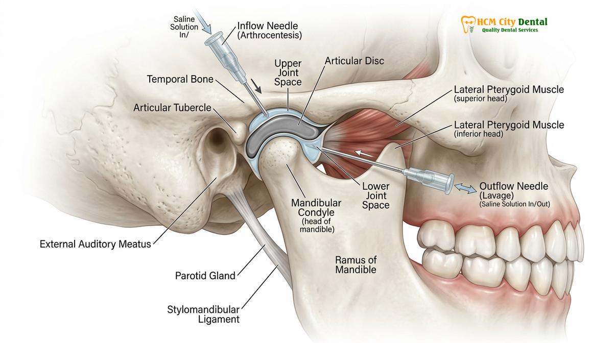
According to Dr. Nguyen Van Cuong, a leading specialist in maxillofacial diagnostics, patient selection is paramount to the success of this procedure. “Arthrocentesis is not a universal cure for all jaw pain,” notes Dr. Cuong. “It is specifically indicated for patients presenting with acute disc displacement without reduction—commonly known as a closed lock—as well as those suffering from severe, localized inflammatory arthralgia that has not responded to a properly calibrated occlusal splint.” By accurately identifying the source of the pathology through comprehensive clinical examination and 3D Cone Beam Computed Tomography (CBCT), specialists can ensure that patients receive the most appropriate level of temporomandibular joint therapy.
“The advent of TMJ arthrocentesis revolutionized maxillofacial surgery by proving that many cases of severe joint dysfunction were not purely mechanical derangements requiring surgical repositioning, but rather biochemical inflammatory states that could be resolved through targeted joint lavage.”
The procedure is typically performed in an outpatient setting under local anesthesia, often supplemented with intravenous conscious sedation to ensure the patient remains completely relaxed. This relaxation is crucial, as involuntary muscle guarding can increase intra-articular pressure and make accessing the joint space more difficult. The surgeon identifies the anatomical landmarks—specifically the Holmlund-Hellsing line, drawn from the middle of the tragus to the outer canthus of the eye—to safely guide the needles into the superior joint space, avoiding the delicate branches of the facial nerve and the superficial temporal vessels.
Washing Away Inflammatory Mediators and Adhesions in the Joint Space
The primary mechanism of joint lavage is the physical removal of pain-inducing cytokines and the hydraulic disruption of fibrous bands that restrict the articular disc.
To fully appreciate the efficacy of a joint fluid wash, one must understand the biochemical environment of a dysfunctional temporomandibular joint. In a healthy TMJ, the synovial fluid provides essential nutrients to the avascular articular cartilage and disc, while also acting as a highly efficient lubricant. However, when the joint is subjected to abnormal loading forces or trauma, a cascade of inflammatory events is triggered. The synovial membrane becomes inflamed (synovitis), leading to localized hypoxia. This oxygen-deprived environment stimulates the release of free radicals and a host of pro-inflammatory cytokines into the synovial fluid.[2]
These inflammatory mediators, particularly Interleukin-1 beta (IL-1β), Interleukin-6 (IL-6), and Tumor Necrosis Factor-alpha (TNF-α), are highly destructive. They not only sensitize the peripheral nerve endings within the joint capsule—resulting in severe, throbbing pain—but they also upregulate the production of matrix metalloproteinases (MMPs). MMPs are enzymes that actively degrade the collagen and proteoglycan matrix of the articular cartilage, accelerating degenerative joint disease (osteoarthritis). Furthermore, the presence of these proteins alters the viscosity of the synovial fluid, reducing its lubricating capacity and increasing mechanical friction during jaw movement.
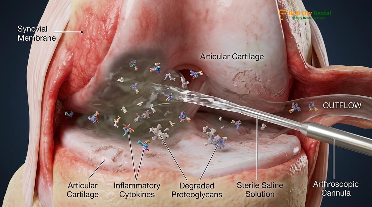
The therapeutic rationale for arthrocentesis is to physically interrupt this destructive biochemical cycle. By flushing the superior joint space with 100 to 300 milliliters of sterile irrigant, the surgeon effectively dilutes and washes away these concentrated inflammatory mediators. This rapid clearance of cytokines leads to a profound and often immediate reduction in joint pain. The table below outlines the primary inflammatory mediators targeted during the lavage process and their pathological effects on the TMJ.
| Inflammatory Mediator | Primary Source in the TMJ | Pathological Effect on Joint Function |
|---|---|---|
| Interleukin-1 Beta (IL-1β) | Inflamed synovial lining cells, macrophages | Stimulates cartilage degradation; induces hyperalgesia (increased pain sensitivity). |
| Interleukin-6 (IL-6) | Chondrocytes, osteoblasts | Promotes chronic inflammation and stimulates osteoclast activity (bone resorption). |
| Tumor Necrosis Factor-alpha (TNF-α) | Macrophages, T-cells | Triggers the release of matrix metalloproteinases (MMPs) which destroy collagen. |
| Prostaglandin E2 (PGE2) | Synovial fibroblasts | Potent vasodilator causing joint effusion (swelling) and severe localized pain. |
Beyond the biochemical benefits, the joint fluid wash also provides a crucial mechanical advantage. In cases of “closed lock” (disc displacement without reduction), the articular disc is often anchored anteriorly by a vacuum effect or by the formation of fine fibrous adhesions between the disc and the articular eminence. The hydraulic pressure generated during arthrocentesis expands the joint capsule, breaking the vacuum seal and physically sweeping away these early adhesions. Once the joint space is expanded and the restrictive bands are lysed, the surgeon can gently manipulate the mandible to help recapture the displaced disc and restore the normal translation of the condyle.
Injection of Hyaluronic Acid for Condylar Lubrication and Disc Movement
Following the lavage, injecting hyaluronic acid into the joint space replenishes depleted synovial fluid, reducing mechanical friction and promoting cartilage regeneration.
While the lavage phase of arthrocentesis is highly effective at removing inflammatory debris and breaking down adhesions, the newly flushed joint space is left temporarily devoid of its natural lubricants. To address this, modern clinical protocols almost universally incorporate viscosupplementation at the conclusion of the procedure. This involves the intra-articular injection of hyaluronic acid (HA), a naturally occurring glycosaminoglycan that is a primary structural component of healthy synovial fluid and articular cartilage.[3]
The hyaluronic acid injection jaw protocol serves multiple vital functions. First, it acts as a boundary lubricant. In a compromised TMJ, the natural HA is often depolymerized by free radicals, losing its high molecular weight and its ability to protect the joint surfaces. By injecting exogenous, high-molecular-weight hyaluronic acid, the surgeon restores the viscoelastic properties of the joint fluid. This viscous layer coats the articular surfaces of the condyle, the eminence, and the disc, significantly reducing mechanical friction during jaw movement. This reduction in friction is essential for preventing the re-adhesion of the disc and facilitating smooth, pain-free translation of the mandible.
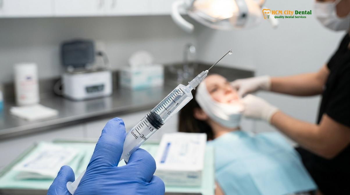
Furthermore, hyaluronic acid exhibits intrinsic anti-inflammatory and analgesic properties. It has been shown to inhibit the chemotaxis of inflammatory cells and block the pain receptors within the synovial membrane. Dr. Nguyen Van Cuong emphasizes the regenerative potential of this step: “Viscosupplementation is not merely a mechanical lubricant; it actively supports the metabolic health of the joint. By providing a protective viscoelastic shield, we allow the damaged chondrocytes the opportunity to synthesize new, healthy matrix proteins, thereby slowing the progression of degenerative joint disease.”
Clinical Contraindications: While TMJ arthrocentesis with hyaluronic acid is highly safe, it is strictly contraindicated in patients presenting with active purulent joint infections (septic arthritis), overlying skin infections at the injection site, or severe bony ankylosis where the joint space is completely obliterated by osseous fusion. Comprehensive imaging is mandatory prior to intervention.
The technique for viscosupplementation is performed immediately after the lavage is complete. The outflow needle is removed, and the syringe containing the sterile irrigant is swapped for a syringe pre-filled with hyaluronic acid. The HA is then slowly injected into the superior joint space through the remaining inflow needle. The surgeon will typically manipulate the jaw through its full range of motion to ensure the even distribution of the viscous fluid across all articular surfaces before removing the final needle and applying a sterile dressing.
Success Rates for Closed Lock TMJ Cases in Saigon
Clinical outcomes for arthrocentesis in treating acute closed lock are highly favorable, with most patients experiencing immediate pain relief and a significant increase in maximal mouth opening.
The clinical efficacy of TMJ arthrocentesis is well-documented in maxillofacial literature, particularly for patients suffering from acute disc displacement without reduction. This condition, characterized by a sudden inability to open the mouth fully (often restricted to less than 25 millimeters) and accompanied by severe pain, is a primary indication for the procedure. When conservative management fails to unlock the jaw, arthrocentesis provides a rapid and minimally invasive solution. Studies indicate that for acute closed lock cases, the success rate of restoring functional mouth opening (greater than 35 millimeters) and significantly reducing Visual Analog Scale (VAS) pain scores exceeds 80%.[4]
In recent years, Ho Chi Minh City has emerged as a leading center for advanced maxillofacial treatments. Patients experiencing severe TMD locking HCMC benefit from the integration of state-of-the-art diagnostic imaging and internationally trained specialists. The success of the procedure is heavily dependent on accurate pre-operative diagnosis. By utilizing high-resolution CBCT scans, surgeons can precisely evaluate the bony architecture of the condyle and the articular eminence, ruling out absolute contraindications such as severe osteophyte formation or bony ankylosis, which would necessitate more invasive surgical approaches.
Clinical Case Review: Acute Closed Lock Resolution
A 34-year-old female patient presented to HCMC Dental Clinic in Ho Chi Minh City with an acute inability to open her mouth wider than 22mm, accompanied by severe pre-auricular pain. She reported a history of morning jaw stiffness and clicking, which suddenly progressed to a “locked” state. After failing to respond to a week of muscle relaxants and a stabilization splint, Dr. Cuong performed a TMJ arthrocentesis with hyaluronic acid viscosupplementation under conscious sedation. Immediately post-procedure, the patient’s maximal incisal opening increased to 41mm. At the three-month follow-up, she maintained a pain-free opening of 44mm and continued nocturnal splint therapy to protect the joint.
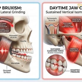
The long-term stability of the results achieved through arthrocentesis is generally excellent, provided that the underlying etiology of the joint dysfunction is addressed. If a patient suffers from severe nocturnal bruxism, the mechanical forces exerted on the joint during sleep can quickly degrade the newly injected hyaluronic acid and trigger a relapse of inflammation. Therefore, post-operative management is just as critical as the surgical procedure itself. Patients are typically prescribed a soft diet for several weeks, instructed in specific jaw physiotherapy exercises to maintain mobility, and fitted with a precisely calibrated occlusal splint to decompress the joint space and stabilize the bite.
“Arthrocentesis provides the critical reset button for an inflamed, locked joint. However, long-term success relies entirely on comprehensive post-operative management, including occlusal stabilization and the mitigation of parafunctional habits like bruxism.”
When to See a Doctor for Jaw Locking and Pain
Temporomandibular joint disorders encompass a wide spectrum of symptoms, ranging from mild, transient clicking to severe, debilitating pain. Because the TMJ is intimately connected to the muscles of mastication, the cervical spine, and the complex trigeminal nerve network, symptoms can often mimic other conditions such as tension headaches, ear infections, or trigeminal neuralgia. It is crucial for patients to recognize the clinical red flags that necessitate immediate evaluation by a qualified maxillofacial specialist or dental professional.
You should seek prompt clinical assessment if you experience a sudden, significant restriction in your ability to open your mouth. A normal maximal incisal opening (the distance between the upper and lower front teeth when the mouth is fully open) is typically between 40 and 55 millimeters. If your opening is suddenly restricted to less than 30 millimeters, or if your jaw deflects sharply to one side upon opening, this strongly suggests a mechanical obstruction within the joint, such as a displaced articular disc. This condition, known as a closed lock, requires timely intervention to prevent the formation of permanent fibrous adhesions within the joint capsule.[5]
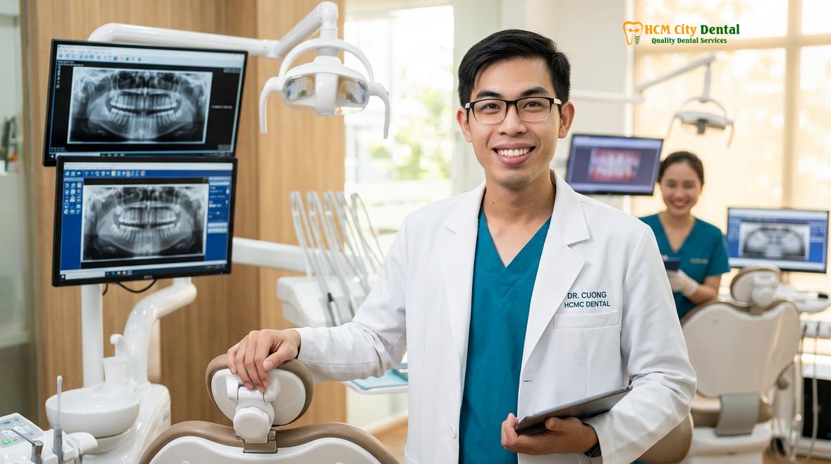
Furthermore, if you experience persistent, severe pain located directly in front of the ear that worsens with chewing or speaking, and this pain does not respond to over-the-counter non-steroidal anti-inflammatory drugs (NSAIDs) or warm compresses, professional diagnostics are required. The presence of loud, grating sounds (crepitus) during jaw movement, especially when accompanied by pain, is indicative of bone-on-bone friction and advanced degenerative joint disease. At HCMC Dental Clinic, our specialists utilize advanced 3D imaging to evaluate the structural integrity of the joint and develop a personalized, evidence-based treatment plan, ensuring that surgical interventions like arthrocentesis are only recommended when clinically necessary and highly likely to succeed.
Frequently Asked Questions
Is TMJ arthrocentesis performed under anesthesia?
Yes, TMJ arthrocentesis is typically performed under local anesthesia, often combined with intravenous conscious sedation to ensure complete patient comfort and prevent involuntary muscle guarding during the joint lavage procedure. The local anesthetic is carefully injected into the pre-auricular region to numb the skin and the joint capsule. The addition of conscious sedation allows the patient to remain relaxed, which lowers intra-articular pressure and makes it significantly easier for the surgeon to manipulate the jaw and break down fibrous adhesions.
How long does the recovery take after joint irrigation?
Initial recovery takes 24 to 48 hours, during which patients may experience mild localized swelling and altered bite sensation, with full functional jaw recovery and pain reduction expected within one to two weeks. Because arthrocentesis is a minimally invasive procedure that does not require incisions or bone removal, the surgical morbidity is very low. Patients are typically advised to adhere to a soft food diet, apply ice packs to minimize swelling, and perform gentle, prescribed jaw stretching exercises to maintain the newly achieved range of motion.
What is the cost of TMJ arthrocentesis in Vietnam?
The estimated cost for TMJ arthrocentesis in Vietnam generally ranges from $300 to $800 per joint, depending on the complexity of the case, imaging requirements, and whether viscosupplementation is utilized. This cost is highly competitive compared to Western countries, making Ho Chi Minh City a preferred destination for patients seeking advanced maxillofacial care. The fee typically encompasses the surgical procedure, the sterile irrigants, and the local anesthesia, though comprehensive pre-operative CBCT imaging and post-operative custom splints may be billed separately.
Can arthrocentesis cure temporomandibular joint osteoarthritis?
While it cannot reverse structural bone degeneration, arthrocentesis effectively manages the clinical symptoms of osteoarthritis by flushing out inflammatory mediators and improving joint mobility, thereby significantly enhancing the patient’s quality of life. Osteoarthritis of the TMJ involves the progressive breakdown of articular cartilage and changes to the underlying bone. Arthrocentesis, particularly when combined with hyaluronic acid injections, provides a highly effective palliative treatment by reducing the biochemical drivers of pain and restoring a protective lubricating layer to the damaged joint surfaces.
Will I still need to wear a bite splint after the procedure?
Yes, maxillofacial specialists strongly recommend continuing the use of a custom occlusal splint post-operatively to protect the newly lubricated joint from nocturnal bruxism forces and to maintain optimal bite stabilization. The splint acts as an orthopedic device, slightly decompressing the joint space and preventing the condyle from seating too deeply into the fossa. This protection is vital during the healing phase, as it prevents the re-accumulation of inflammatory cytokines and protects the injected hyaluronic acid from being prematurely degraded by excessive mechanical loading.
References
- Journal of Oral and Maxillofacial Surgery. The efficacy of TMJ arthrocentesis in the management of temporomandibular disorders: a systematic review. (2021).
- International Journal of Oral and Maxillofacial Surgery. Inflammatory mediators in the synovial fluid of patients with temporomandibular joint disorders. (2019).
- Journal of Cranio-Maxillofacial Surgery. Viscosupplementation with hyaluronic acid following arthrocentesis for TMJ internal derangement. (2020).
- Clinical Oral Investigations. Long-term outcomes of minimally invasive surgical management for closed lock of the temporomandibular joint. (2022).
- Journal of the American Dental Association. Stepwise approach to the management of temporomandibular joint pain and dysfunction. (2018).