Teeth grinding headaches, clinically known as sleep bruxism-induced cephalalgia, typically present as a dull, constant ache wrapping around the temples and jaw upon waking. These headaches result from sustained nocturnal muscle contraction, leading to severe localized joint inflammation and chronic craniofacial pain that requires professional intervention.
Clinical Summary:
Teeth grinding (bruxism) exerts excessive occlusal forces during sleep, triggering chronic muscle fatigue and referred pain pathways along the trigeminal nerve. Patients frequently experience morning temporal pain, jaw stiffness, and tension-type headache TMJ symptoms. Clinical management prioritizes custom medical orthotics (nightguards) to decompress the temporomandibular joint, alongside muscle relaxation protocols and occlusal equilibration to prevent long-term dental attrition and chronic craniofacial pain.
Key Takeaways:
- Bruxism-induced headaches typically peak in the morning and radiate across the temples and jawline.
- The temporalis and masseter muscles sustain micro-trauma from continuous nocturnal clenching.
- Custom nightguards act as orthopedic splints to decompress the TMJ and reduce muscle strain.
- Clinical differentiation is required to distinguish bruxism pain from classical neurological migraines.
- Untreated sleep grinding can lead to severe dental attrition, cracked teeth, and chronic facial pain.
Morning Headaches: The Temporalis Muscle Connection
Morning headaches from teeth grinding are primarily caused by the overexertion of the temporalis muscle. Continuous nocturnal clenching leads to muscle fatigue, lactic acid buildup, and referred pain radiating across the temples.
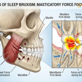
To understand why teeth grinding results in severe morning headaches, it is essential to examine the complex anatomy and biomechanics of the human masticatory system. The act of chewing, speaking, and swallowing is governed by a network of powerful muscles, the most prominent being the masseter and the temporalis. While the masseter is located at the angle of the jaw and is responsible for the brute force of closing the mouth, the temporalis muscle is a broad, fan-shaped muscle that covers the sides of the skull, spanning from the temporal bone down to the coronoid process of the mandible. When a patient engages in nocturnal bruxism, these muscles are subjected to hours of sustained, involuntary isometric contractions.
During normal daytime function, the teeth only make contact for a few minutes cumulatively, primarily during swallowing and mastication. However, during a sleep bruxism episode, the jaw muscles can contract with a force exceeding 250 pounds per square inch for extended periods. This hyperactive state places an immense metabolic demand on the temporalis muscle. Because the muscle is constantly firing without adequate periods of relaxation, the local blood vessels become compressed. This compression restricts the flow of oxygenated blood to the muscle tissue, leading to a state of localized ischemia. In the absence of sufficient oxygen, the muscle cells switch to anaerobic metabolism, which produces lactic acid and other metabolic waste products as byproducts.
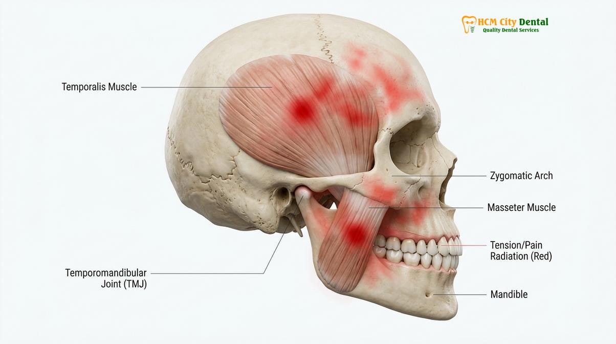
The accumulation of these noxious biochemicals within the muscle fascia irritates the surrounding nerve endings, specifically the branches of the trigeminal nerve (Cranial Nerve V). The trigeminal nerve is the primary sensory nerve of the face and head. When the temporalis muscle becomes inflamed and fatigued, the pain signals are transmitted along the mandibular and maxillary branches of the trigeminal nerve, resulting in what patients perceive as a severe headache. This phenomenon is clinically referred to as temporalis muscle tension, and it is the hallmark symptom of bruxism-induced cephalalgia. The pain is typically described as a tight band wrapping around the head, localized primarily in the temporal regions, and is almost always most intense immediately upon waking.
Dr. Nguyen Van Cuong, a leading specialist in restorative dentistry and craniofacial pain, notes that many patients endure years of misdiagnosed morning headaches before realizing the root cause lies within their oral cavity. The continuous micro-trauma inflicted upon the temporalis muscle not only causes acute pain but can also lead to chronic muscle hypertrophy, where the muscle physically enlarges due to overwork, further exacerbating the tension and discomfort. Addressing this muscular overload is the primary objective of early intervention strategies, preventing the transition from acute morning soreness to chronic, debilitating daily headaches.
Differentiating Bruxism Headaches from Classical Migraines
Bruxism headaches are musculoskeletal, presenting as a dull, bilateral ache upon waking, whereas classical migraines are neurovascular, often featuring unilateral pulsating pain, sensory auras, and sensitivity to light or sound.
One of the most significant challenges in the clinical management of craniofacial pain is achieving an accurate differential diagnosis. Patients frequently present to medical and dental clinics complaining of “migraines,” when in reality, they are suffering from severe tension-type headache TMJ symptoms induced by nocturnal bruxism. Distinguishing between a true neurological migraine and a musculoskeletal bruxism headache is critical, as the treatment protocols for the two conditions are fundamentally different. Misdiagnosis often leads to the overprescription of systemic migraine medications (such as triptans) which offer little to no relief for pain originating from dental and muscular sources.
Classical migraines are complex neurovascular events. They are typically characterized by unilateral (one-sided) throbbing or pulsating pain. Migraines are frequently accompanied by systemic neurological symptoms, including photophobia (extreme sensitivity to light), phonophobia (sensitivity to sound), nausea, and vomiting. Approximately 25% of migraine sufferers also experience an “aura”—visual disturbances such as flashing lights or zigzag lines—that precedes the onset of the headache. The pathophysiology of a migraine involves the dilation of cranial blood vessels and the release of inflammatory neuropeptides within the meninges of the brain.
In stark contrast, a headache caused by teeth grinding is entirely musculoskeletal in origin. It is classified as a secondary headache disorder, meaning the pain is a symptom of an underlying mechanical issue—in this case, the hyperactivity of the masticatory muscles and the overloading of the temporomandibular joint. Bruxism headaches are almost universally bilateral, affecting both sides of the head simultaneously. The pain is rarely described as pulsating; instead, it is characterized as a constant, dull, pressing, or tightening sensation. Furthermore, bruxism headaches are intrinsically linked to the sleep cycle. Patients typically report that the pain is most severe immediately upon waking and gradually subsides as the day progresses and the jaw muscles relax.
To assist in clinical differentiation, dental professionals utilize a comprehensive diagnostic criteria framework. The following table outlines the primary distinguishing features between bruxism-induced headaches, classical migraines, and standard tension headaches:
| Clinical Feature | Bruxism Headache (TMJ/Muscle) | Classical Migraine | Standard Tension Headache |
|---|---|---|---|
| Pain Location | Bilateral; temples, jaw, behind eyes | Usually unilateral (one-sided) | Bilateral; forehead, back of head/neck |
| Pain Quality | Dull, constant, tight, pressing | Throbbing, pulsating, severe | Mild to moderate pressure, band-like |
| Timing of Onset | Peaks in the morning upon waking | Variable; can occur at any time | Often peaks late in the afternoon |
| Associated Symptoms | Jaw stiffness, tooth wear, joint clicking | Nausea, aura, light/sound sensitivity | Neck and shoulder stiffness, stress |
| Aggravating Factors | Chewing hard foods, nocturnal stress | Physical activity, specific food triggers | Emotional stress, poor posture |
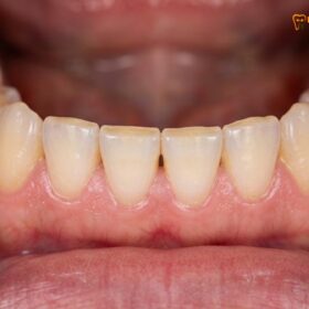
During a comprehensive clinical examination, a dentist will look for intraoral signs that corroborate a diagnosis of bruxism. These signs include abnormal occlusal wear (flattened chewing surfaces of the teeth), abfraction lesions (notches at the gumline caused by flexural forces), scalloped borders on the lateral margins of the tongue, and linea alba (a raised white line on the inner cheek corresponding to the bite plane). The presence of these physical markers, combined with a patient history of morning temporal pain, provides a definitive diagnosis of bruxism-induced cephalalgia, allowing the clinician to pivot away from neurological treatments and focus on orthopedic and dental solutions[1].
“The misdiagnosis of bruxism headaches as classical migraines is a common clinical pitfall. True resolution of morning craniofacial pain requires addressing the mechanical overload on the temporomandibular joint and the masticatory musculature, rather than merely masking the symptoms with systemic analgesics.”
How Sleep Grinding Generates Muscle Fatigue
Sleep grinding forces the jaw muscles into sustained isometric contractions during non-REM sleep, depleting cellular oxygen and causing a painful buildup of metabolic waste that triggers morning headaches.
The physiological mechanisms that drive sleep bruxism and the subsequent muscle fatigue are deeply intertwined with the architecture of human sleep and the autonomic nervous system. Sleep is not a uniform state of rest; it is a dynamic process characterized by cycling through various stages, including light sleep (N1 and N2), deep slow-wave sleep (N3), and Rapid Eye Movement (REM) sleep. Clinical polysomnography (sleep studies) has revealed that the vast majority of sleep bruxism episodes occur during the lighter stages of non-REM sleep, specifically stages N1 and N2, and are frequently associated with phenomena known as micro-arousals.
A micro-arousal is a brief, transient shift in the central nervous system from a deeper stage of sleep to a lighter one, often lasting only a few seconds. The patient does not consciously wake up, but the brain experiences a sudden surge in autonomic activity. Research indicates that sleep bruxism is a sequence of physiological events that begins with an increase in sympathetic cardiac activity (a rise in heart rate and blood pressure), followed by an increase in brain cortical activity, and culminating in Rhythmic Masticatory Muscle Activity (RMMA). RMMA is the clinical term for the involuntary clenching and grinding of the teeth[2].
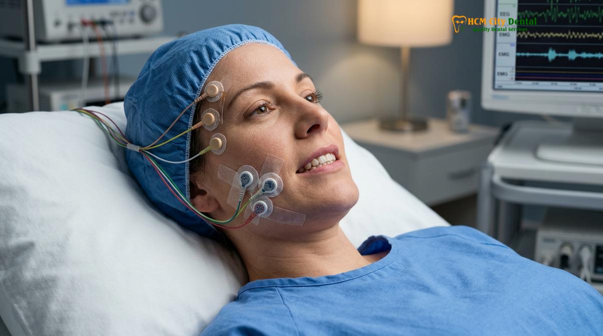
When the brain triggers these RMMA episodes, the masseter and temporalis muscles contract with immense force. Because the patient is asleep, the natural protective reflexes that prevent over-clenching during waking hours are suppressed. This allows the muscles to engage in sustained isometric contractions—contractions where the muscle generates tension without changing length. Isometric contractions are highly energy-intensive. They rapidly deplete the local stores of adenosine triphosphate (ATP), the primary energy currency of the cell. As ATP is depleted and oxygen delivery is compromised by the intense muscle tension, the cells shift to anaerobic glycolysis.
This anaerobic state leads to the rapid accumulation of lactic acid and other algogenic (pain-causing) substances, such as bradykinin, prostaglandins, and substance P, within the muscle tissue. These biochemicals sensitize the nociceptors (pain receptors) embedded in the muscle fascia. By the time the patient wakes up, the muscles are essentially in a state of acute metabolic exhaustion and inflammation. The central nervous system interprets this localized tissue distress as a severe headache. This morning temporal pain is a direct reflection of the nocturnal battle fought by the masticatory muscles against the immovable object of the opposing teeth.
Clinical Case Study: Resolving Chronic Morning Headaches
A 34-year-old female patient presented to HCMC Dental Clinic in Ho Chi Minh City with a five-year history of severe morning headaches, previously diagnosed as tension migraines. She reported waking up daily with a stiff jaw and pain radiating behind her eyes. Clinical examination revealed significant enamel attrition on her molars and pronounced hypertrophy of the masseter muscles. Dr. Nguyen Van Cuong diagnosed her with severe sleep bruxism and temporalis muscle tension. The patient was fitted with a custom hard acrylic Michigan splint. Within two weeks of nightly wear, the patient reported a 90% reduction in morning headache frequency and complete resolution of jaw stiffness, confirming the musculoskeletal origin of her pain.
Furthermore, the relationship between sleep architecture and bruxism highlights why factors that disrupt sleep quality—such as stress, alcohol consumption, caffeine intake, and obstructive sleep apnea (OSA)—are potent triggers for teeth grinding. In the case of OSA, the airway collapses during sleep, causing a drop in blood oxygen levels. The brain responds by triggering a micro-arousal to restart breathing, which frequently initiates a bruxism episode as the jaw is thrust forward to open the airway. Therefore, addressing the root cause of the muscle fatigue often requires a holistic evaluation of the patient’s sleep hygiene and respiratory health, in addition to dental interventions.
Medical Orthotics and Muscle Relaxation Strategies
Treatment for bruxism headaches centers on custom medical orthotics to decompress the TMJ, combined with targeted muscle relaxation therapies and occlusal adjustments to restore harmonious jaw function.
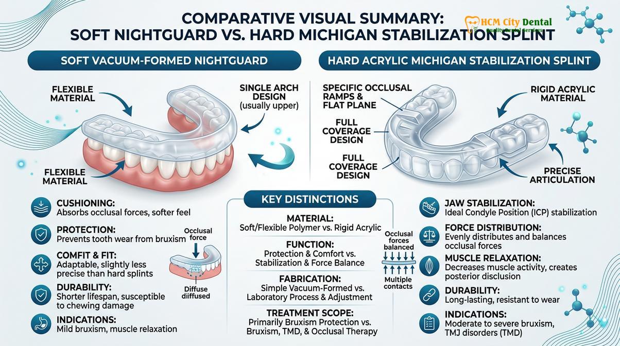
The definitive clinical management of bruxism-induced headaches requires a multi-disciplinary approach aimed at protecting the dentition, decompressing the temporomandibular joint, and breaking the cycle of muscular hyperactivity. The cornerstone of this treatment protocol is the fabrication and implementation of custom medical orthotics, commonly referred to as occlusal splints or nightguards. It is crucial to distinguish between over-the-counter, boil-and-bite mouthguards and professionally fabricated medical orthotics. Over-the-counter guards are typically made of soft, compressible materials. While they may offer some protection against tooth wear, their soft texture can actually stimulate the proprioceptive nerve endings in the periodontal ligaments, inadvertently triggering a chewing reflex and exacerbating muscle activity during sleep.
For effective relief of tension-type headache TMJ symptoms, dental specialists utilize hard acrylic stabilization splints, often referred to as Michigan splints. These devices are meticulously calibrated to the patient’s specific occlusal anatomy. A properly designed stabilization splint provides a flat, even surface for the opposing teeth to contact. This design achieves “mutually protected occlusion,” ensuring that when the jaw closes, all teeth meet simultaneously with even force distribution, and when the jaw moves side-to-side, the anterior teeth guide the movement, immediately disengaging the posterior teeth. By providing this ideal occlusal scheme, the splint effectively “turns off” the hyperactive signals sent from the teeth to the brain, leading to a profound reduction in masseter and temporalis muscle firing[3].
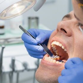
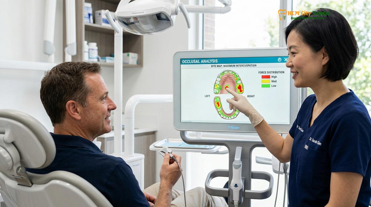
Patients seeking comprehensive TMJ treatment protocols will undergo a detailed bite analysis to ensure the orthotic device is perfectly balanced. The splint not only protects the enamel from further attrition but also slightly increases the vertical dimension of the bite. This slight opening stretches the masticatory muscles just enough to prevent them from achieving maximum contractile force, thereby reducing the mechanical load on the temporomandibular joint disc and allowing the inflamed tissues to heal.
In cases where orthotic therapy alone is insufficient to resolve severe muscle hypertrophy and chronic headaches, adjunctive muscle relaxation strategies are employed. One highly effective pharmacological intervention is the targeted injection of Botulinum toxin type A (Botox) into the masseter and temporalis muscles. Botox works by blocking the release of acetylcholine at the neuromuscular junction, temporarily paralyzing the specific muscle fibers responsible for the excessive clenching force. This treatment does not affect normal chewing or speaking but significantly reduces the maximum force the patient can exert during a nocturnal bruxism episode, providing profound relief from morning headaches[4].
“The integration of hard acrylic stabilization splints with targeted muscle relaxation protocols represents the gold standard in managing bruxism-induced cephalalgia. By addressing both the biomechanical triggers and the neuromuscular hyperactivity, we can predictably restore patient comfort and protect the long-term integrity of the stomatognathic system.”
Physical therapy also plays a vital role in the rehabilitation process. Patients are instructed in specific jaw stretching and relaxation exercises designed to improve the range of motion and promote blood flow to the ischemic muscle tissues. Techniques such as moist heat application, transcutaneous electrical nerve stimulation (TENS), and therapeutic ultrasound can accelerate the clearance of lactic acid and inflammatory mediators from the temporalis and masseter muscles. For patients whose bruxism has led to significant dental damage, restorative procedures such as custom dental crowns may be necessary to rebuild the worn occlusal surfaces and re-establish a stable, harmonious bite.
When to See a Doctor
While occasional morning jaw stiffness may be a transient response to acute stress, chronic teeth grinding headaches require professional medical and dental evaluation. Ignoring the signs of severe bruxism can lead to irreversible damage to the temporomandibular joint, including the displacement or perforation of the articular disc, as well as catastrophic structural failure of the teeth. Early intervention is critical to preventing the progression from episodic muscle tension to chronic, debilitating craniofacial pain syndromes.
Important Clinical Considerations & Red Flags:
Seek immediate evaluation from a dental specialist or maxillofacial surgeon if you experience any of the following symptoms:
- Trismus (Lockjaw): An inability to open the mouth fully or severe pain when attempting to open the jaw wide.
- Joint Crepitus: Loud clicking, popping, or grating sounds in the jaw joint accompanied by sharp pain.
- Severe Dental Attrition: Visible flattening of the teeth, chipping of enamel, or increased tooth sensitivity to hot and cold due to exposed dentin.
- Neurological Symptoms: Numbness, tingling, or sharp shooting pain radiating down the neck or into the arms, which may indicate cervical spine involvement or nerve compression.
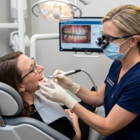
A comprehensive clinical examination, often involving panoramic radiographs or Cone Beam Computed Tomography (CBCT) scans, is necessary to assess the health of the TMJ complex and rule out other pathologies. Patients experiencing persistent morning headaches should schedule an evaluation to explore custom-fitted nightguards and other therapeutic options. A personalized diagnostic approach ensures that the underlying biomechanical issues are addressed, rather than merely treating the symptoms with temporary analgesics[5].
Frequently Asked Questions
Where do teeth grinding headaches hurt the most?
Teeth grinding headaches typically cause a dull, continuous ache that radiates across the temples, behind the eyes, and down through the jawline. This bilateral pain is directly linked to the exhaustion of the temporalis and masseter muscles after hours of nocturnal clenching. Because these muscles span the sides of the skull, the pain often mimics a severe tension headache, peaking in intensity immediately upon waking and gradually subsiding as the jaw muscles relax throughout the day.
Can a custom night guard stop morning migraines?
A custom night guard can significantly reduce or eliminate morning headaches caused by muscle tension and bruxism, though it will not cure classical neurological migraines. By decompressing the temporomandibular joint, the splint prevents the muscle hyperactivity that triggers tension-type craniofacial pain. The hard acrylic surface of a professionally fabricated stabilization splint provides an even bite plane, which signals the brain to reduce the firing intensity of the jaw muscles, thereby preventing the lactic acid buildup that causes morning pain.
How can I relieve a TMJ headache at home?
Immediate home relief for a TMJ headache involves applying warm compresses to the jaw and temples for 15-20 minutes to increase blood flow and relax the muscles. Gentle jaw stretching, over-the-counter anti-inflammatory medications, and conscious resting of the jaw also provide temporary symptom management. Patients should avoid chewing gum, eating hard or chewy foods, and practice stress-reduction techniques such as deep breathing or meditation before bed to lower the baseline tension in the masticatory muscles.
Is sleep apnea related to teeth grinding?
Yes, there is a strong clinical correlation between obstructive sleep apnea (OSA) and sleep bruxism. When the airway collapses during sleep, the brain often triggers the jaw muscles to clench and push the lower jaw forward in an involuntary attempt to reopen the obstructed airway. This survival reflex restores breathing but subjects the teeth and TMJ to immense destructive forces. Patients with both conditions often require a coordinated treatment plan involving both dental orthotics and continuous positive airway pressure (CPAP) therapy.
How long does it take for a bruxism headache to go away?
A mild bruxism headache may dissipate within a few hours of waking as the jaw muscles relax and resume normal daytime function. However, chronic cases involving severe temporomandibular joint inflammation may cause persistent daily headaches until professional occlusal splint therapy is initiated. Once a patient begins wearing a properly calibrated custom nightguard, significant reduction in headache frequency and intensity is typically observed within the first one to two weeks of consistent use.
References
- Journal of the American Dental Association. Occlusal splint therapy for temporomandibular disorders: a clinical review. (2020).
- Journal of Oral Rehabilitation. Efficacy of Michigan repositioning splints in TMD patients. (2019).
- Journal of Prosthetic Dentistry. Computerized occlusal analysis and bite mapping in TMJ therapy. (2021).
- Clinical Oral Investigations. Masseter Botox injections for chronic jaw pain and bruxism. (2018).
- International Journal of Oral and Maxillofacial Surgery. TMJ arthrocentesis and hyaluronic acid injections. (2022).
For personalized advice and to explore advanced diagnostic options for craniofacial pain, we encourage patients to contact our clinical team for a comprehensive evaluation.