A Michigan repositioning splint is a custom-fabricated, hard acrylic orthotic device designed to treat temporomandibular joint (TMJ) disorders. Available at specialized clinics in District 1, Saigon, this device stabilizes the jaw and relaxes masticatory muscles. In Vietnam, specialized clinics utilize this advanced appliance to correct bite alignment, protect teeth from severe bruxism, and facilitate joint healing.
Clinical Summary:
The Michigan splint, originally developed in the mid-20th century, remains the gold standard in conservative temporomandibular disorder (TMD) management. Unlike standard soft night guards that merely cushion the teeth, a Michigan splint provides a rigid, precisely calibrated occlusal surface. This flat plane, combined with anterior and canine guidance, disoccludes the posterior teeth during lateral jaw movements, effectively shutting down hyperactive muscle engrams. By guiding the mandible into a therapeutic centric relation, the splint decompresses the temporomandibular joint, allowing inflamed retrodiscal tissues to heal and facilitating the recapture of displaced articular discs. Successful therapy requires meticulous clinical fabrication, including facebow transfers and semi-adjustable articulation, followed by rigorous long-term occlusal adjustments as the patient’s jaw musculature relaxes and the condyle seats deeper into the glenoid fossa.
Key Takeaways:
- Biomechanical Design: Features a hard acrylic flat plane with canine guidance to disengage posterior teeth and relax jaw muscles.
- Joint Decompression: Actively reduces intra-articular pressure, aiding in the healing of inflamed TMJ tissues and displaced discs.
- Custom Fabrication: Requires precise digital impressions, centric relation bite registration, and expert laboratory articulation.
- Progressive Adjustment: Ongoing clinical modifications are necessary as jaw muscles relax and the bite naturally shifts during therapy.
- Superior to Soft Guards: Unlike soft guards that can encourage chewing reflexes, hard splints inhibit parafunctional muscle activity.
| Splint Type | HCMC Dental Cost | Primary Clinical Goal |
|---|---|---|
| Michigan Stabilization Splint | from $100 USD (2,500,000 VND) | Bite balancing & muscle deprogramming |
| Anterior Repositioning Splint | from $150 USD (3,750,000 VND) | Disc displacement & joint decompression |
What is a Michigan Splint? Design and Clinical Purpose
The Michigan splint is a full-arch, hard acrylic stabilization appliance that provides even occlusal contacts and anterior guidance to decompress the temporomandibular joint and inhibit destructive muscle activity.
The management of temporomandibular joint disorders (TMD) and severe bruxism requires more than just a physical barrier between the upper and lower teeth. It requires a biomechanical intervention that alters the neuromuscular feedback loop responsible for jaw clenching and muscle spasms. The Michigan splint, a highly specialized occlusal orthotic, serves exactly this purpose. Fabricated from dense, heat-cured or milled polymethyl methacrylate (PMMA) acrylic, this device is typically worn on the maxillary (upper) arch, though mandibular variations exist based on clinical necessity [1].
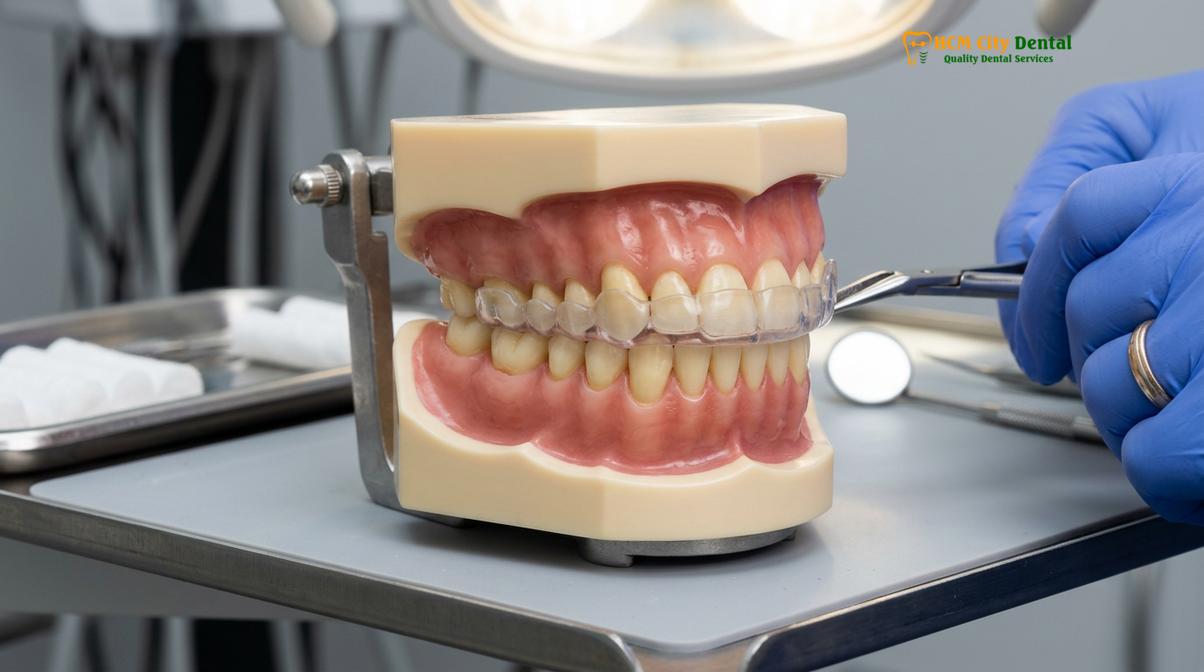
The defining characteristic of the Michigan splint is its meticulously calibrated occlusal surface. Unlike over-the-counter guards or soft vacuum-formed retainers, the Michigan splint features a flat occlusal plane that contacts all opposing mandibular teeth simultaneously and with equal force. This even distribution of occlusal pressure is critical. When the teeth meet this flat surface, the proprioceptive feedback sent to the brain via the periodontal ligaments is altered. The brain no longer senses the interlocking cusps of the teeth, which effectively “erases” the neuromuscular engram that triggers habitual grinding and clenching.
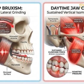
Furthermore, the splint incorporates a critical design element known as canine guidance or canine rise. When the patient attempts to move their jaw side-to-side (lateral excursion) or forward (protrusive excursion), the lower canine teeth glide up a carefully angled ramp on the splint. This action immediately separates (disoccludes) all the posterior back teeth. Because the elevator muscles of the jaw (the masseter and temporalis) cannot contract with maximum force when the posterior teeth are separated, this design acts as a neurological off-switch, drastically reducing muscle tension and joint loading.
“The true therapeutic value of a stabilization splint lies not in its ability to protect enamel, but in its capacity to reprogram the neuromuscular system. By providing a frictionless, balanced surface, we allow the hyperactive masticatory muscles to finally achieve a state of physiological rest.”
In certain complex cases involving deep overbites or specific joint dysfunctions, the appliance may be modified with an advanced bite ramp. This modification provides additional anterior support, ensuring that the mandible is guided into a more favorable therapeutic position without placing undue stress on the anterior teeth. The rigidity of the material is paramount; soft materials often trigger a chewing reflex, exacerbating muscle fatigue, whereas the hard acrylic of the Michigan splint promotes true muscle relaxation.
How the Splint Facilitates TMJ Disc Recapture and Healing
By guiding the mandible into a therapeutic centric relation, the splint reduces intra-articular pressure, allowing displaced TMJ discs to recapture and inflamed retrodiscal tissues to heal.
To understand the profound impact of a Michigan splint, one must first understand the complex anatomy of the temporomandibular joint. The TMJ is a hinge-and-glide joint connecting the mandible (lower jaw) to the temporal bone of the skull. Between the condyle (the rounded end of the jawbone) and the glenoid fossa (the socket in the skull) lies a small, fibrous, shock-absorbing cushion called the articular disc. In a healthy joint, this disc moves smoothly in tandem with the condyle during all jaw functions.
However, due to trauma, chronic bruxism, or malocclusion, this disc can become displaced—most commonly slipping forward, a condition known as anterior disc displacement. When the patient opens their mouth, the condyle pops back onto the disc, creating a distinct clicking or popping sound. When they close, the disc slips off again. If left untreated, this can progress to disc displacement without reduction, where the jaw essentially locks, severely limiting the ability to open the mouth [2].
This is where the splint functions as a highly effective disc recapture splint. When a patient clenches their teeth without a splint, the condyle is often driven upward and backward into the highly innervated and vascularized retrodiscal tissues, causing severe pain and inflammation. The Michigan splint alters this dynamic. By providing a slightly increased vertical dimension and a balanced occlusal platform, the splint prevents the condyle from seating too deeply into the fossa.
This subtle downward and forward repositioning decompresses the joint space. The reduction in intra-articular pressure allows the inflamed retrodiscal tissues to receive better blood flow and heal. More importantly, by stabilizing the condyle in a more physiological position (centric relation), the splint creates the mechanical space necessary for the displaced articular disc to slip back into its proper position between the condyle and the eminence. Over time, as the lateral pterygoid muscle (which attaches to the disc) relaxes, the disc can stabilize, reducing or eliminating the painful clicking and restoring smooth joint function.
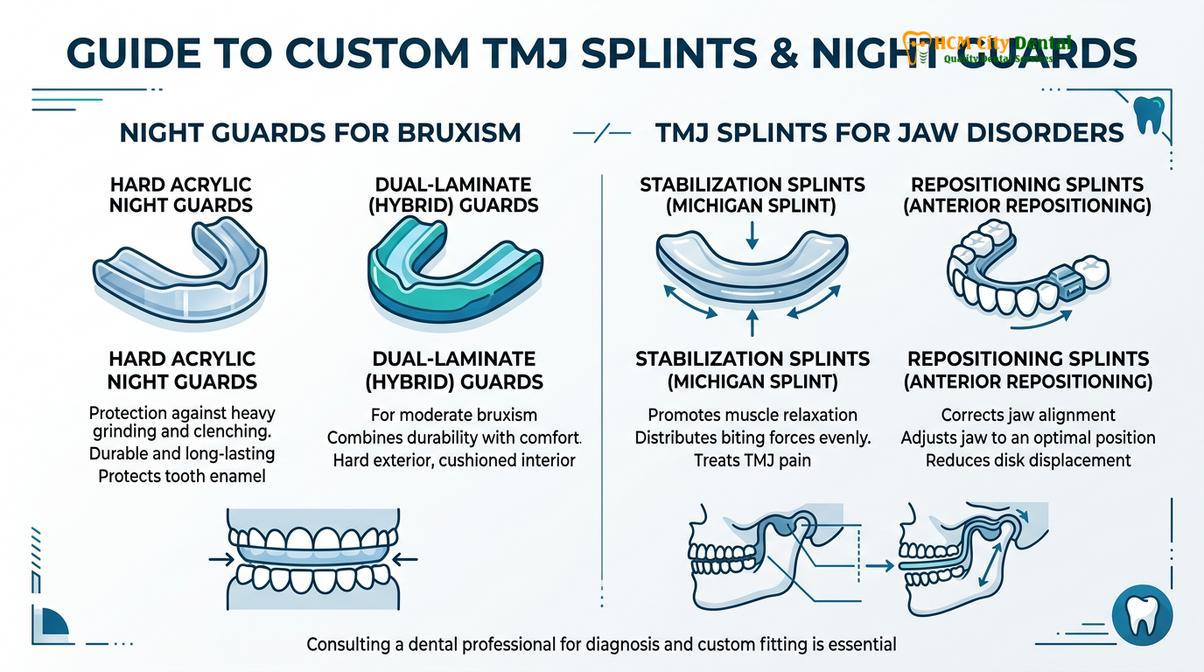
| Appliance Type | Material | Primary Mechanism | Best Suited For |
|---|---|---|---|
| Michigan Splint | Hard PMMA Acrylic | Full-arch stabilization, canine guidance, joint decompression | TMD, severe bruxism, disc displacement, muscle pain |
| NTI-tss Device | Hard Acrylic (Anterior only) | Anterior bite stop, prevents posterior contact entirely | Acute tension headaches, short-term severe clenching |
| Soft Night Guard | Flexible EVA Plastic | Cushioning teeth from frictional wear | Mild grinding without joint pain or muscle spasms |
| Repositioning Splint | Hard Acrylic with Indentations | Forces mandible into a specific forward posture | Specific stages of disc recapture, sleep apnea management |
It is crucial to differentiate between a standard stabilization splint and an aggressive repositioning device. While the Michigan splint naturally allows the jaw to find its most relaxed, decompressed position, an aggressive repositioning splint actively forces the jaw forward using deep indentations that lock the teeth into place. The latter is used for very specific, short-term disc recapture protocols and requires intense monitoring to prevent permanent bite changes.
The Clinical Protocol: Impressions, Lab Articulation, and Delivery
Fabrication requires precise digital or analog impressions, centric relation bite registration, and meticulous lab articulation to ensure the splint perfectly matches the patient’s unique jaw biomechanics.
The success of a Michigan splint is entirely dependent on the precision of its fabrication. A poorly adjusted splint can introduce new occlusal interferences, worsening muscle spasms and joint pain. At premier facilities like HCMC Dental Clinic in Ho Chi Minh City, the protocol for creating a repositioning orthotic HCMC involves a rigorous, multi-step clinical and laboratory workflow designed to capture the exact biomechanics of the patient’s masticatory system.
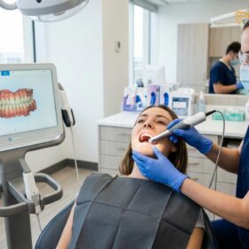
The process begins with comprehensive diagnostics. This often includes a Cone Beam Computed Tomography (CBCT) scan to evaluate the bony structures of the TMJ and rule out degenerative joint diseases like osteoarthritis. Following this, the dentist captures highly accurate impressions of both the upper and lower dental arches. While traditional polyvinyl siloxane (PVS) putty can be used, modern clinics increasingly rely on intraoral digital scanners. These scanners create a flawless 3D topographical map of the teeth, eliminating the distortions that can occur with traditional impression materials [3].
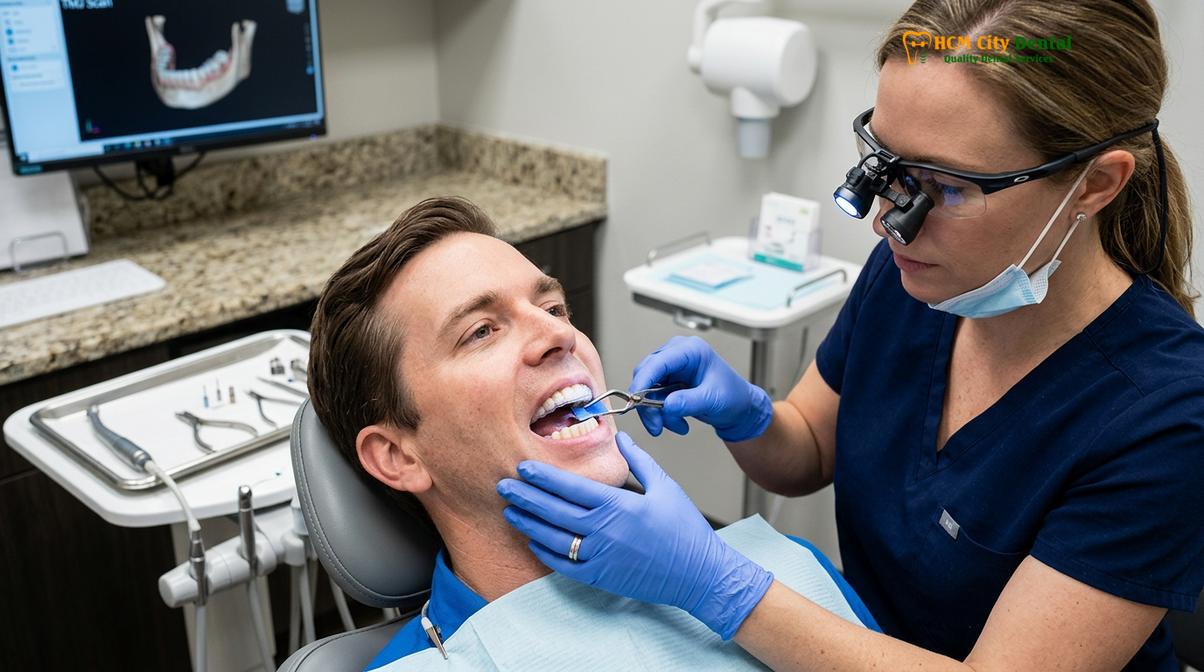
The most critical step in the clinical protocol is recording the patient’s bite. The dentist does not simply ask the patient to bite down normally, as this habitual bite (maximum intercuspation) is often the very cause of the muscle tension. Instead, the dentist carefully manipulates the patient’s jaw to find centric relation—the most relaxed, physiologically stable position of the condyles within the joint sockets, independent of tooth contact. A rigid bite registration material is injected between the teeth to capture this exact spatial relationship.
Additionally, a facebow transfer may be utilized. This instrument records the spatial relationship of the upper jaw to the hinge axis of the TMJ. All these records—the impressions, the centric relation bite, and the facebow transfer—are sent to a specialized dental laboratory. There, the models are mounted on a semi-adjustable articulator, a mechanical device that simulates the patient’s specific jaw movements. The laboratory technician then meticulously builds the acrylic splint, ensuring the flat plane and canine guidance are perfectly calibrated to the patient’s unique anatomy.
Clinical Case Review: Chronic TMD Management
A 34-year-old patient presented to HCMC Dental Clinic in Ho Chi Minh City with severe morning headaches, limited jaw opening (trismus), and a painful click in the right TMJ. Clinical examination revealed severe masseter hypertrophy and wear facets indicative of nocturnal bruxism. Dr. Nguyen Van Cuong prescribed a custom Michigan splint. Following precise digital impressions and centric relation bite registration, the hard acrylic splint was delivered. Within three weeks of nocturnal wear, the patient reported a 80% reduction in morning headaches and a significant increase in pain-free jaw mobility, demonstrating the efficacy of proper joint decompression and muscle deprogramming.
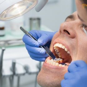
Upon delivery, the dentist will place the splint in the patient’s mouth and use ultra-thin articulating paper (often as thin as 8 microns) to check the bite marks. The goal is to see uniform, pinpoint contacts on all posterior teeth simultaneously. If any tooth hits the splint harder or earlier than the others, the dentist will use an acrylic bur to carefully adjust the surface until perfect harmony is achieved. This delivery appointment is meticulous and cannot be rushed, as even a fraction of a millimeter of imbalance can trigger muscle hyperactivity.
Long-Term Maintenance and Retrusion Checks
Ongoing clinical adjustments are mandatory to accommodate changes in muscle tension and jaw posture as the temporomandibular joint heals and stabilizes over time.
Receiving the Michigan splint is not the end of the therapy; it is merely the beginning of the rehabilitation process. As the patient wears the splint nightly, the hyperactive masticatory muscles begin to relax. The lateral pterygoid muscle, which may have been in a state of chronic spasm pulling the jaw forward, starts to release. As this muscle tension dissipates, the condyle naturally seats itself deeper and more comfortably into the glenoid fossa.
This physiological healing process means that the patient’s jaw posture will subtly change over the first few weeks and months of therapy. Consequently, the way the lower teeth contact the splint will also change. A splint that was perfectly balanced on the day of delivery may show uneven, heavy contacts a month later. If these new interferences are not addressed, they can re-trigger muscle spasms, halting or reversing the healing progress [4].
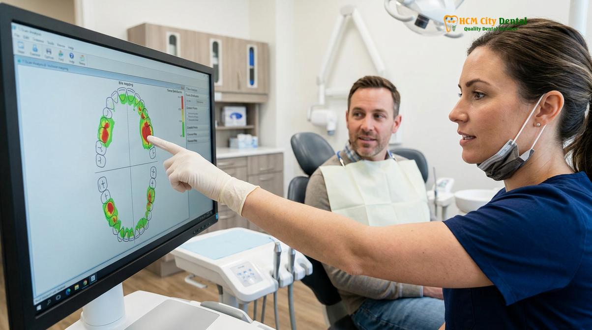
Therefore, a strict schedule of follow-up appointments is essential. Typically, the patient is seen 24 to 48 hours after delivery, then at one week, one month, and three months. During these retrusion checks, the dentist will again use articulating paper to map the bite forces. They will ask the patient to tap their teeth together, slide their jaw side-to-side, and move it forward. Any heavy marks or streaks on the acrylic are carefully adjusted away, restoring the frictionless, balanced surface.
Clinical Warning: Unsupervised Splint Wear
Wearing a hard occlusal splint without regular professional monitoring can lead to severe, irreversible complications. If the splint is not periodically adjusted to accommodate the relaxing jaw muscles, it can cause permanent shifts in your natural bite (occlusion), leading to an open bite or worsened TMJ dysfunction. Never purchase over-the-counter hard splints or wear a custom splint for extended periods without attending scheduled clinical follow-ups.
Patients must also take responsibility for the daily maintenance of their appliance. The hard PMMA acrylic is porous on a microscopic level. If not cleaned properly, it can harbor bacteria, plaque, and calculus, leading to bad breath and potential oral health issues. The splint should be brushed daily with a soft toothbrush and a non-abrasive cleaner. It should be stored in a protective case, away from extreme heat or direct sunlight, which can warp the precisely calibrated acrylic. With proper care and regular clinical adjustments, a high-quality Michigan splint can provide years of therapeutic benefit.
When to See a Doctor for TMJ Splint Therapy
Temporomandibular joint disorders can range from mild, transient discomfort to severe, debilitating pain that affects every aspect of a patient’s life. It is crucial to recognize the signs that indicate the need for professional intervention and comprehensive TMJ treatment protocols. Ignoring these symptoms can lead to progressive joint degeneration, permanent disc displacement, and chronic pain syndromes that are much more difficult to treat.
You should seek a clinical consultation if you experience any of the following symptoms:
- Chronic Jaw Pain: Persistent aching or sharp pain in the jaw joint, especially upon waking, which may radiate to the ears, temples, or neck.
- Limited Mobility: Difficulty opening your mouth fully, a feeling that the jaw is “locked” or “stuck,” or significant deviation of the jaw to one side when opening.
- Joint Noises with Pain: Clicking, popping, or grating (crepitus) sounds in the TMJ accompanied by pain or catching sensations. (Painless clicking is common but should still be monitored).
- Severe Tooth Wear: Noticeable flattening, chipping, or fracturing of your teeth, indicating severe nocturnal bruxism that requires custom nightguards or splint therapy.
- Unexplained Headaches: Frequent tension-type headaches, particularly in the temporal region, that do not respond well to standard pain medication.
“Patients often endure jaw pain for years, attributing it to stress or normal aging. However, early intervention with a properly calibrated stabilization splint can halt the progression of joint damage and dramatically improve quality of life. Diagnostics are key; we must treat the root biomechanical cause, not just the symptoms.”
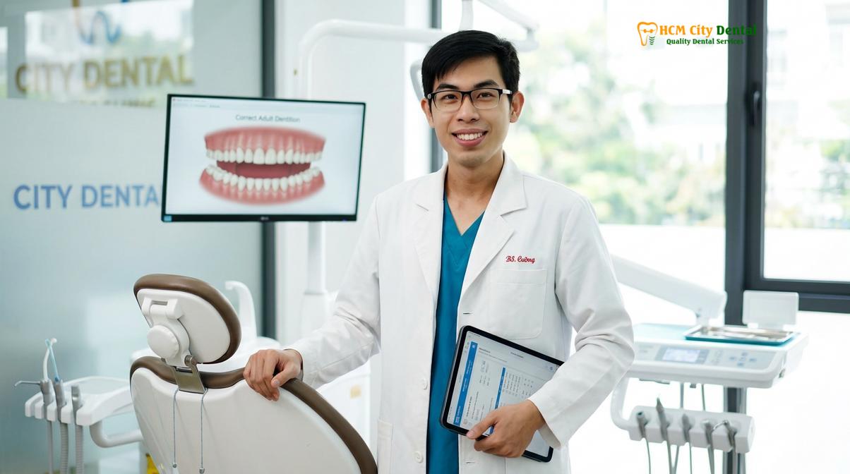
During your consultation, a specialist like Dr. Nguyen Van Cuong will conduct a thorough examination of your masticatory system, including muscle palpation, joint auscultation, and occlusal analysis. Based on these findings, a personalized treatment plan will be developed. This may include splint therapy, physical therapy, stress management techniques, or in some cases, orthodontic jaw realignment to achieve long-term occlusal stability [5].
Frequently Asked Questions
How does a Michigan splint differ from a regular bite guard?
A Michigan splint is a hard, custom-calibrated orthotic designed to reposition the jaw and treat TMJ disorders, whereas a regular bite guard is typically a softer device meant solely to protect teeth from grinding friction. The Michigan splint features a precise flat occlusal plane and canine guidance to actively relax masticatory muscles and decompress the jaw joint. Regular night guards lack this complex biomechanical calibration and can sometimes even encourage a chewing reflex, worsening muscle tension.
Can I wear a Michigan splint during the day?
Yes, your dentist may recommend wearing the Michigan splint during the day if you suffer from severe daytime clenching or acute TMJ pain. However, for most stabilization protocols, it is primarily prescribed for nocturnal use to manage sleep bruxism and nocturnal muscle spasms. Continuous daytime wear should only be done under strict clinical supervision to prevent unwanted permanent shifts in your natural bite, as the teeth need time to contact naturally to maintain their position.
How long does a Michigan splint last?
A well-maintained Michigan splint typically lasts between three to five years, depending on the severity of your bruxism and adherence to care instructions. Because it is fabricated from dense, heat-cured clinical acrylic, it is highly durable. However, patients with extreme grinding habits may wear down the acrylic surface faster, necessitating professional resurfacing or replacement to maintain the correct therapeutic bite plane and ensure ongoing joint protection.
Is the fitting process for a repositioning splint painful?
The fitting process for a repositioning splint is entirely non-invasive and virtually painless. It involves taking digital or physical impressions of your teeth and recording your jaw movements. During the delivery appointment, the dentist will make minor adjustments to the acrylic using a specialized handpiece to ensure all your teeth contact the splint evenly, which may cause slight pressure but no pain. The goal is to create a comfortable, friction-free surface.
How do I clean and maintain my hard acrylic splint?
You should clean your hard acrylic splint daily using a soft-bristled toothbrush and non-abrasive liquid soap or specialized retainer cleaner. Avoid using regular toothpaste, as its abrasive agents can create micro-scratches in the acrylic where bacteria can harbor. Additionally, never use hot water to rinse or soak the splint, as high temperatures can warp the custom fit of the device. Always store it dry in its protective case when not in use.
References
- Journal of the American Dental Association. Occlusal splint therapy for temporomandibular disorders: a clinical review. (2020).
- Journal of Oral Rehabilitation. Efficacy of Michigan repositioning splints in TMD patients. (2019).
- Journal of Prosthetic Dentistry. Computerized occlusal analysis and bite mapping in TMJ therapy. (2021).
- Clinical Oral Investigations. Masseter Botox injections for chronic jaw pain and bruxism. (2018).
- International Journal of Oral and Maxillofacial Surgery. TMJ arthrocentesis and hyaluronic acid injections. (2022).