Bruxism symptoms and signs primarily include involuntary teeth grinding and jaw clenching, leading to flattened enamel, morning jaw soreness, and chronic facial pain. Identifying these clinical indicators early is crucial to preventing severe dental fractures, temporomandibular joint (TMJ) disorders, and irreversible tooth wear.
Clinical Summary:
Bruxism manifests through both visual dental damage and physical discomfort. Key signs include tooth wear facets, chipped enamel, and linea alba on the inner cheeks. Patients frequently experience morning jaw soreness, tension headaches, and myofascial pain due to excessive masticatory muscle activity. Clinical intervention, often involving custom nightguards and stress management, is essential to halt progressive structural damage like abfraction lesions and tooth fractures.
Key Takeaways:
- Bruxism involves involuntary grinding or clenching, occurring during sleep or waking hours.
- Visual signs include flattened chewing surfaces, chipped enamel, and a raised white line (linea alba) inside the cheek.
- Physical symptoms often present as morning jaw soreness, dull temporal headaches, and unexplained earaches.
- Prolonged grinding forces can cause abfraction lesions at the gumline and severe tooth fractures.
- Early diagnosis and intervention with custom nightguards can protect dentition and alleviate myofascial pain.
What is Bruxism? Understanding Teeth Grinding and Clenching
Bruxism is a repetitive jaw-muscle activity characterized by the involuntary clenching or grinding of teeth, which can occur during sleep or while awake.
Bruxism is a complex, multifactorial condition that extends far beyond a simple bad habit. Clinically, it is classified into two distinct categories: sleep bruxism and awake bruxism. While both involve the hyperfunction of the masticatory system, their underlying triggers and physiological manifestations differ significantly. Sleep bruxism is considered a sleep-related movement disorder, often associated with micro-arousals in the central nervous system, sleep apnea, and altered neurotransmitter function [1]. Awake bruxism, conversely, is typically a subconscious reaction to emotional stress, anxiety, or deep concentration, manifesting primarily as sustained jaw clenching rather than rhythmic grinding.
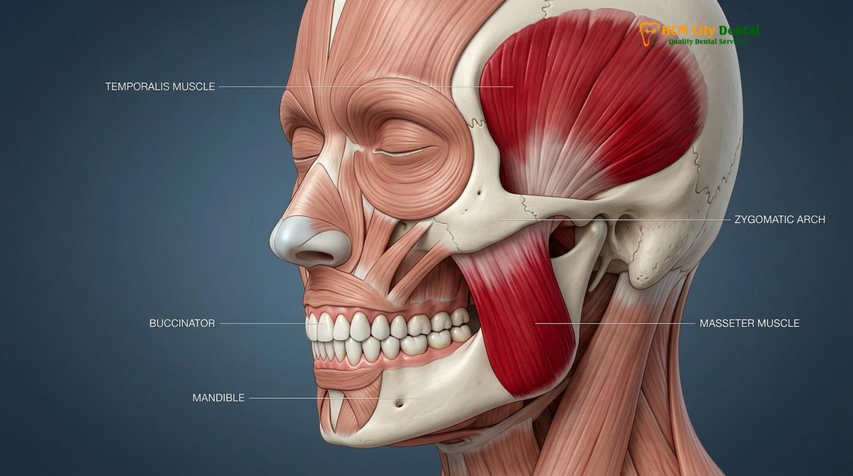
The pathophysiology of bruxism involves the trigeminal nerve, which controls the powerful muscles of mastication—namely the masseter, temporalis, and medial pterygoid muscles. During a bruxism episode, these muscles contract with a force that can exceed normal chewing pressure by up to ten times. This immense, unregulated force is directed entirely onto the teeth, the periodontium, and the temporomandibular joints. Because the protective reflexes that normally prevent us from biting down too hard are suppressed during sleep, nocturnal grinding can cause catastrophic damage to the oral structures before the patient even realizes there is a problem [2].
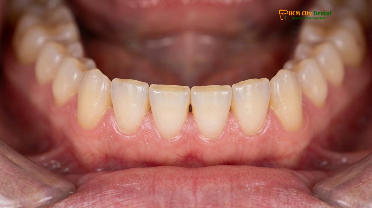
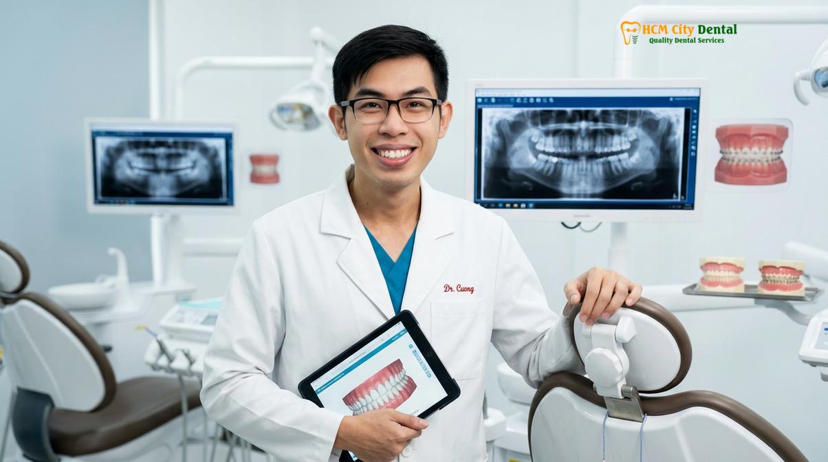
According to Dr. Nguyen Van Cuong, a leading specialist in restorative dentistry, the prevalence of stress-induced bruxism has seen a sharp increase in recent clinical observations. “Patients often present with advanced dental attrition and chronic facial pain, completely unaware that their nocturnal grinding is the root cause,” notes Dr. Cuong. Understanding the mechanics of this condition is the first step toward effective management. The continuous friction between the upper and lower arches strips away the protective enamel, while the sustained muscular contraction leads to a buildup of lactic acid and inflammatory mediators in the facial muscles.
Furthermore, systemic factors play a significant role in the exacerbation of bruxism symptoms and signs. The consumption of central nervous system stimulants, such as caffeine and certain medications (including selective serotonin reuptake inhibitors, or SSRIs), has been clinically proven to increase the frequency and intensity of grinding episodes. Alcohol consumption and tobacco use are also strong risk factors, as they disrupt normal sleep architecture and lower the threshold for motor neuron excitability. Recognizing these contributing factors is essential for a comprehensive diagnostic approach, allowing dental professionals to tailor treatment plans that address both the symptoms and the underlying causes.
Visual Indicators: Wear Facets, Chipped Enamel, and Linea Alba
Dentists diagnose bruxism visually by identifying flattened tooth surfaces, micro-fractures in the enamel, and a distinct white line of calloused tissue inside the cheek.
The most definitive evidence of chronic bruxism is written directly on the surfaces of the teeth and the surrounding soft tissues. Because patients are often unaware of their grinding habits, dental professionals rely heavily on these visual indicators during routine examinations. The primary hallmark of this condition is the presence of tooth wear facets. These are distinct, flattened areas on the occlusal (chewing) surfaces of the posterior teeth or the incisal edges of the anterior teeth. Unlike normal physiological wear, which occurs gradually over a lifetime, tooth wear facets caused by bruxism are accelerated, highly polished, and often match perfectly when the patient’s upper and lower jaws are manipulated into a grinding position [3].
As the grinding continues, the hardest substance in the human body—dental enamel—begins to fail. This leads to chipped enamel, particularly on the delicate edges of the incisors and canines. The micro-trauma from constant lateral forces causes the enamel prisms to fracture and flake away, leaving jagged, uneven edges. Once the enamel is breached, the underlying dentin is exposed. Dentin is significantly softer and wears away at a much faster rate, creating cupped-out craters on the chewing surfaces. This exposure not only accelerates structural loss but also leads to severe thermal and tactile sensitivity.
“The progressive loss of enamel due to bruxism is not merely an aesthetic concern; it fundamentally alters the occlusal vertical dimension, leading to a collapsed bite and profound functional impairment of the entire stomatognathic system.”
Beyond the hard tissues, the soft tissues of the mouth provide crucial diagnostic clues. One of the most common soft-tissue signs is linea alba. This is a raised, horizontal white line that runs along the buccal mucosa (the inner lining of the cheek), directly parallel to the occlusal plane where the upper and lower teeth meet. Linea alba is a form of hyperkeratosis—a calloused thickening of the tissue—resulting from the chronic friction, suction, and negative pressure generated during intense clenching and grinding episodes. While painless and benign, its presence is a definitive red flag for parafunctional jaw activity.
Another frequent soft-tissue indicator is a scalloped tongue, also known as crenated tongue. This occurs when the tongue is persistently pressed against the lingual surfaces of the teeth during clenching, leaving distinct indentations along its lateral borders. Together, tooth wear facets, chipped enamel, linea alba, and a scalloped tongue paint a clear clinical picture of a masticatory system under severe, chronic stress. Identifying these visual signs early allows for conservative interventions, such as the fabrication of custom nightguards, before irreversible damage necessitates complex restorative procedures like full-mouth rehabilitation.
Physical Symptoms: Morning Soreness, Earaches, and Myofascial Pain
Beyond dental damage, bruxism causes significant physical discomfort, most notably morning jaw stiffness, referred ear pain, and chronic myofascial tension.
While the visual signs of bruxism are evident to a dental professional, the physical symptoms are what typically drive a patient to seek medical attention. The most pervasive and immediate symptom reported by patients is morning jaw soreness. This discomfort is the direct result of the masticatory muscles—specifically the masseter and temporalis—being overworked throughout the night. Just as a heavy weightlifting session leaves skeletal muscles fatigued and aching due to micro-tears and lactic acid accumulation, hours of nocturnal clenching leave the jaw muscles stiff, tender, and exhausted upon waking [4].
This muscular fatigue often radiates beyond the jaw, manifesting as tension headaches. These headaches are typically described as a dull, constant ache wrapping around the sides of the head, localized in the temporal regions. Because the temporalis muscle fans out across the side of the skull, its hypercontraction during bruxism directly triggers these tension-type headaches. Patients frequently misdiagnose these as standard migraines or stress headaches, unaware that the root cause lies in their dental occlusion and nocturnal habits.
Another perplexing symptom of bruxism is the presence of unexplained earaches or tinnitus (ringing in the ears). The temporomandibular joint (TMJ) is located mere millimeters from the auditory canal. When the jaw muscles are in spasm and the joint is subjected to immense compressive forces from clenching, the resulting inflammation can easily refer pain to the ear. Patients often visit otolaryngologists (ENTs) complaining of ear pain, only to find that their ears are perfectly healthy and the true culprit is referred myofascial pain from the masticatory system.
| Symptom Category | Sleep Bruxism Presentation | Awake Bruxism Presentation |
|---|---|---|
| Muscular Pain | Severe morning jaw soreness; stiffness upon waking. | Progressive fatigue and soreness worsening by late afternoon. |
| Headaches | Temporal headaches present immediately in the morning. | Tension headaches developing during periods of high stress. |
| Dental Damage | Rapid progression of tooth wear facets and enamel chipping. | Slower wear, primarily localized to clenching surfaces. |
| Awareness | Unconscious; often reported by a sleep partner hearing grinding. | Subconscious; patient may catch themselves clenching. |
Chronic bruxism inevitably leads to broader temporomandibular disorders (TMD). The constant pressure degrades the articular disc within the TMJ, leading to clicking, popping, or grating sounds (crepitus) when opening or closing the mouth. In severe cases, the muscles may go into a protective spasm, resulting in trismus—a condition where the patient is physically unable to open their mouth fully. Addressing these physical symptoms requires a comprehensive approach that goes beyond protecting the teeth, focusing on neuromuscular relaxation and joint stabilization.
The Danger of Abfraction Lesions and Tooth Fracture
Unrelenting occlusal forces from bruxism can cause the teeth to flex, leading to wedge-shaped defects at the gumline and catastrophic structural fractures.
One of the most insidious and structurally damaging consequences of chronic bruxism is the development of abfraction lesions. Unlike tooth decay caused by bacteria, or abrasion caused by aggressive tooth brushing, abfraction is a biomechanical failure of the tooth structure. When the jaws clench and grind, immense lateral and oblique forces are applied to the teeth. Because teeth are not perfectly rigid, these forces cause the tooth to flex microscopically. The stress of this flexion concentrates at the cervical margin—the narrowest point of the tooth, right at the gumline [5].
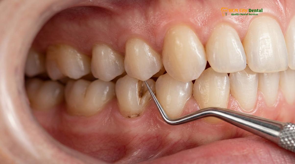
Over time, this concentrated stress causes the chemical bonds between the enamel prisms at the cervical margin to break down. The enamel micro-fractures and flakes away, creating deep, sharp, wedge-shaped notches at the gumline. These abfraction lesions expose the underlying dentin, leading to severe hypersensitivity to cold, hot, and sweet stimuli. Furthermore, the loss of cervical tooth structure compromises the structural integrity of the entire tooth, making it highly susceptible to decay and further mechanical failure. If left untreated, these lesions can progress deep into the tooth, eventually encroaching upon the dental pulp and necessitating root canal therapy.
Clinical Warning: Ignoring the early signs of abfraction lesions and tooth wear facets can lead to irreversible structural damage. Once the enamel is lost at the cervical margin, the tooth becomes highly vulnerable to catastrophic vertical root fractures, which almost always require complete tooth extraction.
Beyond cervical defects, the sheer force of bruxism puts patients at a high risk for catastrophic tooth fractures. A tooth that has been weakened by large fillings, root canal treatments, or extensive wear is particularly vulnerable. The constant grinding can induce “cracked tooth syndrome,” characterized by incomplete fractures that cause sharp, shooting pain upon biting release. As the bruxism continues, these micro-cracks propagate, eventually leading to a split tooth or a fractured cusp. Repairing such damage often requires extensive restorative work, such as dental crowns, to hold the remaining tooth structure together.
The biomechanical toll of bruxism highlights the critical need for early intervention. By identifying abfraction lesions and micro-cracks early, dental professionals can implement protective strategies. Occlusal adjustments, the use of rigid acrylic splints, and the restoration of damaged areas with composite resins or ceramics are vital steps in halting the progression of this destructive cycle and preserving the natural dentition for the long term.
When to See a Doctor: Clinical Considerations and Workflows
Recognizing the bruxism symptoms and signs is only the first step; seeking timely professional evaluation is critical to preventing irreversible damage. You should schedule a clinical consultation if you experience persistent morning jaw soreness, notice visible flattening or chipping of your teeth, or if a sleep partner informs you that you make grinding noises during the night. Additionally, unexplained earaches, chronic temporal headaches, and increased tooth sensitivity—especially near the gumline where abfraction lesions form—are strong indicators that your masticatory system is under pathological stress.
Clinical Case Review: A 34-year-old patient visited HCMC Dental Clinic in Ho Chi Minh City complaining of severe morning jaw soreness and hypersensitivity. Clinical examination revealed advanced tooth wear facets on the lower incisors and deep abfraction lesions on the premolars. Following a comprehensive occlusal analysis, Dr. Cuong prescribed a custom hard-acrylic nightguard and performed conservative composite restorations. Within two weeks, the patient reported a complete cessation of myofascial pain and protected enamel integrity.
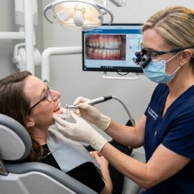
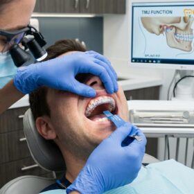
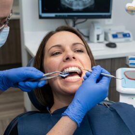
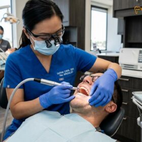
During a professional evaluation, a dentist will conduct a thorough clinical examination. This includes palpating the masseter and temporalis muscles to check for hypertrophy (enlargement) and tenderness, evaluating the temporomandibular joints for clicking or restricted movement, and performing a detailed occlusal analysis to map out the bite forces. In complex cases, especially where sleep apnea is suspected as a co-morbidity, a referral for polysomnography (a sleep study) may be recommended to monitor muscle activity and airway function during sleep.
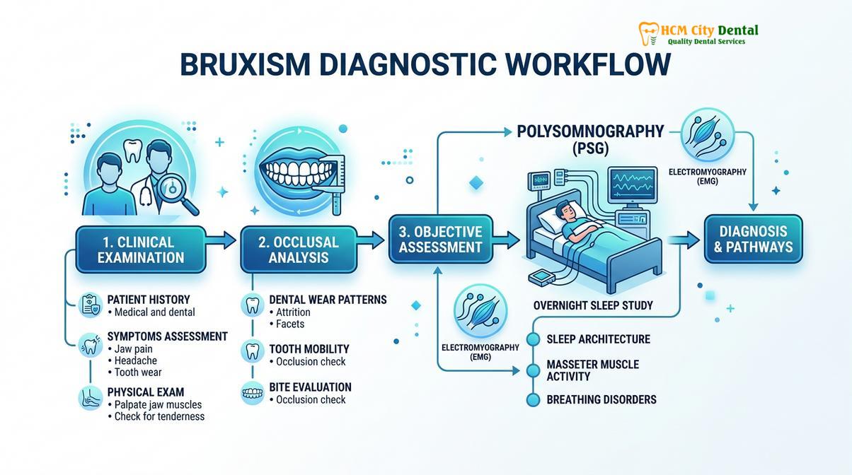
Treatment protocols are highly personalized based on clinical diagnostics. The first line of defense is typically the fabrication of a custom occlusal splint or nightguard. Unlike over-the-counter boil-and-bite guards, which can sometimes exacerbate muscle activity due to their soft, chewy texture, a custom hard-acrylic guard provides a stable, frictionless surface that protects the teeth and helps deprogram the jaw muscles. For patients experiencing severe myofascial pain, targeted Botox injections into the masseter muscles may be recommended to temporarily reduce muscle contractility without affecting normal chewing function. For a comprehensive overview of advanced interventions, patients can explore specialized temporomandibular joint (TMJ) treatments.
“Effective management of bruxism requires a dual approach: protecting the structural integrity of the teeth with precision-milled orthotics, while simultaneously addressing the neuromuscular hyperactivity through behavioral and clinical therapies.”
Ultimately, managing bruxism is an ongoing process. Regular dental check-ups are essential to monitor the condition of the teeth, adjust occlusal splints as needed, and ensure that the chosen therapies remain effective. If you suspect you are suffering from teeth grinding, do not wait for a tooth to fracture or for chronic pain to set in. Contact the specialists at HCMC Dental Clinic to schedule a comprehensive evaluation and take the first step toward protecting your smile and restoring your comfort.
Frequently Asked Questions
How do I know if I grind my teeth in my sleep?
You may know you grind your teeth in your sleep if you wake up with morning jaw soreness, dull headaches, or if a sleep partner hears grinding noises. A clinical dental examination can confirm this by identifying tooth wear facets, chipped enamel, and localized muscle hypertrophy. Because sleep bruxism occurs unconsciously, many patients remain unaware until a dentist points out the physical damage during a routine check-up.
Can teeth grinding cause gum recession?
Yes, chronic teeth grinding can contribute to gum recession. The excessive occlusal forces cause the teeth to flex at the cervical margin, leading to abfraction lesions and bone loss, which subsequently causes the overlying gingival tissue to recede and expose the sensitive tooth roots. This biomechanical stress compromises the periodontal attachment, making the gums more susceptible to inflammation and further recession.
What is linea alba inside the cheek?
Linea alba is a raised, horizontal white line of calloused tissue on the inner mucosal lining of the cheek. It forms parallel to the biting surfaces of the teeth as a hyperkeratotic response to chronic friction, suction, or accidental biting associated with bruxism. While it is completely benign and painless, it serves as a clear visual indicator to dentists that a patient is engaging in parafunctional clenching or grinding.
Are morning headaches a definitive sign of bruxism?
While morning headaches are a strong indicator of sleep bruxism, they are not definitive on their own. They must be evaluated alongside other clinical signs such as tooth wear facets, masseter muscle tenderness, and TMJ clicking to rule out other conditions like sleep apnea or migraines. The headaches associated with bruxism are typically tension-type headaches localized in the temporal regions, caused by the overworking of the temporalis muscle during the night.
How do custom nightguards help with bruxism symptoms?
Custom nightguards help by providing a protective acrylic barrier between the upper and lower teeth, preventing enamel attrition and distributing occlusal forces evenly. They also help deprogram the masticatory muscles, reducing morning jaw soreness and preventing the progression of abfraction lesions. By establishing a stable, frictionless occlusal plane, a custom guard allows the jaw to rest in a neutral position, significantly alleviating myofascial tension and protecting the temporomandibular joint.
References
- Journal of the American Dental Association. Pathophysiology and clinical management of sleep bruxism. (2020).
- Journal of Oral Rehabilitation. Neuromuscular mechanisms and masticatory muscle hyperactivity in bruxism. (2019).
- Journal of Prosthetic Dentistry. Clinical evaluation of tooth wear facets and occlusal vertical dimension. (2021).
- Clinical Oral Investigations. Myofascial pain and temporomandibular disorders associated with clenching. (2018).
- International Journal of Oral and Maxillofacial Surgery. Biomechanical analysis of abfraction lesions and cervical stress. (2022).