A TMJ MRI scan in Vietnam is widely regarded as the gold-standard imaging technique used to diagnose complex temporomandibular joint disorders. By providing high-resolution views of the articular disc, ligaments, and joint fluid, this non-invasive scan allows specialists to accurately identify the root cause of jaw pain and dysfunction. For patients suffering from chronic facial pain, unexplained headaches, or severe limitations in jaw movement, understanding the internal mechanics of the jaw joint is often the first critical step toward effective rehabilitation. Unlike traditional dental X-rays that only show bone, an MRI reveals the intricate soft tissue environment where most temporomandibular disorders originate.
Clinical Summary:
Magnetic Resonance Imaging (MRI) is the definitive diagnostic tool for evaluating the soft tissues of the temporomandibular joint. Unlike standard X-rays or 3D cone-beam scans, an MRI accurately visualizes disc displacement, joint effusion, and retrodiscal inflammation. In Vietnam, top-tier dental clinics collaborate with advanced imaging centers to integrate these high-resolution scans into comprehensive treatment plans, ensuring precise, evidence-based interventions ranging from conservative splint therapy to advanced joint management.
Key Takeaways:
- MRI is the only non-invasive modality that clearly visualizes the TMJ articular disc and surrounding soft tissues.
- It is essential for diagnosing disc displacement with or without reduction and detecting inflammatory joint effusion.
- While CBCT evaluates bone structures, MRI is required for comprehensive soft tissue and ligament analysis.
- Clinical signs like severe jaw locking, sudden bite changes, and chronic pain often necessitate an MRI referral.
- HCMC Dental Clinic coordinates seamlessly with specialized imaging centers in Saigon for accurate, timely diagnostics.
Understanding Soft Tissues: Why MRI is Critical for TMJ Discs
Magnetic Resonance Imaging is the premier diagnostic method for the TMJ because it captures detailed images of the articular disc, ligaments, and synovial fluid that standard radiographs cannot detect.
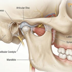
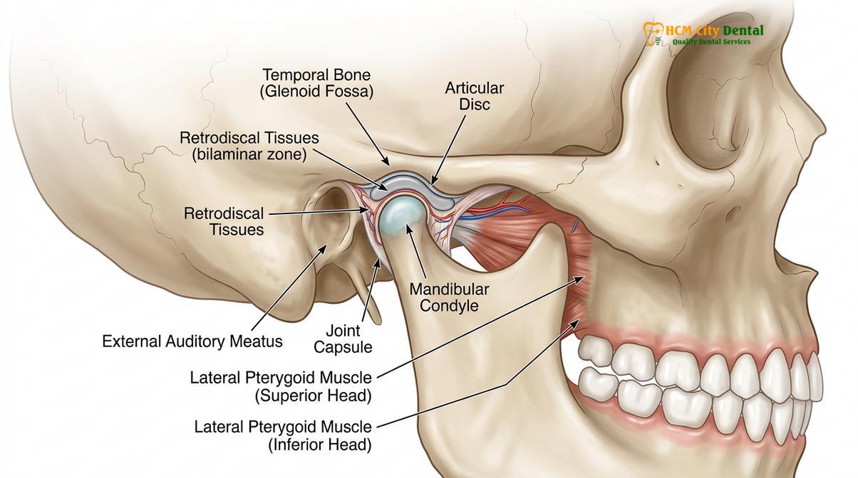
The temporomandibular joint is one of the most complex and frequently used joints in the human body. Unlike a simple hinge joint, the TMJ allows for both rotational (hinging) and translational (sliding) movements. This intricate biomechanics is made possible by a critical piece of soft tissue: the articular disc. Composed of dense, avascular fibrocartilage, this disc acts as a shock absorber and a smooth gliding surface between the mandibular condyle (the ball of the jaw) and the articular eminence of the temporal bone (the socket). Because standard dental X-rays and even advanced 3D scans rely on ionizing radiation that only reflects dense structures like bone, they render the articular disc completely invisible to the clinician.
Magnetic Resonance Imaging utilizes a powerful magnetic field and radiofrequency pulses to excite protons within the body’s water molecules. Because soft tissues like the articular disc, the highly vascularized retrodiscal tissues, and the synovial fluid contain varying amounts of water, an MRI can differentiate between them with astonishing clarity. In clinical practice, a disc displacement MRI is considered the definitive test for diagnosing internal derangements of the jaw joint[1]. When a patient presents with a clicking jaw, it is almost always an indication that the articular disc is slipping out of place—a condition known as disc displacement with reduction. While clinical examination can suggest this diagnosis, only an MRI can confirm the exact morphological changes of the disc, such as whether it has become thinned, folded, or permanently deformed over time.
Furthermore, the retrodiscal tissue—a highly innervated and vascularized pad of tissue located behind the condyle—is often the primary source of TMJ pain. This area, also known as the bilaminar zone, consists of a superior lamina rich in elastin and an inferior lamina composed of collagen. When the articular disc is displaced forward, the condyle can compress this sensitive retrodiscal tissue against the temporal bone during chewing. This compression leads to severe inflammation, swelling, and pain. An MRI, specifically utilizing T2-weighted sequences, is highly sensitive to the presence of edema (swelling) and joint effusion (excess fluid in the joint space)[2]. Identifying this inflammation is crucial because it dictates the urgency and type of intervention required to help prevent long-term joint degradation.
“The ability to visualize the exact position and morphology of the articular disc in both open and closed mouth positions makes MRI an indispensable tool in modern temporomandibular joint diagnostics, shifting treatment from guesswork to precision medicine.”
MRI vs. CBCT: Hard Tissue vs. Soft Tissue Imaging
While Cone Beam Computed Tomography (CBCT) excels at revealing bony architecture and degenerative joint disease, MRI is exclusively required to assess the health, position, and integrity of the soft tissues.
In the realm of maxillofacial diagnostics, patients are often confused about why multiple types of scans might be recommended by their specialist. The distinction between Cone Beam Computed Tomography (CBCT) and Magnetic Resonance Imaging (MRI) is fundamental to understanding modern jaw diagnostics. CBCT is an advanced form of X-ray that creates a highly detailed, three-dimensional reconstruction of the craniofacial skeleton. It is unparalleled in its ability to detect hard tissue abnormalities. If a clinician suspects osteoarthritis of the TMJ, condylar flattening, osteophyte (bone spur) formation, or subchondral cysts, a CBCT is the imaging modality of choice[3]. It provides sub-millimeter accuracy of the bony surfaces, allowing the dentist to see exactly how the bone is remodeling under mechanical stress.
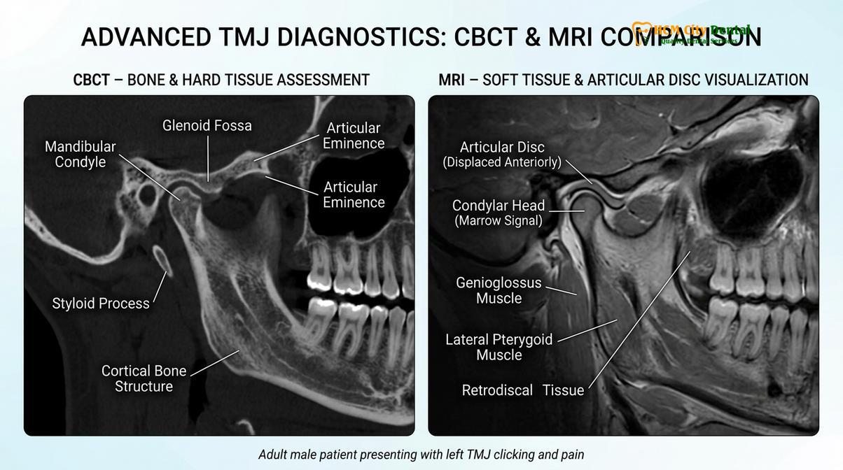
However, CBCT has a significant limitation: it cannot image soft tissue. The articular disc, the joint capsule, the lateral pterygoid muscle, and the synovial fluid all appear as a uniform, empty black space on a CBCT scan. Therefore, if the bone appears perfectly healthy on a CBCT, but the patient is still experiencing severe locking or pain, the problem lies within the invisible soft tissues. Recent comparative studies emphasize that while CBCT is superior for osseous changes, MRI remains the gold standard for evaluating the articular disc and joint effusion[5]. It does not use ionizing radiation; instead, it provides a vivid map of the joint’s internal soft structures, allowing for a comprehensive evaluation of the joint’s functional mechanics.
| Diagnostic Feature | CBCT (Cone Beam CT) | MRI (Magnetic Resonance) |
|---|---|---|
| Primary Focus | Hard tissues (Bone, Teeth) | Soft tissues (Disc, Muscles, Fluid) |
| Radiation Exposure | Low-dose ionizing radiation | None (Magnetic fields & radio waves) |
| Articular Disc Visibility | Invisible | Highly detailed and precise |
| Joint Effusion (Inflammation) | Cannot be detected | Clearly visible on T2 sequences |
| Bone Degeneration (Arthritis) | Excellent, high-resolution detail | Moderate detail, secondary to soft tissue |
In many comprehensive TMJ treatment protocols, these two imaging modalities are used synergistically. A CBCT might be taken first in the dental office to rule out gross bony pathology, tumors, or severe arthritic degeneration. If the CBCT is clear but symptoms of internal derangement persist, an MRI is ordered to evaluate the disc position and check for joint effusion. This dual-imaging approach ensures that no aspect of the joint’s anatomy is overlooked, providing a complete picture of both the hard and soft tissue environments.
Clinical Symptoms That Warrant a TMJ MRI Scan
An MRI is clinically indicated when patients present with progressive jaw locking, severe unremitting pain, suspected joint effusion, or sudden alterations in their dental occlusion.
Not every patient who experiences a clicking jaw or mild muscle tension requires an MRI. In fact, asymptomatic clicking is relatively common in the general population and often does not require aggressive intervention. However, there are specific clinical thresholds and symptom progressions that strongly indicate the need for advanced soft tissue imaging. Recognizing these signs early can help prevent irreversible damage to the joint structures and surrounding musculature.
When to See a Doctor: Important Clinical Notes
One of the most critical symptoms warranting an MRI is a condition known as a “closed lock,” clinically referred to as disc displacement without reduction. In this scenario, the articular disc slips forward and becomes trapped in front of the condyle. When the patient attempts to open their mouth, the condyle cannot glide past the bunched-up disc. The result is a sudden, severe limitation in mouth opening—often restricted to less than 25 millimeters, whereas a normal opening is typically 40 to 50 millimeters. The jaw may also deflect to the affected side upon opening. An MRI is urgently needed in these cases to confirm the position of the disc, assess its shape, and determine if it can be recaptured through conservative therapies.
Important Note: Sudden inability to open the mouth wider than 25 millimeters, accompanied by sharp pain in front of the ear, is a hallmark sign of acute disc displacement without reduction. You should see a dental specialist immediately for clinical evaluation to prevent permanent disc deformation.

Another significant symptom is the presence of severe, unremitting joint pain that does not respond to conservative therapies like muscle relaxants, soft diet, or basic physical therapy. This type of pain often indicates joint effusion—a buildup of inflammatory fluid within the joint capsule. Effusion is a sign of active tissue distress, often resulting from the condyle compressing the highly innervated retrodiscal tissues. Because joint effusion can only be definitively diagnosed via the T2-weighted sequences of an MRI, obtaining this scan is crucial for formulating an effective anti-inflammatory treatment plan[4]. Furthermore, if a patient experiences a sudden change in their bite—such as their back teeth no longer touching on one side—this can be a sign of acute joint effusion pushing the condyle downward, making an MRI an essential diagnostic step.
Preparing for Your TMJ MRI Scan: What to Expect
For many patients, undergoing an MRI can be a source of anxiety, particularly if they are unfamiliar with the procedure. Understanding what to expect can significantly alleviate these concerns and ensure a smooth diagnostic process. A TMJ MRI is a specialized scan that requires specific protocols to capture the intricate movements of the jaw joint accurately. Unlike a standard brain MRI, the TMJ protocol involves capturing images in multiple jaw positions to assess the dynamic relationship between the condyle and the articular disc.
When you arrive at the imaging center, you will be asked to remove all metal objects, including jewelry, hairpins, glasses, and removable dental appliances. Because the MRI uses a powerful magnet, any ferromagnetic materials can distort the images or pose a safety risk. If you have permanent metal dental work, such as crowns, bridges, or titanium dental implants, these are generally safe and will not prevent you from having the scan, though they may cause minor localized artifacts on the images. Patients with certain medical devices, such as pacemakers or specific types of cochlear implants, must notify the technician beforehand, as these may be contraindicated for MRI.
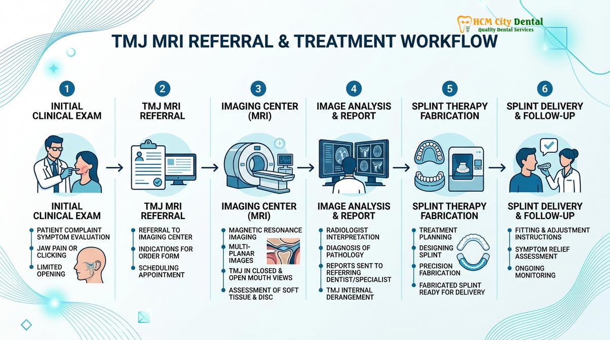
During the scan, you will lie comfortably on a padded table that slides into the MRI machine. A specialized surface coil will be placed near your jaw to capture high-resolution images of the TMJ. The machine will make loud knocking or thumping noises as the magnetic gradients switch on and off; you will be provided with earplugs or headphones to minimize the noise. The technician will instruct you to hold your jaw in specific positions—typically fully closed, partially open, and maximally open. To help you maintain the open positions without straining your muscles, the technician may provide a small, sterile bite block to rest your teeth against. The entire procedure usually takes between 30 to 45 minutes, and it is crucial to remain as still as possible to ensure the images are sharp and diagnostically useful.
Interpreting the MRI Results: What Your Dental Specialist Looks For
Once the MRI scan is complete, the digital images are analyzed by a specialized radiologist and your dental specialist. Interpreting a TMJ MRI requires a deep understanding of maxillofacial anatomy and joint biomechanics. The clinician will evaluate several key factors across different imaging sequences to build a comprehensive diagnostic picture, often referencing national standards such as those outlined by the Vietnam Odonto-Stomatology Association (VOSA) for imaging protocols[6].
First, the specialist will assess the position of the articular disc in the closed-mouth position. In a healthy joint, the posterior band of the disc should sit directly above the condyle, often described as the “12 o’clock position.” If the disc is located anteriorly (forward) or medially (inward) to the condyle, it indicates disc displacement. Next, the clinician will examine the open-mouth images to see if the disc “reduces” or snaps back into its proper position between the condyle and the articular eminence. If it does, the diagnosis is disc displacement with reduction. If it remains trapped in front of the condyle, it is diagnosed as disc displacement without reduction.
“The presence of a hyperintense signal within the joint space on a T2-weighted MRI sequence is the definitive marker for joint effusion, guiding the clinician to prioritize anti-inflammatory protocols before attempting structural rehabilitation.”
Beyond disc position, the morphology or shape of the disc is carefully evaluated. A healthy disc is biconcave, resembling a bowtie. In chronic TMJ disorders, the disc may become thickened, folded, or completely flattened due to prolonged mechanical stress. The specialist will also look for signs of joint effusion, which appears as a bright white signal on T2-weighted images, indicating active inflammation and fluid accumulation. Finally, the integrity of the lateral pterygoid muscle and the presence of any bone marrow edema within the condyle are assessed, providing a complete overview of the joint’s health and guiding the subsequent treatment strategy.
Case Study: Resolving Chronic Jaw Pain in Ho Chi Minh City
To illustrate the clinical value of advanced imaging, consider the case of a 34-year-old patient visiting HCMC Dental Clinic in Ho Chi Minh City. The patient presented with a two-year history of worsening left-sided facial pain, frequent headaches, and a recent inability to open her mouth wide enough to eat comfortably. Previous treatments at other facilities, which relied solely on standard panoramic X-rays, had diagnosed her with simple muscle tension and prescribed basic painkillers, offering only temporary relief.
Dr. Nguyen Van Cuong, a leading specialist at HCMC Dental Clinic, emphasizes that accurate diagnostics are the foundation of successful TMJ therapy. Dr. Cuong routinely reviews complex MRI reports to differentiate between muscular spasms and true internal derangements. His evidence-based approach ensures that patients receive targeted interventions rather than generic treatments. Upon clinical examination, Dr. Cuong noted a maximum opening of only 28 millimeters and significant tenderness over the left joint capsule. Suspecting a soft tissue derangement, he immediately referred the patient for a TMJ MRI scan.

The MRI results were definitive: the patient had an anterior disc displacement without reduction on the left side, accompanied by moderate joint effusion (inflammation) in the superior joint space. Armed with this precise diagnosis, the clinical team developed a targeted treatment plan. The initial phase focused on reducing the inflammation using specific anti-inflammatory protocols and a soft diet. Once the effusion subsided, the patient was fitted with custom nightguards designed to decompress the joint space and prevent nocturnal bruxism from further aggravating the retrodiscal tissues. Within three months, the patient’s pain had significantly decreased, and her jaw mobility improved to a functional 42 millimeters, highlighting the critical role of MRI in guiding effective therapy.
How HCMC Dental Facilitates MRI Referrals in Saigon
Our clinic utilizes a streamlined referral protocol, partnering with advanced imaging centers to ensure patients receive prompt, high-resolution TMJ MRI scans for accurate diagnosis.
Navigating the healthcare system for specialized imaging can be daunting for patients experiencing severe jaw pain. At HCMC Dental Clinic in Ho Chi Minh City, we have developed a seamless, patient-centric workflow to facilitate TMJ MRI scans. The process begins with a comprehensive clinical evaluation. During this initial consultation, the specialist will meticulously palpate the muscles of mastication, measure the maximum interincisal opening, evaluate lateral jaw excursions, and listen for joint sounds using a stethoscope or Doppler ultrasound. If the clinical findings suggest an internal derangement or joint effusion, an MRI referral is generated.
We collaborate closely with top-tier medical imaging centers across Saigon that are equipped with state-of-the-art 1.5 Tesla and 3.0 Tesla MRI machines. Our referral includes specific instructions for the radiologist, detailing the required open and closed mouth sequences to ensure the resulting images provide the exact diagnostic information needed. Once the scan is completed, the digital files are sent securely to our clinic, where our specialists review them in conjunction with your clinical symptoms to formulate a personalized, evidence-based treatment plan.
For detailed information on diagnostics and custom splint therapy options, visit our comprehensive TMJ & Jaw Pain Treatment Ho Chi Minh City guidelines.
References
- Journal of Oral and Maxillofacial Surgery. Magnetic resonance imaging of the temporomandibular joint: a clinical review.
- International Journal of Oral Science. Diagnostic accuracy of MRI in temporomandibular joint disorders.
- Journal of Dentistry. Correlation between clinical findings and MRI in TMJ internal derangement.
- Clinical Oral Investigations. The role of joint effusion in temporomandibular disorders: an MRI study.
- American Journal of Orthodontics and Dentofacial Orthopedics. CBCT versus MRI in the assessment of the temporomandibular joint.
- Vietnam Odonto-Stomatology Association (VOSA). National Guidelines on Temporomandibular Joint Disorders and Imaging Protocols.