TMJ palpation pain points are localized areas of extreme muscle tenderness and hyperirritability evaluated by dentists to diagnose temporomandibular disorders (TMD). By applying controlled, standardized pressure to specific masticatory muscles, clinicians can identify active trigger points that refer pain to the jaw, teeth, or head, thereby guiding targeted therapeutic interventions and restorative treatments.
Clinical Summary:
Temporomandibular joint (TMJ) palpation is a foundational diagnostic protocol used to differentiate between muscular (myofascial) and structural (articular) jaw disorders. During a clinical examination, a dentist systematically applies measured pressure to the masseter, temporalis, and pterygoid muscles to locate taut bands and hyperirritable nodules known as trigger points. These pain points are often the result of chronic bruxism, malocclusion, or stress-induced clenching, leading to localized ischemia and the accumulation of metabolic waste within the muscle tissue. Identifying these specific pain referral patterns allows clinicians to map the exact source of orofacial pain—which frequently mimics toothaches or ear infections—and develop an evidence-based treatment plan. Management typically involves custom occlusal splint therapy, such as nightguards, combined with physical therapy and neuromuscular re-education to decompress the joint, restore normal muscle resting length, and alleviate chronic discomfort.
Key Takeaways:
- Palpation is a standardized diagnostic tool used to measure muscle tenderness and locate hyperirritable nodules in the jaw.
- Trigger points in the masseter and temporalis muscles frequently refer pain to the teeth, mimicking severe dental infections.
- Spasms in the lateral pterygoid muscle are a primary biomechanical cause of anterior disc displacement and jaw clicking.
- Pain mapping helps clinicians differentiate between myofascial muscle pain and structural joint inflammation (arthralgia).
- Custom nightguards and occlusal splints are frontline therapies to disengage the bite and allow overworked masticatory muscles to relax.
What is TMJ Palpation? How Dentists Test Muscle Tenderness
TMJ palpation is a diagnostic technique where a dentist applies specific, measured pressure to the jaw muscles and joints to identify inflammation, muscle spasms, and trigger points.
The clinical evaluation of temporomandibular disorders relies heavily on a systematic and highly calibrated physical examination. TMJ palpation is not merely the act of pressing on the face; it is a precise diagnostic protocol designed to evaluate the health, tone, and reactivity of the masticatory musculature and the temporomandibular joint capsule itself. When a patient presents with chronic jaw pain, headaches, or unexplained toothaches, the dentist must act as a clinical detective, using palpation to trace the symptoms back to their anatomical origin. This process is governed by established diagnostic criteria, which dictate the exact amount of pressure, the duration of pressure, and the specific anatomical landmarks to be tested.[1]
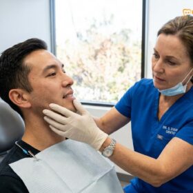
During a standardized palpation exam, the clinician applies approximately 1 kilogram (about 2.2 pounds) of pressure to the extraoral muscles, such as the masseter and temporalis, and approximately 0.5 kilograms (about 1.1 pounds) of pressure to the joint capsule and intraoral structures. This measured force is critical; applying too little pressure may result in a false negative, missing deep-seated trigger points, while applying too much pressure can elicit a normal pain response in healthy tissue, leading to a false positive. The dentist will typically use the pads of their index or middle fingers, employing either a flat palpation technique (pressing the muscle against underlying bone) or a pincer palpation technique (grasping the muscle belly between the thumb and fingers) depending on the muscle being examined.
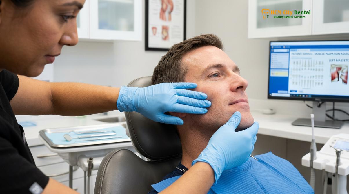
The primary goal of this examination is to identify TMJ palpation pain points, which manifest as localized areas of tenderness, taut bands of muscle fibers, or active trigger points. When the clinician presses on these specific areas, they are assessing the patient’s pain response, which is often categorized on a scale from mild tenderness to severe pain that causes the patient to flinch or pull away (a positive “jump sign”). Furthermore, the dentist is evaluating whether the pain remains localized to the area being pressed or if it radiates to other regions of the head and neck, a phenomenon known as referred pain.
“Standardized palpation pressure is the cornerstone of diagnosing myofascial temporomandibular disorders. Without calibrated force, the distinction between normal tissue sensitivity and pathological hyperirritability becomes entirely subjective, compromising the efficacy of subsequent treatment plans.”
At HCMC Dental Clinic in Ho Chi Minh City, comprehensive TMJ evaluations are conducted with meticulous attention to these diagnostic protocols. Dr. Nguyen Van Cuong emphasizes that a thorough palpation exam must also include a bilateral comparison. By palpating the left and right sides simultaneously or in rapid succession, the clinician can detect asymmetries in muscle volume (hypertrophy), muscle tone, and pain thresholds. This bilateral assessment is crucial because TMD often presents asymmetrically, with one side bearing the brunt of parafunctional habits like unilateral chewing or uneven teeth grinding.
Beyond the muscles, the palpation exam extends to the temporomandibular joint itself. The dentist will palpate the lateral pole of the condyle (the outer aspect of the jaw joint) while the patient’s mouth is closed, and then palpate the posterior attachment tissues by placing a finger slightly inside the ear canal while the patient opens and closes their mouth. This allows the clinician to feel for joint effusions, capsular inflammation, and the mechanical clicking or popping associated with internal derangements. The integration of muscle and joint palpation provides a holistic view of the patient’s orofacial health, setting the stage for accurate diagnosis and effective intervention.
Locating Trigger Points in the Masseter and Temporalis Muscles
Trigger points in the masseter and temporalis are hyperirritable nodules within taut muscle bands that cause localized tenderness and often refer pain to the teeth, ears, or forehead.
The masseter and temporalis muscles are the primary engines of mastication, responsible for the immense force required to close the jaw and chew food. Due to their continuous use and their susceptibility to stress-induced parafunctional habits like bruxism (teeth grinding) and clenching, these muscles are the most common sites for the development of myofascial trigger points. Understanding the anatomy and the specific pain referral patterns of these muscles is essential for both the clinician diagnosing the condition and the patient seeking relief from chronic orofacial pain.[2]
The masseter is a thick, powerful, rectangular muscle that connects the zygomatic arch (cheekbone) to the angle and lower border of the mandible (lower jaw). It consists of two distinct layers: a superficial belly and a deep belly. When a patient suffers from chronic masseter muscle pain, the clinician will systematically palpate both layers. Trigger points in the superficial layer of the masseter frequently refer pain to the lower jaw, the lower molar teeth, and along the eyebrow. In contrast, trigger points in the deep layer of the masseter tend to refer pain deep into the ear, often mimicking the symptoms of otitis media (an ear infection) or causing a sensation of tinnitus (ringing in the ears). This complex referral pattern is why many patients undergo unnecessary dental treatments or visit ENT specialists before finally being diagnosed with a muscular TMJ disorder.
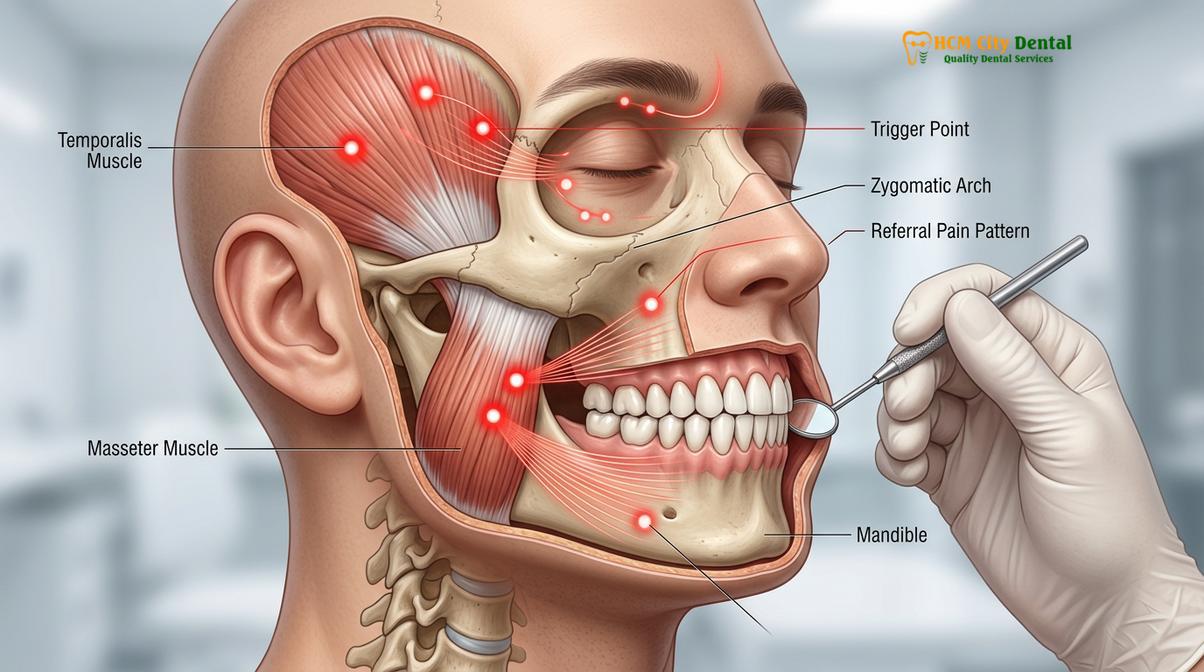
The temporalis muscle is a large, fan-shaped muscle that covers the side of the head, originating from the temporal fossa and inserting onto the coronoid process of the mandible. It is divided into three functional regions: the anterior (vertical) fibers, the middle (oblique) fibers, and the posterior (horizontal) fibers. Temporalis tenderness is a hallmark sign of TMD and is frequently the underlying cause of what patients perceive as chronic tension headaches. When the dentist palpates the anterior fibers of the temporalis, trigger points here often refer pain to the upper incisors and the supraorbital ridge (above the eye). Trigger points in the middle fibers refer pain to the upper premolars and molars, while those in the posterior fibers refer pain backward toward the occiput or deep into the temporomandibular joint itself.
The biochemical mechanism behind these trigger points involves the “energy crisis” theory. When a muscle is subjected to sustained, low-level contractions—such as during nocturnal clenching—the local blood vessels become compressed. This localized ischemia (lack of blood flow) deprives the muscle fibers of oxygen and ATP (cellular energy). Without ATP, the myosin cross-bridges within the muscle fibers cannot detach, leading to a sustained, involuntary localized contraction—the taut band. This metabolic crisis results in the accumulation of inflammatory byproducts, such as substance P, bradykinin, and lactic acid, which sensitize the local nociceptors (pain nerves), creating a highly irritable trigger point that is exquisitely painful upon palpation.
| Muscle Examined | Location of Trigger Point | Primary Referred Pain Zone | Common Misdiagnosis |
|---|---|---|---|
| Masseter (Superficial) | Lower angle of the jaw | Lower molars, lower jaw, eyebrow | Mandibular tooth decay, sinusitis |
| Masseter (Deep) | Just below the cheekbone | Deep within the ear, TMJ area | Ear infection (Otitis Media), Tinnitus |
| Temporalis (Anterior) | Temple region, above the eye | Upper incisors, forehead | Maxillary anterior pulpitis, Migraine |
| Temporalis (Middle) | Side of the head, above the ear | Upper premolars and molars | Maxillary posterior toothache |
The phenomenon of referred pain is mediated by the trigeminal nerve, the primary sensory nerve of the face and mouth. The sensory inputs from the masticatory muscles, the teeth, and the TMJ all converge on the same pool of neurons in the trigeminal subnucleus caudalis in the brainstem. When a trigger point in the masseter sends a barrage of pain signals to this nucleus, the brain can misinterpret the origin of the signal, projecting the sensation of pain onto the teeth rather than the muscle. This convergence-projection mechanism underscores the absolute necessity of a thorough palpation exam; without it, a clinician might mistakenly perform a root canal on a perfectly healthy tooth when the true culprit is a hyperactive muscle.
Lateral Pterygoid Spasm: The Source of Anterior Disc Displacement
Spasms in the lateral pterygoid muscle pull the temporomandibular joint’s articular disc forward, leading to anterior disc displacement, clicking sounds, and restricted jaw movement.
While the masseter and temporalis are easily accessible for extraoral palpation, the pterygoid muscles—specifically the lateral and medial pterygoids—are situated deep within the facial structure, making them more challenging to evaluate but no less critical to TMJ health. The lateral pterygoid muscle, in particular, plays a pivotal and highly specialized role in the biomechanics of the temporomandibular joint. It is the primary muscle responsible for opening the mouth, protruding the jaw forward, and moving the jaw from side to side. When this muscle becomes dysfunctional, it sets off a cascade of mechanical failures within the joint itself.[3]
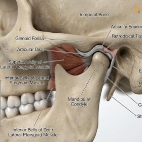
The lateral pterygoid consists of two distinct heads: the inferior head and the superior head. The inferior head attaches to the neck of the mandibular condyle and is active during mouth opening. The superior head, however, is unique; it attaches directly to the articular disc—the cartilaginous cushion that sits between the condyle and the skull—and is active during mouth closing, stabilizing the disc against the articular eminence. A pterygoid spasm, particularly in the superior head, creates a continuous, abnormal forward pull on the articular disc.
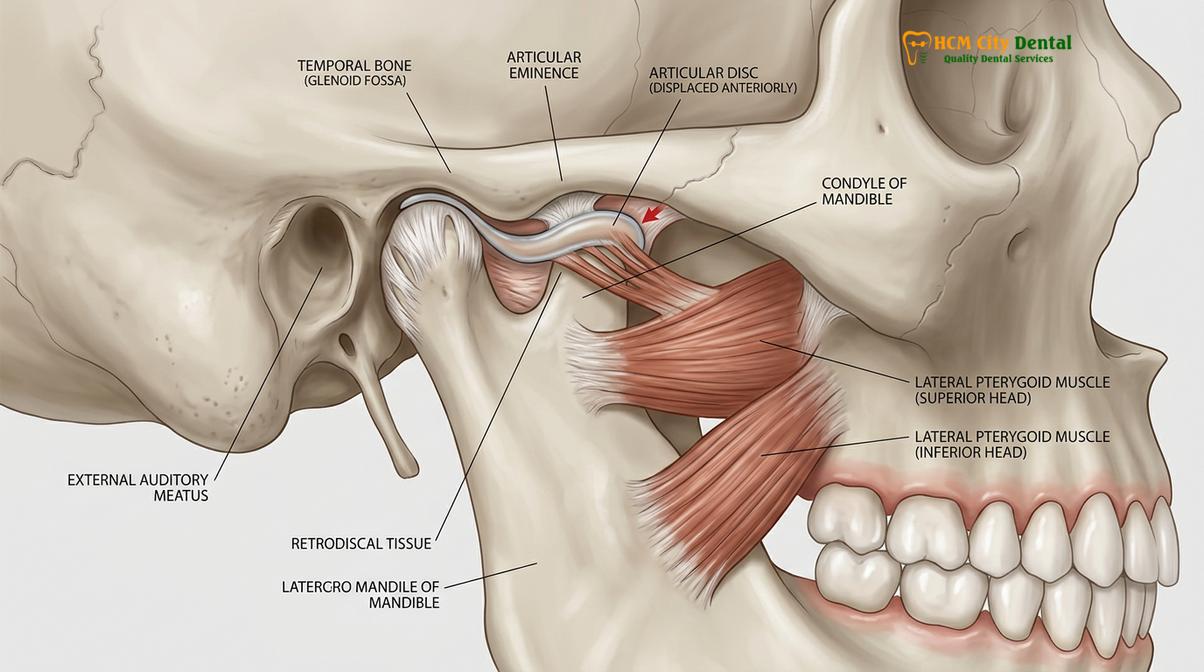
Under normal circumstances, the articular disc moves synchronously with the condyle as the mouth opens and closes. However, when chronic clenching or a misaligned bite causes the superior head of the lateral pterygoid to go into spasm, the muscle loses its ability to relax. This sustained contraction pulls the disc anteriorly (forward) and medially (inward), displacing it from its normal position atop the condyle. When the patient attempts to open their mouth, the condyle must translate forward and “jump” back onto the displaced disc, producing the characteristic clicking or popping sound associated with anterior disc displacement with reduction. If the spasm is severe and chronic, the disc may become permanently displaced (anterior disc displacement without reduction), resulting in a “closed lock” where the patient cannot open their mouth more than a few centimeters.
Clinical Case Study: Resolving Pterygoid Spasm
A 34-year-old patient visited HCMC Dental Clinic in Ho Chi Minh City complaining of severe pain in front of the ear, a loud clicking sound when chewing, and an inability to fully open her mouth. Previous treatments had focused solely on the joint capsule. Dr. Nguyen Van Cuong performed a functional manipulation test, identifying severe lateral pterygoid spasms. By implementing a targeted treatment plan involving an anterior repositioning splint to decompress the joint and intraoral myofascial release techniques, the muscle spasm was resolved. Within six weeks, the patient’s jaw range of motion returned to normal, and the painful clicking was eliminated, demonstrating the critical link between muscle tension and joint mechanics.
Because the lateral pterygoid is located deep behind the maxilla, direct palpation is notoriously difficult and often uncomfortable for the patient. Attempting to reach the muscle by sliding a finger behind the upper molars (the maxillary tuberosity) often elicits a pain response simply due to the pressure on the sensitive mucosal tissues, leading to false-positive diagnoses. Therefore, modern clinical protocols rely heavily on functional manipulation to test the lateral pterygoid. The dentist will ask the patient to protrude their jaw against resistance (the dentist pushing back on the chin) or to open their mouth against resistance. If these resisted movements elicit sharp pain in the TMJ area, it is a strong indicator of lateral pterygoid dysfunction and spasm.
The medial pterygoid, the counterpart to the lateral pterygoid, acts synergistically with the masseter to elevate the mandible and close the mouth. It forms a muscular “sling” around the angle of the jaw. Trigger points in the medial pterygoid refer pain deep into the mouth, the back of the throat, and the base of the tongue. Patients with medial pterygoid spasms often complain of pain when swallowing or a sensation of a “lump in the throat.” Evaluating these deep muscles requires a high degree of clinical expertise, as their symptoms frequently mimic pharyngeal infections or glossopharyngeal neuralgia.
Pain Mapping: Differentiating Muscular TMD from Joint Arthralgia
Pain mapping involves systematically isolating symptoms to determine whether the discomfort originates from myofascial trigger points or structural inflammation within the temporomandibular joint itself.
One of the most critical challenges in managing orofacial pain is determining the precise anatomical source of the patient’s symptoms. Temporomandibular disorders are broadly classified into two main categories: myogenous TMD (originating from the muscles) and arthrogenous TMD (originating from the joint structures). Because the muscles and the joint operate as a single, interdependent biomechanical unit, dysfunction in one inevitably affects the other. However, the treatment protocols for muscle pain are vastly different from those for joint inflammation. Therefore, dentists utilize a process called pain mapping to differentiate between muscular myofascial pain and structural joint arthralgia.[4]
Pain mapping begins with a detailed patient history and the standardized palpation exam described earlier. The clinician maps the exact locations of tenderness, noting whether the pain is localized to the muscle bellies (indicating myalgia) or if it is centered directly over the TMJ capsule (indicating arthralgia). If palpation of the masseter and temporalis reproduces the patient’s chief complaint—such as their familiar headache or toothache—the primary diagnosis leans heavily toward a myogenous origin.
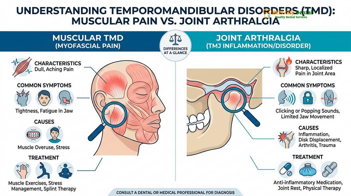
To further differentiate the source of pain, the dentist will perform joint loading tests. Joint loading involves applying superior and posterior pressure to the mandible, effectively compressing the condyle against the articular disc and the glenoid fossa. If this compression elicits sharp, localized pain within the joint space, it suggests the presence of capsulitis (inflammation of the joint capsule), synovitis (inflammation of the synovial lining), or retrodiscal tissue compression. The retrodiscal tissues, located behind the condyle, are highly vascularized and heavily innervated. If the articular disc is displaced anteriorly, the condyle may press directly onto these sensitive tissues during chewing, causing severe, sharp pain that is distinctly different from the dull, aching pain characteristic of muscle trigger points.
“The clinical distinction between myogenous and arthrogenous pain is paramount. Prescribing a muscle relaxant for an inflamed joint capsule is as ineffective as performing joint surgery for a masseter trigger point. Accurate pain mapping dictates the trajectory of successful therapy.”
Another crucial diagnostic tool in pain mapping is the use of diagnostic nerve blocks or local anesthetic infiltrations. If a patient presents with severe, radiating pain that makes palpation inconclusive, the dentist may inject a small amount of local anesthetic directly into a suspected muscle trigger point. If the pain—including the referred pain to the teeth or head—completely resolves within minutes, the diagnosis of myofascial pain is confirmed. Conversely, if the muscle is anesthetized but the pain persists during jaw movement, the source is likely intra-articular (within the joint), necessitating a different therapeutic approach, such as joint arthrocentesis or the use of anti-inflammatory medications.
Furthermore, the dentist will evaluate the patient’s range of motion. A normal, healthy jaw should open between 40 to 50 millimeters (measured between the upper and lower incisors). If the opening is restricted (trismus), the clinician must determine if the restriction is due to a “soft end-feel” or a “hard end-feel.” A soft end-feel, where the jaw can be gently stretched open a bit further despite pain, typically indicates muscle spasms (the muscles are guarding and refusing to stretch). A hard end-feel, where the jaw hits a solid mechanical block and cannot be forced open further, strongly indicates a structural joint issue, such as an anteriorly displaced disc without reduction blocking the path of the condyle.
Clinical Workflows: From Diagnosis to Nightguards and Therapy
Following a precise palpation diagnosis, treatment typically involves custom occlusal splints, such as nightguards, combined with physical therapy to decompress the joint and relax hyperactive muscles.
Once the specific TMJ palpation pain points have been identified and the pain has been accurately mapped to either the musculature or the joint capsule, the clinician can develop a targeted, evidence-based treatment plan. The primary objective of TMD therapy is to reduce pain, restore normal jaw function, and prevent further damage to the dental and articular structures. Because the majority of TMD cases have a significant muscular component—driven by nocturnal bruxism or daytime clenching—the cornerstone of conservative management is occlusal splint therapy.[5]
A custom-fitted nightguard (also known as an occlusal stabilization splint) is a hard acrylic device worn over the upper or lower teeth, typically during sleep. Unlike soft, over-the-counter mouthguards, a clinical-grade nightguard is precisely calibrated to the patient’s unique bite. The therapeutic mechanism of a nightguard is multifaceted. First, it provides a smooth, flat surface for the opposing teeth to glide against. When a patient attempts to grind their teeth, the canine guidance built into the splint causes the posterior teeth to separate (disocclude). This immediate separation shuts down the neurological feedback loop that tells the masseter and temporalis muscles to contract with maximum force. By reducing the intensity of muscle contractions, the nightguard allows the overworked muscles to rest, facilitating the healing of trigger points and the clearance of metabolic waste.
Clinical Warning: Over-the-Counter Mouthguards
Patients experiencing TMJ muscle pain should strictly avoid soft, “boil-and-bite” mouthguards sold in pharmacies. The spongy texture of these devices often encourages the jaw muscles to chew on them continuously throughout the night, exacerbating muscle spasms, increasing trigger point irritability, and potentially worsening the underlying temporomandibular disorder.
In addition to reducing muscle hyperactivity, a properly adjusted nightguard slightly increases the vertical dimension of the bite. This slight opening of the jaw decompresses the temporomandibular joint, relieving pressure on the highly innervated retrodiscal tissues and allowing the articular disc to rest in a more physiological position. For patients diagnosed with severe lateral pterygoid spasms and anterior disc displacement, a specialized anterior repositioning splint may be utilized temporarily to guide the mandible forward, recapturing the disc and allowing the retrodiscal ligaments to heal and tighten.
Beyond splint therapy, comprehensive management often involves a multidisciplinary approach. Physical therapy plays a crucial role in treating myofascial pain. Techniques such as ischemic compression (applying sustained pressure directly to the trigger point to force blood out, followed by a rush of fresh, oxygenated blood upon release), spray and stretch techniques, and ultrasound therapy can rapidly deactivate trigger points. The dentist may also prescribe short-term pharmacological interventions, including non-steroidal anti-inflammatory drugs (NSAIDs) to reduce joint inflammation, or muscle relaxants to break the cycle of severe acute spasms.
For patients whose TMJ pain is linked to severe malocclusion or missing teeth, restorative dentistry or orthodontic intervention may be necessary to establish a stable, harmonious bite. By ensuring that the teeth meet evenly and that the forces of mastication are distributed properly, the clinician can prevent the neuromuscular system from constantly compensating for bite discrepancies, thereby reducing the long-term risk of muscle fatigue and trigger point formation. Through a comprehensive approach that begins with precise palpation and culminates in targeted therapy, patients can achieve lasting relief from the debilitating effects of temporomandibular disorders.
When to See a Doctor for Jaw Pain
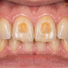
While occasional jaw tension or mild muscle fatigue after chewing tough foods is common and often resolves on its own, certain symptoms indicate a more serious underlying temporomandibular disorder that requires professional clinical evaluation. Ignoring persistent jaw pain can lead to chronic muscle contractures, permanent joint damage, and severe wear on the dental enamel.

You should seek a comprehensive evaluation from a qualified dental professional if you experience any of the following red flag symptoms:
- Inability to Open or Close the Mouth: If your jaw becomes “locked” in an open or closed position, or if your opening is severely restricted (less than the width of two fingers), this indicates a significant mechanical obstruction or severe muscle spasm requiring immediate attention.
- Pain that Disrupts Sleep or Daily Activities: Chronic, throbbing pain in the jaw, temples, or ears that does not respond to over-the-counter pain relievers or interferes with your ability to eat, speak, or sleep.
- Sudden Changes in Your Bite: If your upper and lower teeth suddenly feel like they do not fit together properly, or if you can only make contact on one side of your mouth, this may indicate acute joint inflammation or a displaced articular disc.
- Loud, Painful Clicking or Grating Sounds: While painless clicking is common, clicking or popping accompanied by sharp pain, or a grinding (crepitus) sound that indicates bone-on-bone friction, requires clinical imaging and diagnosis.
- Unexplained Toothaches or Earaches: If you experience severe pain in multiple teeth or deep in the ear, but your dentist or ENT physician finds no signs of infection or decay, the pain may be referred from active TMJ trigger points.
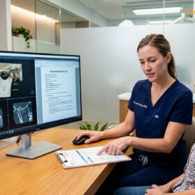
A thorough clinical examination, including standardized TMJ palpation and potentially advanced imaging such as a CBCT scan or MRI, is essential to accurately diagnose the source of the pain. Based on clinical diagnostics, a personalized treatment plan can be developed to manage the condition safely and effectively. If you are experiencing these symptoms, consider scheduling a consultation at a specialized facility, such as comprehensive temporomandibular joint therapy centers, to prevent further progression of the disorder.
Frequently Asked Questions
Why does it hurt when the doctor presses on my jaw muscle?
It hurts because the doctor is pressing on a hyperirritable trigger point within a tense muscle band. When jaw muscles are overworked from clenching or grinding, localized areas of inflammation and metabolic waste accumulation develop, making the tissue highly sensitive to palpation pressure. This pressure temporarily restricts blood flow to an already oxygen-deprived area, triggering a sharp pain response from the local nerve endings. The dentist uses this specific pain response to map the extent of the muscle dysfunction and determine the most effective course of treatment.
What is a trigger point?
A trigger point is a localized, hyperirritable nodule found within a taut band of skeletal muscle. In the context of TMJ disorders, these microscopic areas of sustained muscle contraction restrict blood flow, cause localized tenderness, and frequently refer pain to surrounding teeth, ears, or the head. They are essentially small muscle cramps that have become stuck in a contracted state due to an energy crisis within the muscle fibers. Releasing these points through physical therapy or splinting is crucial for restoring normal muscle function.
How do you relieve spasms in the pterygoid muscle?
Relieving pterygoid spasms typically involves a combination of intraoral physical therapy, warm compresses, and custom occlusal splint therapy. A dentist may also prescribe muscle relaxants or recommend targeted stretching exercises to decompress the temporomandibular joint and restore normal muscle resting length. Because the pterygoid muscles are located deep within the jaw, direct massage is difficult; therefore, therapies that disengage the bite, such as wearing a nightguard, are highly effective in allowing these deep muscles to relax passively over time.
Can TMJ muscle pain feel like a toothache?
Yes, TMJ muscle pain frequently mimics a severe toothache due to a phenomenon known as referred pain. Trigger points in the masseter and temporalis muscles share nerve pathways with the teeth via the trigeminal nerve, causing the brain to misinterpret muscle tension as dental pain. This is why patients often complain of throbbing pain in their upper or lower molars, even when dental x-rays show no signs of cavities or infection. Accurate palpation by a dentist is required to differentiate between true dental pathology and referred muscular pain.
How does a nightguard help with TMJ palpation pain?
A custom nightguard helps by providing a smooth, even biting surface that prevents the upper and lower teeth from locking together. This disocclusion reduces the neurological feedback that triggers intense muscle contractions, allowing the masseter and temporalis muscles to relax and heal. By decreasing the force exerted during nocturnal teeth grinding, the nightguard prevents the formation of new trigger points, reduces the accumulation of inflammatory byproducts in the muscle tissue, and significantly lowers the pain experienced during clinical palpation.
References
- Journal of the American Dental Association. Diagnostic Criteria for Temporomandibular Disorders (DC/TMD) Clinical Protocols. (2014).
- Journal of Oral Rehabilitation. Myofascial pain of the jaw muscles: comparison of palpation techniques. (2018).
- International Journal of Prosthodontics. The role of the lateral pterygoid muscle in TMJ disc displacement. (2019).
- Journal of Orofacial Pain. Referred pain patterns of the masseter and temporalis muscles in TMD patients. (2020).
- Clinical Oral Investigations. Efficacy of stabilization splints in the management of masticatory muscle pain. (2021).
For a personalized evaluation of jaw pain, trigger points, or to discuss custom occlusal therapy, please contact our clinical team or explore our general dentistry services to ensure your bite and joint mechanics are functioning harmoniously.