Black gums are primarily caused by physiological melanin pigmentation, smoker’s melanosis, amalgam tattoos, or certain systemic medications. While often a harmless cosmetic variation, sudden gingival darkening requires clinical evaluation to rule out underlying medical conditions or acute periodontal infections.
Clinical Summary:
Gingival hyperpigmentation, commonly referred to as dark or black gums, manifests due to a variety of endogenous and exogenous factors. The most prevalent etiology is physiological melanin production, a benign genetic trait. Reactive causes include smoker’s melanosis, where nicotine stimulates melanocyte activity, and iatrogenic factors such as amalgam tattoos from embedded dental materials. Systemic conditions, including Addison’s disease and Peutz-Jeghers syndrome, as well as certain medications like minocycline, can also induce pathological darkening of the oral mucosa. Diagnosis relies on a comprehensive clinical examination, patient history, and occasionally histological biopsy to differentiate benign pigmentation from malignant lesions like oral melanoma. For patients seeking aesthetic improvement, laser gum depigmentation offers a highly effective, minimally invasive treatment modality with predictable outcomes and rapid tissue recovery.
Key Takeaways:
- Physiological melanin production is the most common, entirely benign cause of naturally dark gums.
- Tobacco use triggers smoker’s melanosis, a reactive pigmentation that is often reversible upon cessation.
- Amalgam tattoos present as localized, harmless metallic stains near areas of previous dental restorations.
- Systemic conditions like Addison’s disease can manifest early through oral mucosal hyperpigmentation.
- Laser depigmentation provides a precise, minimally invasive solution for aesthetic gingival correction.
What Causes Dark or Black Gums?
Dark gums result from an accumulation of melanin, exogenous pigments like metal particles, or systemic health factors. A thorough clinical examination is essential to determine the exact etiology and rule out pathology.
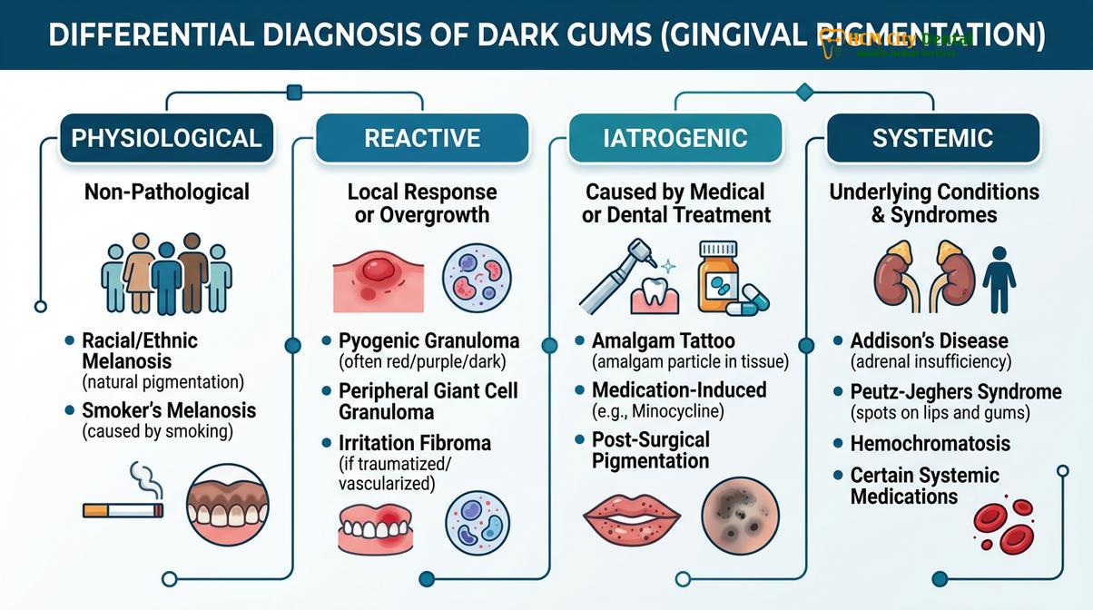
The color of healthy gingival tissue varies significantly among individuals, ranging from pale pink to deep purple or black. This variation is primarily dictated by the vascular supply, the thickness of the overlying epithelium, the degree of keratinization, and, most importantly, the presence of pigment-producing cells. When patients inquire about the black gums causes, dental professionals must conduct a systematic evaluation to categorize the pigmentation as physiological, reactive, iatrogenic, or pathological. Understanding the underlying mechanism is the critical first step in determining whether the discoloration is a mere cosmetic concern or a symptom of a broader medical issue.
In clinical practice, the differential diagnosis of gingival pigmentation requires a detailed patient history, including genetic background, lifestyle habits, medication usage, and the timeline of the color change. Dr. Nguyen Van Cuong emphasizes that while the vast majority of dark gum cases are completely benign, any rapid, asymmetrical, or symptomatic change in oral pigmentation warrants immediate investigation. The diagnostic workflow often begins with a visual inspection under high-illumination magnification, followed by palpation, periodontal probing, and radiographic imaging if exogenous materials are suspected.
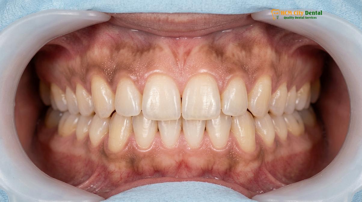
Histological Basis of Gingival Color
To fully comprehend why gums turn black, one must look at the histological architecture of the oral mucosa. The gingiva is composed of a stratified squamous epithelium overlying a dense connective tissue stroma known as the lamina propria. Within the basal layer of this epithelium reside melanocytes—specialized dendritic cells responsible for synthesizing melanin. Melanin is packaged into intracellular organelles called melanosomes, which are then transferred to adjacent keratinocytes. The density, size, and distribution of these melanosomes dictate the clinical color of the gums. Interestingly, the absolute number of melanocytes is relatively constant across different ethnicities; it is the functional activity of these cells that creates the visual spectrum of gingival pigmentation[1].
The Importance of Clinical Evaluation
When a patient presents with concerns about their gingival aesthetics, a comprehensive assessment is mandatory. Dentists will evaluate the distribution pattern of the pigment—whether it is diffuse and bilateral, which typically suggests a physiological or systemic cause, or localized and unilateral, which may point to an exogenous tattoo or a neoplastic process. For patients undergoing routine dental examinations, documenting the baseline color of the gingiva is a standard protocol. This baseline allows clinicians to monitor any subtle changes over time, ensuring that any pathological shifts are detected early. If the pigmentation is deemed purely cosmetic, the discussion can then shift toward aesthetic management options.
Physiological Melanin Pigmentation (Natural Dark Gums)
Natural melanin pigmentation is a benign, genetically determined condition where melanocytes produce excess pigment in the gingival tissues. It is completely harmless and requires no medical treatment.
Physiological melanin pigmentation is the most frequent cause of dark or black gums globally. It is an entirely normal, non-pathological condition that reflects an individual’s genetic makeup. Just as melanin determines the color of one’s skin, hair, and eyes, it also dictates the hue of the oral mucosa. This type of pigmentation is typically present from childhood and progresses gradually as the individual matures. It is most commonly observed in individuals of African, Asian, Mediterranean, and Middle Eastern descent, though it can occur in any demographic.
Clinically, physiological pigmentation presents as a diffuse, bilateral, and symmetrical darkening of the attached gingiva. The color can range from light brown to deep black, and the pigmentation usually spares the marginal gingiva (the collar of tissue immediately surrounding the teeth) and the mucogingival junction. The tissue itself remains perfectly healthy, exhibiting normal stippling (a slight texture similar to an orange peel), firm consistency, and an absence of bleeding or inflammation upon probing.
“Physiological gingival pigmentation is a reflection of genetic diversity rather than a disease process. The functional hyperactivity of melanocytes in the basal layer provides a natural, benign coloration that requires intervention only if the patient desires aesthetic modification.”
Genetic and Environmental Interplay
The synthesis of melanin is a complex biochemical pathway regulated by the enzyme tyrosinase, which converts the amino acid tyrosine into melanin. Genetic factors dictate the baseline expression of tyrosinase, but environmental factors can also influence its activity. For instance, mechanical stimulation from aggressive tooth brushing or chronic friction can sometimes induce a mild increase in localized melanin production as a protective cellular response. However, the primary driver remains genetic inheritance. Because this condition is physiological, it does not predispose the patient to periodontal disease or oral cancer. Patients seeking specialized gingival care for physiological pigmentation are doing so strictly for cosmetic reasons, aiming to achieve a uniform pink smile.
Smoker’s Melanosis and Nicotine Effects
Nicotine stimulates melanocytes to produce excess melanin, resulting in diffuse brown or black patches on the gums known as smoker’s melanosis. Quitting smoking can gradually reverse this discoloration.
Tobacco use is a profound environmental trigger for gingival discoloration, leading to a condition clinically diagnosed as smoker’s melanosis. When an individual smokes, the oral mucosa is continuously exposed to thousands of chemical compounds, including nicotine, benzopyrenes, and reactive oxygen species. Nicotine, in particular, has a direct stimulating effect on the melanocytes located in the basal layer of the gingival epithelium. In an attempt to protect the mucosal tissues from the toxic and thermal insults of cigarette smoke, these melanocytes upregulate their production of melanin, resulting in a visible darkening of the gums.
Smoker’s melanosis typically presents as diffuse, irregular brown or black patches, most prominently located on the anterior labial gingiva (the gums covering the front teeth) of both the upper and lower jaws. The severity and extent of the pigmentation are directly correlated with the duration and frequency of tobacco use; heavier smokers generally exhibit darker and more widespread discoloration. Interestingly, this pigmentation can also extend to the buccal mucosa (inside of the cheeks) and the palate, particularly in individuals who practice reverse smoking or use pipe tobacco.
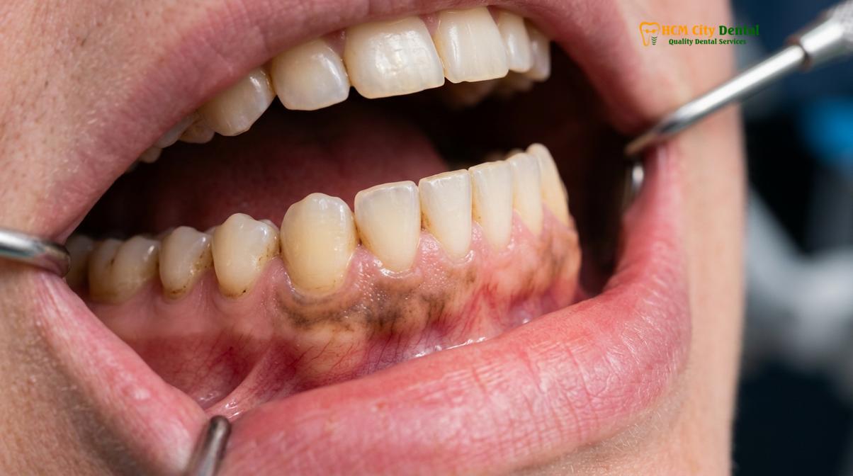
Clinical Warning: While smoker’s melanosis itself is a benign reactive process, tobacco use is a primary risk factor for severe periodontal disease and oral squamous cell carcinoma. Patients exhibiting smoking-induced pigmentation should undergo rigorous oral cancer screenings and periodontal evaluations.
Pathophysiology and Reversibility
The biochemical mechanism behind smoker’s melanosis involves the binding of polycyclic amines from tobacco smoke to melanin. Melanin acts as a defense mechanism, binding to these toxic compounds to prevent them from causing deeper cellular DNA damage. While the pigmentation serves a protective role, it is often aesthetically displeasing to the patient. The most encouraging aspect of smoker’s melanosis is its potential for reversibility. Upon complete cessation of tobacco use, the melanocytes gradually return to their baseline level of activity. Over a period of several months to a few years, the excess melanin is naturally exfoliated along with the normal turnover of epithelial cells, leading to a spontaneous lightening of the gums[2]. Dentists often use this potential for aesthetic improvement as a motivational tool to encourage patients to quit smoking during preventative dental maintenance visits.
Amalgam Tattoo: Metallic Staining on Gums
An amalgam tattoo is a localized, harmless blue, grey, or black spot caused by silver filling particles becoming embedded in the gum tissue during dental procedures.
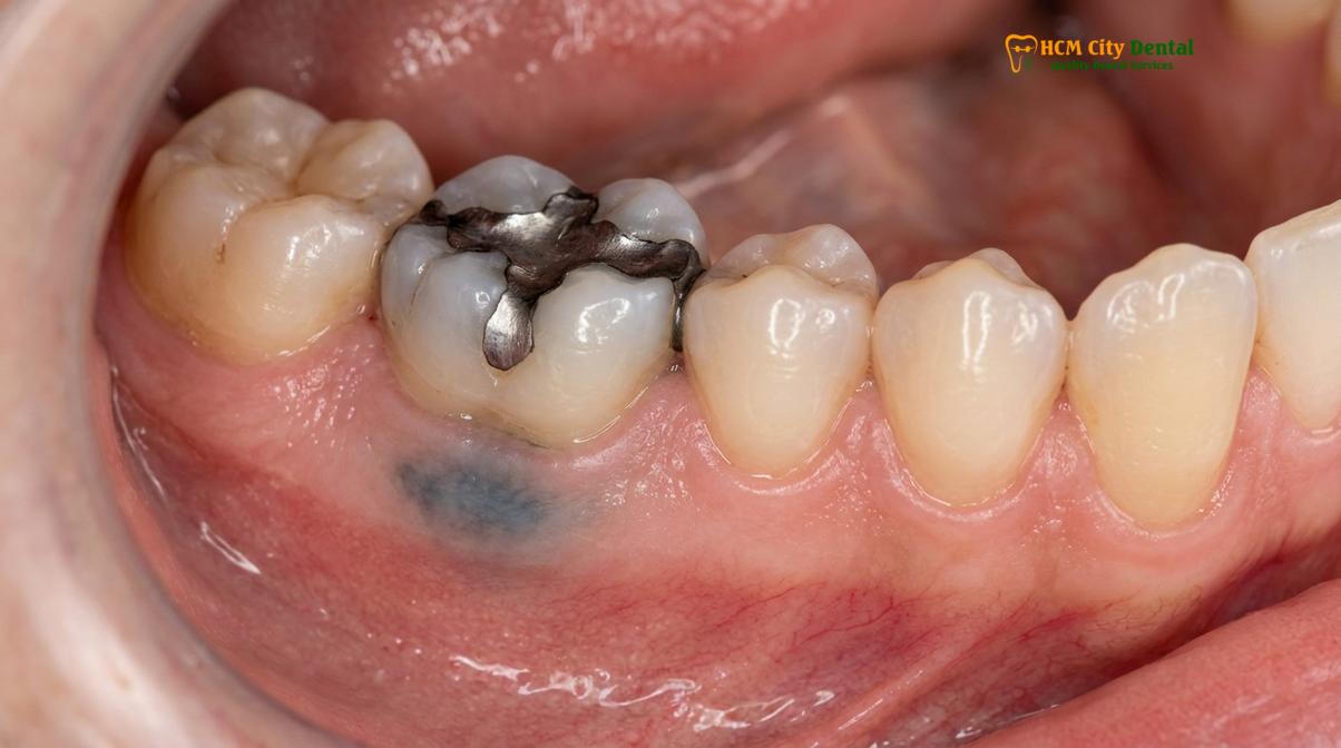
Not all dark spots on the gums are caused by melanin. An amalgam tattoo is a common iatrogenic (treatment-induced) condition resulting from the accidental implantation of dental amalgam particles into the oral soft tissues. Dental amalgam, traditionally used for filling cavities, is an alloy composed of silver, tin, copper, and mercury. During the placement, polishing, or removal of an amalgam restoration, microscopic metallic fragments can become dislodged. If the adjacent gingival tissue is abraded or ulcerated—often due to the friction of a high-speed dental bur or the aggressive use of dental floss immediately after placement—these particles can become permanently embedded in the lamina propria.
Clinically, an amalgam tattoo appears as a distinct, flat, asymptomatic macule that is blue, grey, or black in color. It is almost exclusively found adjacent to a tooth that currently has, or previously had, an amalgam filling. The alveolar mucosa and the gingiva are the most common sites. Because the metallic particles are inert, they do not elicit a significant inflammatory response, and the surrounding tissue remains perfectly healthy.
Diagnostic Differentiation
While an amalgam tattoo is entirely harmless, its clinical appearance can sometimes mimic more sinister lesions, such as an oral melanoma or a blue nevus. Therefore, accurate diagnosis is crucial. Dentists typically confirm the presence of an amalgam tattoo by taking a localized periapical radiograph. Because the amalgam particles are radiopaque (dense enough to block X-rays), they often appear as tiny, bright white specks on the X-ray film, confirming the metallic nature of the stain[3]. If the radiograph is clear but the clinical suspicion of a tattoo remains high, a biopsy may be performed to definitively rule out malignancy. Histologically, an amalgam tattoo shows fine, dark, granular pigment dispersed along collagen bundles and around blood vessels, with minimal to no inflammatory infiltrate. No treatment is required for an amalgam tattoo unless the patient requests removal for aesthetic purposes, which can be achieved through minor surgical excision or laser ablation.
Medical Conditions and Medications Linking to Dark Gums
Systemic diseases like Addison’s disease, Peutz-Jeghers syndrome, and certain medications such as minocycline can induce pathological gingival hyperpigmentation.
When a patient asks, “why are my gums turning black,” especially if the change is rapid or occurs in adulthood without a history of smoking, clinicians must consider systemic etiologies. The oral cavity often serves as a mirror for systemic health, and changes in gingival pigmentation can be the first clinical sign of an underlying endocrine disorder, genetic syndrome, or adverse pharmacological reaction. Identifying these systemic links is a critical component of comprehensive medical-dental care.
One of the most notable endocrine disorders associated with oral hyperpigmentation is Addison’s disease (primary adrenal insufficiency). In this condition, the adrenal glands fail to produce adequate levels of cortisol and aldosterone. In response, the pituitary gland overproduces adrenocorticotropic hormone (ACTH) in an attempt to stimulate the failing adrenal glands. ACTH shares a precursor molecule with melanocyte-stimulating hormone (MSH). The elevated levels of MSH inadvertently stimulate melanocytes across the body, leading to a generalized bronzing of the skin and distinct, patchy brown or black hyperpigmentation on the gingiva, buccal mucosa, and lips. Recognizing these oral signs can lead to a life-saving diagnosis, as untreated Addison’s disease can result in a fatal adrenal crisis.
Syndromic and Pharmacological Causes
Peutz-Jeghers syndrome is a rare, autosomal dominant genetic disorder characterized by the development of benign hamartomatous polyps in the gastrointestinal tract and distinct mucocutaneous pigmentation. Patients with this syndrome typically present with multiple dark blue, brown, or black freckle-like macules on the lips, perioral skin, and oral mucosa, including the gums. These pigmented spots usually appear in early childhood and are a crucial diagnostic marker for the syndrome, prompting necessary gastrointestinal screening for polyp-related complications.
Medication-induced gingival pigmentation is another significant category. Several classes of drugs can cause the gums to turn dark, either by stimulating melanin production or through the direct deposition of drug metabolites into the tissue. Minocycline, a broad-spectrum tetracycline antibiotic frequently prescribed for severe acne or rheumatoid arthritis, is a well-documented culprit. Prolonged use of minocycline can lead to a blue-grey or black discoloration of the alveolar bone, which shows through the translucent gingiva, giving the gums a dark appearance. Other medications associated with oral pigmentation include antimalarial drugs (such as chloroquine), certain antipsychotics, and heavy metal exposure (lead, bismuth)[4].
| Condition / Medication | Clinical Presentation | Underlying Mechanism |
|---|---|---|
| Addison’s Disease | Patchy brown/black macules on gums and buccal mucosa | Elevated ACTH and MSH stimulating melanocyte activity |
| Peutz-Jeghers Syndrome | Multiple dark freckles on lips and oral mucosa | Genetic mutation (STK11) causing mucocutaneous melanosis |
| Minocycline Therapy | Blue-grey discoloration of underlying bone and gums | Chelation of drug metabolites with iron/calcium in tissues |
| Antimalarial Drugs | Diffuse blue-black pigmentation on hard palate and gums | Accumulation of drug complexes in the mucosal stroma |
Clinical Treatment: Laser Gum Depigmentation (Gum Bleaching)
Laser gum depigmentation is a highly effective, minimally invasive procedure that targets and vaporizes melanin-producing cells to restore a natural pink gingival appearance.
For patients whose dark gums are a source of aesthetic dissatisfaction, modern dentistry offers highly effective solutions. The most advanced and preferred method for dark gums treatment is laser gum depigmentation, colloquially known as gum bleaching. Despite the term “bleaching,” no chemical whitening agents are used. Instead, the procedure utilizes focused light energy to precisely ablate the melanin-rich epithelial layers, allowing new, unpigmented tissue to regenerate.
The physics of dental lasers make them exceptionally suited for this procedure. Clinicians typically utilize diode lasers (ranging from 810 nm to 980 nm) or Erbium lasers (Er:YAG or Er,Cr:YSGG). Diode lasers are highly absorbed by melanin and hemoglobin, making them incredibly efficient at targeting the pigmented cells while simultaneously coagulating blood vessels to ensure a virtually bloodless surgical field. Erbium lasers, on the other hand, are highly absorbed by water, allowing for precise, cold ablation of the tissue with minimal thermal damage to the underlying connective tissue[5]. The choice of laser depends on the patient’s gingival biotype and the depth of the pigmentation.
Clinical Case Study: A 28-year-old patient visited HCMC Dental Clinic in Ho Chi Minh City presenting with severe physiological melanin pigmentation, expressing reluctance to smile. After a thorough periodontal assessment, Dr. Nguyen Van Cuong performed a diode laser depigmentation procedure under local anesthesia. The targeted ablation successfully removed the hyperpigmented epithelium. Within 14 days, the patient exhibited complete re-epithelialization, revealing healthy, uniformly pink gingiva and reporting a significant boost in self-confidence.
The Surgical Protocol
The clinical workflow for laser gum depigmentation is straightforward and patient-friendly. First, a topical anesthetic is applied, followed by the administration of a local anesthetic to ensure the patient is entirely comfortable. The dentist then uses the laser handpiece in a continuous, sweeping motion across the pigmented areas. The laser energy vaporizes the superficial epithelial cells containing the melanin. Because the laser seals nerve endings and blood vessels as it cuts, patients experience minimal intraoperative bleeding and significantly reduced postoperative discomfort compared to traditional scalpel techniques. The procedure typically takes 30 to 60 minutes per arch, depending on the extent of the discoloration. For patients seeking gum bleaching saigon, this laser-assisted approach has become the gold standard due to its precision and rapid recovery profile.
Aesthetic Outcomes and Recovery After Depigmentation
Patients typically experience rapid healing within a few days after laser depigmentation, achieving stable, long-lasting aesthetic results with minimal postoperative discomfort.
The success of laser gum depigmentation is measured not only by the immediate removal of pigment but also by the quality of tissue healing and the longevity of the aesthetic result. Following the procedure, the gingival tissue undergoes a predictable sequence of wound healing: hemostasis, inflammation, proliferation, and remodeling. Because the laser causes minimal collateral thermal damage, the inflammatory phase is brief. Within the first 24 to 48 hours, a thin, whitish fibrinous slough may form over the treated area; this is a normal biological bandage protecting the underlying regenerating tissue.
Re-epithelialization occurs rapidly. By the end of the first week, the fibrinous layer sloughs off, revealing immature, pink gingiva. Complete tissue maturation and the return of normal gingival stippling typically occur within three to four weeks. Postoperative care is relatively simple. Patients are advised to maintain meticulous oral hygiene using a soft-bristled toothbrush, avoid spicy or highly acidic foods that could irritate the healing tissue, and abstain from smoking, which can delay healing and trigger repigmentation. Analgesics like ibuprofen or acetaminophen are usually sufficient to manage any mild tenderness experienced during the first few days.
“The transition from scalpel abrasion to laser-assisted depigmentation has revolutionized periodontal aesthetics. Lasers provide unparalleled precision, ensuring that only the targeted melanocytes are ablated while preserving the structural integrity of the underlying lamina propria, resulting in faster healing and superior patient comfort.”
Long-Term Stability and Repigmentation Risks
One of the most common questions regarding laser depigmentation is the permanence of the results. While the procedure is highly effective, it is important to understand that it does not alter the patient’s underlying genetics. The melanocytes in the adjacent untreated areas or deeper tissue layers can eventually migrate and resume melanin production. Clinical studies indicate that the results of laser depigmentation are generally stable for several years, but mild repigmentation can occur in some patients after 2 to 5 years. The rate of repigmentation varies widely and is influenced by genetic predisposition, hormonal changes, and environmental factors like sun exposure and tobacco use. If repigmentation does occur, a minor touch-up laser procedure can easily restore the pink aesthetic. Regular follow-ups as part of advanced gum disease treatments and aesthetic monitoring ensure that the gingival contours and color remain optimal.
When to See a Doctor
While the majority of black gums causes are benign, certain clinical presentations require immediate professional evaluation. The oral cavity is susceptible to malignant melanoma, a highly aggressive form of cancer that arises from melanocytes. Although oral melanoma is rare, it often presents as a rapidly growing, asymmetrical, dark brown or black macule with irregular borders. If you notice a new pigmented lesion that changes in size, shape, or color, or if the pigmentation is accompanied by swelling, ulceration, or spontaneous bleeding, you must seek a diagnostic biopsy without delay.
Additionally, if the darkening of the gums is associated with severe pain, a foul odor, and the blunting of the gingival papillae (the triangular tissue between teeth), it may indicate Acute Necrotizing Ulcerative Gingivitis (ANUG), also known as trench mouth. This is a severe, rapidly progressive bacterial infection that causes tissue necrosis, leading to a grey or black pseudomembrane over the gums. ANUG requires urgent clinical intervention, including mechanical debridement and antibiotic therapy.
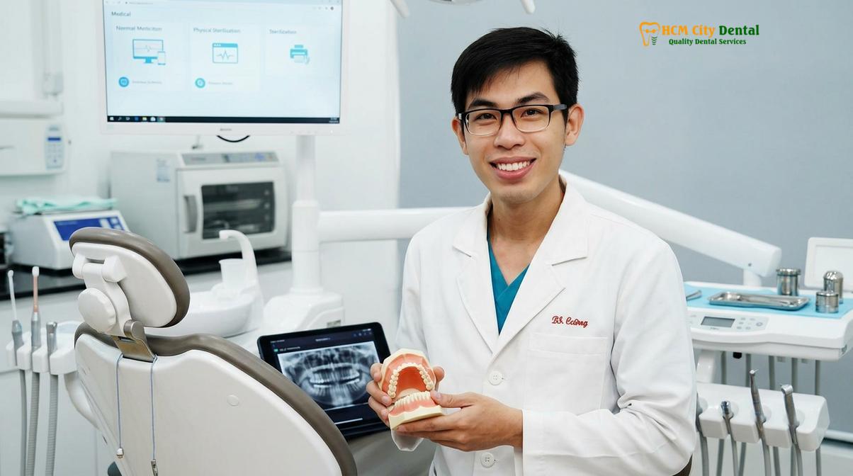
For any concerns regarding changes in your oral health, or if you are considering aesthetic improvements, scheduling a comprehensive evaluation is essential. The clinical team at HCMC Dental Clinic in Ho Chi Minh City utilizes advanced diagnostic tools to accurately determine the etiology of gingival discoloration. Whether you require reassurance regarding physiological pigmentation, management of a systemic condition, or wish to explore cosmetic laser depigmentation, a personalized consultation will provide the clarity and care you need. We encourage patients to prioritize their oral health by seeking professional oral health assessments whenever unexplained changes occur.
Frequently Asked Questions
Why are my gums turning black or dark?
Gums typically turn black due to an increase in melanin production, tobacco use, medication side effects, or embedded metal particles from dental fillings. In most cases, this discoloration is a harmless cosmetic issue rather than a disease. However, sudden changes in gingival pigmentation should be evaluated by a dental professional to rule out underlying systemic conditions or localized infections.
Is having black gums dangerous?
Having black gums is generally not dangerous, especially if the pigmentation has been present since childhood as a natural physiological trait. However, if the darkening occurs rapidly, is accompanied by pain, bleeding, or ulceration, it could indicate an acute infection like trench mouth or a systemic issue such as Addison’s disease, requiring immediate medical attention.
How do dentists fix black gums?
Dentists primarily treat black gums using laser gum depigmentation, a minimally invasive procedure that vaporizes excess melanin in the gingival tissue. Other methods include surgical scalpel technique, electrosurgery, or rotary abrasion. The choice of treatment depends on the patient’s specific gingival biotype, the extent of the pigmentation, and the underlying cause of the discoloration.
Does laser gum depigmentation hurt?
Laser gum depigmentation involves minimal discomfort, as the procedure is performed under local anesthesia to ensure the patient feels no pain during the treatment. Post-operatively, patients may experience mild tenderness or sensitivity for a few days, which is typically well-managed with over-the-counter analgesics and standard oral hygiene care.
Can amalgam fillings turn gums black?
Yes, amalgam fillings can cause localized black or blue-grey spots on the gums, a condition clinically known as an amalgam tattoo. This occurs when microscopic silver or metallic particles become dislodged during a dental procedure and embed themselves into the adjacent gingival tissue, creating a permanent but completely harmless stain.
References
- Journal of Periodontology. Gingival pigmentation: causes, treatment, and clinical considerations. (2021).
- International Journal of Dentistry. Smoker’s melanosis and its reversibility upon cessation. (2019).
- Journal of Oral and Maxillofacial Pathology. Amalgam tattoo: a clinical and histopathological review. (2020).
- Clinical Oral Investigations. Systemic diseases manifesting as oral hyperpigmentation. (2018).
- Lasers in Medical Science. Efficacy of diode lasers in gingival depigmentation procedures. (2022).