Dry socket (alveolar osteitis) is a painful dental condition occurring when the protective blood clot at a tooth extraction site fails to form or dislodges. Treatment requires professional socket irrigation and the application of medicated dressings to soothe exposed nerves and promote secondary healing.
Clinical Summary:
Alveolar osteitis, commonly known as dry socket, is a localized post-extraction complication characterized by the premature loss or degradation of the intra-alveolar blood clot. This exposes the underlying alveolar bone and nerve endings to the oral environment, resulting in severe, radiating pain that typically manifests 48 to 96 hours after surgery. Clinical management focuses on palliative care rather than curative surgical intervention. The standard protocol involves gentle debridement via sterile irrigation to remove necrotic debris and food particles, followed by the insertion of an obtundent medicated dressing (often containing eugenol) to shield the bone and provide immediate analgesic relief. While the condition is self-limiting and will eventually heal through secondary intention, professional intervention is highly recommended to mitigate the intense discomfort and facilitate an optimal healing environment.
Key Takeaways:
- Onset Timeline: Symptoms typically begin 2 to 4 days after a tooth extraction, not immediately.
- Primary Indicator: Severe, throbbing pain that radiates to the ear, eye, or neck, often unresponsive to standard painkillers.
- Visual Presentation: An empty-looking socket with visible whitish bone, lacking the normal dark blood clot.
- Clinical Treatment: Involves professional irrigation and the placement of a soothing, medicated intra-alveolar dressing.
- Prevention: Strictly avoiding smoking, spitting, and using straws is critical to protecting the fragile blood clot.
What is Dry Socket (Alveolar Osteitis)?
Dry socket is a localized inflammation of the jawbone following a tooth extraction, occurring when the essential blood clot fails to form, dissolves, or is mechanically dislodged prematurely.
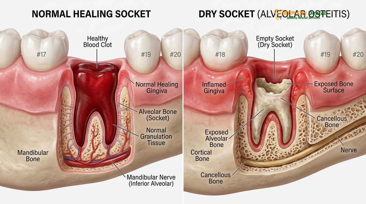
To fully comprehend the pathology of a dry socket, it is essential to understand the normal biological sequence of wound healing following a dental extraction. When a tooth is removed from the jawbone, the body immediately initiates a hemostatic response. Blood fills the empty alveolar socket and coagulates, forming a robust blood clot. This clot serves multiple critical functions: it acts as a physical biological bandage protecting the underlying bone and nerve endings, it provides a structural scaffold for the infiltration of fibroblasts and endothelial cells, and it releases growth factors necessary for the generation of new granulation tissue[1].
In the case of alveolar osteitis, this physiological process is disrupted. The blood clot either fails to form adequately, is mechanically dislodged, or undergoes premature lysis (breakdown) before the underlying bone has been covered by a protective layer of healing epithelium. This phenomenon is clinically referred to as fibrinolysis, where plasminogen is activated into plasmin, an enzyme that aggressively breaks down the fibrin network of the blood clot[2]. Once the clot is lost, the bony walls of the socket—specifically the lamina dura—are left completely exposed to the harsh oral environment.
The exposed bone is highly innervated and extremely sensitive to temperature changes, mechanical stimulation from the tongue or food, and chemical irritation from oral fluids and bacterial byproducts. This exposure triggers a localized inflammatory response within the bone marrow spaces, leading to the intense pain characteristic of the condition. It is important to note that alveolar osteitis is not a primary bacterial infection (like an abscess), but rather a localized inflammatory osteitis resulting from the loss of the protective clot.
Incidence and Prevalence
While dry socket can occur after the extraction of any tooth, its incidence varies significantly depending on the anatomical location and the complexity of the surgery. In general routine extractions, the incidence rate is relatively low, ranging from 1% to 5%. However, the risk increases dramatically following the surgical removal of impacted mandibular third molars (lower wisdom teeth), where incidence rates can reach up to 30% in certain high-risk populations[3]. The dense, relatively avascular nature of the mandibular bone, combined with the surgical trauma required to remove impacted teeth, creates an environment highly susceptible to clot failure.
Key Symptoms and How to Identify Dry Socket
The hallmark of a dry socket is severe, radiating pain that begins a few days post-extraction, accompanied by an empty-looking socket and often a foul taste or odor.
Differentiating between normal post-operative discomfort and the onset of alveolar osteitis is a critical skill for both patients and clinicians. Normal extraction pain typically peaks within the first 24 to 48 hours and gradually subsides, responding well to prescribed or over-the-counter analgesics. In stark contrast, the pain associated with a dry socket follows a delayed trajectory, usually erupting suddenly between the second and fourth day following the procedure.
Detailed Clinical Presentation
The symptomatology of this condition is highly specific. Patients frequently describe the pain as a deep, dull, throbbing ache that is exceptionally severe. Because the trigeminal nerve innervates the jaw, the pain frequently radiates along the nerve pathways. A patient with a lower molar dry socket will often complain of pain shooting up into the ear, the temple, the eye, or down into the neck on the affected side. This radiating neuralgia is one of the most definitive dry socket signs and is often what drives the patient to seek emergency dental care.
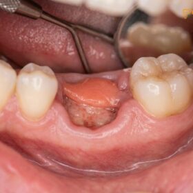
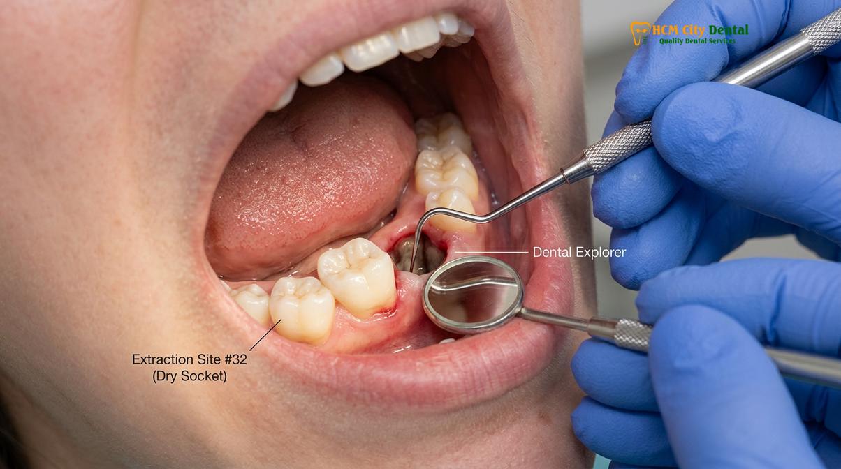
Upon visual inspection, the clinical signs are equally distinct. A healthy healing socket will contain a dark red, gelatinous blood clot, or later, a grayish-white layer of fibrin and early granulation tissue. In a dry socket, the alveolus appears empty. The clinician or patient may look into the extraction site and see a dark void, or they may see the exposed, whitish-yellow alveolar bone at the base and walls of the socket. The surrounding gingival tissue may appear inflamed, erythematous (red), and swollen, though massive facial swelling is typically absent unless a secondary infection is present.
Furthermore, the accumulation of food debris and the proliferation of oral bacteria within the unprotected socket often lead to secondary symptoms. Patients frequently report a persistent, foul taste in their mouth and noticeable halitosis (bad breath). This is due to the fermentation of trapped food particles and the breakdown of necrotic tissue within the bony crypt.
| Clinical Feature | Normal Extraction Healing | Dry Socket (Alveolar Osteitis) |
|---|---|---|
| Pain Onset & Duration | Peaks at 24-48 hours, then steadily decreases. | Starts or severely worsens 2-4 days post-extraction. |
| Pain Characteristics | Localized soreness, manageable with standard analgesics. | Severe, throbbing, radiating to ear/neck, highly resistant to standard painkillers. |
| Visual Appearance | Socket filled with a dark red clot or grayish healing tissue. | Empty socket, visible whitish bone, lack of a blood clot. |
| Odor and Taste | Normal oral environment. | Frequent bad breath (halitosis) and a foul, metallic taste. |
| Systemic Signs | None. | Usually none (fever indicates a different complication like infection). |
Why Dry Socket Occurs: Causes and Risk Factors
Dry socket is caused by the premature breakdown or dislodgement of the blood clot, driven by a combination of biological fibrinolysis, mechanical trauma, and specific patient behaviors.
The exact etiology of alveolar osteitis is considered multifactorial. It is rarely the result of a single event but rather a combination of biological predispositions, surgical factors, and post-operative patient compliance. The core mechanism, as previously established, is the failure of the blood clot to survive the initial healing phase. Understanding why this happens requires examining several distinct risk categories.
Biological and Chemical Factors
At the biochemical level, the premature dissolution of the blood clot is driven by excessive fibrinolytic activity. Certain oral bacteria, particularly anaerobic strains like Treponema denticola, produce enzymes that activate plasminogen into plasmin, accelerating the breakdown of the fibrin clot[4]. Patients with pre-existing periodontal disease or poor oral hygiene have higher bacterial loads, thereby increasing this enzymatic activity. Additionally, localized inflammation resulting from surgical trauma releases tissue activators that further promote fibrinolysis.
Hormonal fluctuations also play a significant role. Clinical studies have consistently shown that women taking oral contraceptives (birth control pills) have a statistically higher incidence of dry socket after extraction. The elevated estrogen levels associated with these medications are known to enhance systemic fibrinolytic activity, making the blood clot more fragile and susceptible to premature degradation.
Mechanical and Behavioral Risk Factors
Patient behavior in the immediate post-operative period is perhaps the most controllable risk factor. Any action that creates negative pressure (suction) within the oral cavity can physically dislodge the fragile clot from the socket walls. This includes drinking through a straw, aggressive spitting, or vigorous rinsing. Once the clot is mechanically pulled from the socket, the bone is instantly exposed.
Smoking is widely recognized as one of the most profound risk factors for developing a dry socket. The act of inhaling smoke creates the aforementioned negative pressure, but the chemical composition of tobacco smoke is equally detrimental. Nicotine is a potent vasoconstrictor, meaning it narrows the blood vessels. This reduces the localized blood supply to the extraction site, depriving the area of the oxygen and nutrients necessary for robust clot formation and tissue regeneration. Furthermore, the carbon monoxide in smoke decreases tissue oxygenation, and the heat from the smoke can physically damage the healing cells.
“In clinical practice, we observe that the vast majority of alveolar osteitis cases occur in patients who smoke within the first 48 hours after surgery. The combination of physical suction and chemical vasoconstriction creates an incredibly hostile environment for a fragile blood clot to survive.”
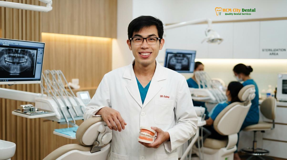
— Dr. Nguyen Van Cuong, Clinical Director
Surgical Complexity
The nature of the extraction itself heavily influences the risk. Simple extractions of single-rooted teeth rarely result in dry sockets. However, surgical extractions—particularly those involving impacted wisdom teeth, dense surrounding bone, or the need for bone removal and tooth sectioning—cause greater localized trauma. This trauma triggers a stronger inflammatory response, which in turn increases the release of tissue-derived fibrinolytic agents. Furthermore, if the surrounding bone is highly sclerotic (dense with poor blood supply), the initial bleeding into the socket may be insufficient to form a substantial clot in the first place.
Professional Treatments and Dressing Changes
Professional treatment involves thoroughly irrigating the socket to remove debris and placing a medicated, obtundent dressing to soothe the exposed bone and provide immediate pain relief.
When a patient presents with symptoms indicative of a dry socket, prompt professional intervention is required. The goal of treatment is not to surgically recreate a blood clot, but rather to provide palliative care—managing the severe pain and protecting the exposed bone while the body heals via secondary intention (healing from the base of the socket upwards).
The Clinical Workflow
The treatment protocol is highly standardized and generally provides rapid relief for the patient. The procedure typically follows these clinical steps:
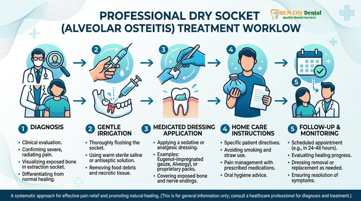
1. Diagnostic Assessment: The dentist will first review the patient’s medical history and the timeline of the pain. A visual examination of the socket is performed to confirm the absence of the clot and the presence of exposed bone. In some cases, a periapical radiograph (X-ray) may be taken to rule out other complications, such as retained root fragments, foreign bodies, or an underlying bone infection (osteomyelitis).
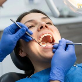
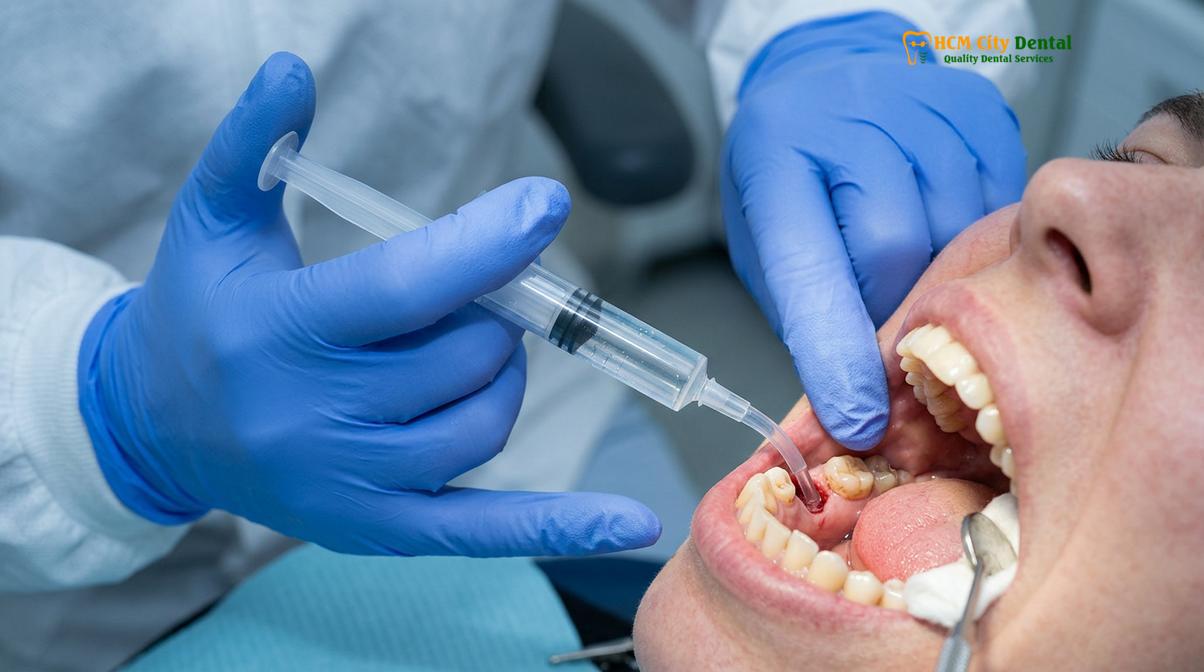
2. Socket Irrigation: The first active step in treatment is the meticulous debridement of the socket. The clinician will use a curved, blunt-tipped plastic syringe to gently flush the alveolus with a sterile solution—typically warm saline or a chlorhexidine gluconate antimicrobial rinse. This irrigation removes accumulated food debris, necrotic tissue, and bacterial biofilm that are contributing to the inflammation and pain. The irrigation must be gentle to avoid further traumatizing the sensitive bone.
3. Application of Medicated Dressing: Once the socket is clean and isolated, the dentist will place an intra-alveolar medicated dressing. These dressings are specifically formulated to be obtundent (soothing to the nerves) and antimicrobial. The most common active ingredient is eugenol (derived from clove oil), which has profound topical anesthetic and antibacterial properties. Products like Alvogyl (a fibrous paste containing eugenol, butamben, and iodoform) or zinc oxide eugenol (ZOE) paste carried on a small strip of sterile gauze are frequently utilized[5]. The dressing acts as a physical barrier, replacing the lost blood clot, and immediately begins to numb the exposed nerve endings.
4. Pharmacological Management: While the medicated dressing provides localized relief, systemic pain management is often necessary. The dentist may recommend a specific regimen of non-steroidal anti-inflammatory drugs (NSAIDs), such as ibuprofen, to manage the deep bone inflammation. In severe cases, a short course of stronger prescription analgesics may be required. Antibiotics are generally not prescribed for a standard dry socket unless there is clear evidence of a spreading secondary infection, as alveolar osteitis is primarily an inflammatory condition, not an infectious one.
Clinical Case Review: Managing Severe Post-Extraction Pain
A 24-year-old patient presented to HCMC Dental Clinic in Ho Chi Minh City complaining of excruciating, radiating jaw pain three days after the surgical extraction of a lower right impacted wisdom tooth. The patient reported that standard ibuprofen was entirely ineffective. Clinical examination by Dr. Nguyen Van Cuong revealed an empty socket devoid of a blood clot, with visible alveolar bone and localized gingival erythema. The diagnosis of alveolar osteitis was confirmed. The socket was gently irrigated with warm sterile saline to remove trapped food debris, and an Alvogyl medicated dressing was carefully packed into the bony crypt. The patient reported a significant reduction in pain within 15 minutes of the dressing placement. They were instructed on proper home care and returned 48 hours later for a dressing change, at which point early granulation tissue was visible, and the pain had largely subsided.
Follow-Up and Dressing Changes
The medicated dressing is not a permanent solution. Depending on the material used, it may dissolve on its own, or it may need to be manually removed and replaced. Patients are typically scheduled for a follow-up appointment every 48 to 72 hours. During these visits, the dentist will remove the old dressing, re-irrigate the socket, and assess the healing progress. If the bone is still exposed and sensitive, a fresh dressing is applied. This process is repeated until the patient’s pain is manageable without the dressing and a layer of healing epithelium has begun to cover the bone.
Safe Self-Care and Home Pain Relief
While awaiting professional care, patients can manage dry socket pain at home using over-the-counter anti-inflammatories, cold compresses, and gentle saline rinses, but must avoid irritating the site.
It is crucial to understand that there is no definitive dry socket home remedy that can cure the condition or replace the need for professional medicated dressings. However, if a patient develops symptoms over a weekend or in the middle of the night, certain safe self-care strategies can help mitigate the intense discomfort until they can reach a dental clinic.
Palliative Home Management Strategies
Over-the-Counter Analgesics: The first line of defense at home is systemic pain relief. Non-steroidal anti-inflammatory drugs (NSAIDs) like ibuprofen or naproxen are generally more effective for dental bone pain than acetaminophen, as they actively reduce the localized inflammation driving the pain. Patients should follow the dosage instructions on the packaging and consult a pharmacist if they have underlying health conditions. It is vital to swallow these medications; placing an aspirin directly on the extraction site will cause a severe chemical burn to the tissues and worsen the condition.
Cold Therapy: Applying a cold compress or ice pack to the outside of the face over the affected area can help numb the region and reduce any associated inflammatory swelling. The cold should be applied in intervals—15 minutes on, 15 minutes off—to prevent thermal damage to the skin.
Gentle Saline Rinses: Keeping the socket clean is essential, but it must be done with extreme care. Patients can mix half a teaspoon of salt into a glass of warm water to create a soothing, mildly antiseptic rinse. The key is to avoid vigorous swishing. Instead, the patient should take a mouthful of the warm saline, gently tilt their head to let the liquid pool over the extraction site, and then passively let the water fall out of their mouth into the sink. This helps dislodge loose food particles without creating the negative pressure that could further damage the healing tissues.
Hydration and Diet: Maintaining hydration is important for overall healing, but patients must strictly avoid using straws. The diet should be restricted to very soft, bland foods that require minimal chewing, such as yogurt, applesauce, mashed potatoes, and lukewarm broths. Hot, spicy, acidic, or crunchy foods must be entirely avoided, as they will cause excruciating pain if they contact the exposed bone.
Clinical Warning: Dangerous Home Remedies
Patients must never attempt to pack the socket themselves with cotton, tissue, or unverified household substances. Introducing non-sterile materials into an open bony wound can lead to severe, deep-space infections. Furthermore, never use toothpicks, tweezers, or water flossers to probe or clean the socket, as this will cause immense pain and further traumatize the exposed bone and nerves. Always seek professional evaluation for severe post-extraction pain.
How to Prevent Dry Socket After Extraction
Preventing dry socket requires strict adherence to post-operative instructions, particularly avoiding smoking, straws, and vigorous rinsing to protect the fragile blood clot.
Because the treatment of alveolar osteitis is primarily palliative, the clinical focus is heavily weighted toward prevention. A successful extraction outcome relies just as much on the patient’s post-operative compliance as it does on the surgeon’s skill. Dentists employ several strategies before, during, and after the procedure to minimize the risk of clot failure.
Clinical Prophylaxis
In patients identified as high-risk—such as those with a history of dry socket, heavy smokers, or those undergoing difficult mandibular third molar impaction surgeries—the dentist may utilize prophylactic measures. This often includes prescribing a pre-operative and post-operative antimicrobial mouth rinse, such as 0.12% chlorhexidine gluconate. Reducing the overall bacterial load in the oral cavity significantly decreases the enzymatic breakdown of the blood clot. In some surgical cases, the clinician may place a resorbable gelatin sponge (like Gelfoam) or platelet-rich fibrin (PRF) into the socket and suture the gingiva over it to provide a stable scaffold for clot formation.
Strict Post-Operative Guidelines
The most critical aspect of prevention is patient education. Following an extraction, patients are provided with a strict set of guidelines designed to protect the blood clot during its most vulnerable phase (the first 72 hours).
- Avoid Negative Pressure: Patients must absolutely avoid using straws, smoking, or forceful spitting. The suction created in the mouth can easily dislodge the clot.
- Smoking Cessation: Smoking must be suspended for a minimum of 48 to 72 hours post-surgery. The combination of suction and the vasoconstrictive effects of nicotine is the leading behavioral cause of dry socket.
- Dietary Modifications: A soft food diet is mandatory. Patients should chew on the opposite side of the mouth and avoid hard, crunchy foods (like chips or nuts) that can fragment and become lodged in the surgical site.
- Modified Oral Hygiene: While keeping the mouth clean is vital, patients must avoid brushing directly over the extraction site for the first few days. Vigorous rinsing with mouthwash should be replaced with gentle, passive warm salt water soaks starting 24 hours after the surgery.
“Prevention is always superior to treatment. We spend significant time educating our patients that the blood clot is their best defense against pain. Following the post-operative instructions meticulously for the first three days drastically reduces the incidence of complications.”
— Dr. Nguyen Van Cuong, Clinical Director
When to See a Doctor
Immediate dental evaluation is required if post-extraction pain worsens after a few days, is unresponsive to medication, or is accompanied by signs of infection like fever or swelling.
While dry socket is a highly painful condition, it is generally not life-threatening. However, it is crucial to distinguish between the localized inflammation of alveolar osteitis and a true bacterial infection of the surgical site or the underlying bone. Patients should not attempt to endure severe pain under the assumption that it is a normal part of the healing process.
You should seek immediate clinical evaluation if you experience any of the following red flag symptoms:
- Pain that suddenly intensifies 2 to 4 days after the extraction and is not relieved by prescribed or over-the-counter analgesics.
- Visible exposed bone in the socket or the clear absence of a dark blood clot.
- The onset of a fever, chills, or systemic malaise.
- Progressive swelling of the face, jaw, or neck that worsens after the third post-operative day.
- The presence of purulent discharge (pus) oozing from the extraction site.
- Difficulty swallowing or breathing (which requires immediate emergency medical attention).
If you are experiencing severe post-operative complications, prompt intervention is essential. We encourage patients to seek Emergency Dental Care for a comprehensive diagnostic evaluation and immediate palliative treatment. Based on clinical diagnostics, our team can safely manage the complication and guide the socket back to a healthy healing trajectory.
Frequently Asked Questions
What does dry socket pain feel like?
Dry socket pain is typically a severe, throbbing, and deep ache that radiates from the extraction site to the ear, eye, temple, or neck on the same side of the face. Unlike normal post-operative soreness that gradually improves, this pain sharply intensifies a few days after the extraction and is often resistant to standard over-the-counter pain medications. The exposed bone is also exquisitely sensitive to cold air and liquids.
When does dry socket start and end?
The onset of dry socket usually occurs between two to four days after a tooth extraction. With prompt professional treatment, the severe pain can be significantly reduced within hours, and the condition generally resolves entirely within seven to ten days as new tissue forms over the exposed bone. Without treatment, the severe pain can persist for a week or longer, significantly delaying the overall healing process.
Can dry socket heal on its own without treatment?
Yes, a dry socket can eventually heal on its own as the body slowly generates new granulation tissue to cover the exposed bone. However, because the pain is excruciating and the exposed socket is highly susceptible to food impaction and delayed healing, professional palliative treatment is strongly recommended to manage discomfort and prevent complications. Enduring the pain without clinical help is unnecessary and highly distressing.
How does a dentist treat dry socket?
A dentist treats a dry socket by gently flushing the area with a sterile saline or chlorhexidine solution to remove trapped food and debris. Following irrigation, they pack the socket with a medicated dressing—often containing eugenol—which provides immediate, soothing relief to the exposed bone and nerve endings. This dressing may need to be changed every few days until the socket begins to heal naturally.
What can I eat if I have dry socket?
If you have a dry socket, you should consume a strict soft-food diet consisting of items like yogurt, applesauce, mashed potatoes, smooth soups, and scrambled eggs. You must avoid hard, crunchy, spicy, or highly acidic foods that can irritate the exposed bone, and never use a straw, as the suction can further disrupt the healing process. Ensure all foods are lukewarm, as extreme temperatures will trigger severe pain.
References
- Journal of Oral and Maxillofacial Surgery. Pathogenesis and management of alveolar osteitis. (2020).
- International Journal of Oral and Maxillofacial Surgery. Risk factors associated with dry socket. (2019).
- Cochrane Database of Systematic Reviews. Local interventions for the management of alveolar osteitis. (2022).
- Journal of the American Dental Association. Efficacy of chlorhexidine in preventing dry socket. (2021).
- British Journal of Oral and Maxillofacial Surgery. Clinical evaluation of intra-alveolar dressings. (2018).