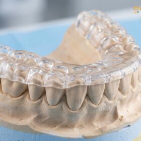
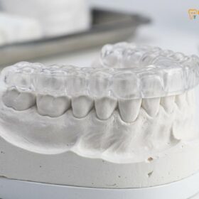
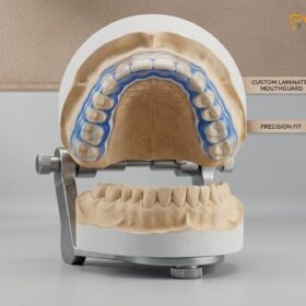
Bruxism symptoms and warning signs primarily include morning jaw pain, unexplained tooth sensitivity, flattened enamel, and chronic tension headaches. Recognizing these early indicators of teeth grinding or jaw clenching is critical to preventing irreversible dental damage and temporomandibular joint (TMJ) disorders before they require complex restorative treatments.
Clinical Summary:
Bruxism is a prevalent but often subconscious condition characterized by the involuntary grinding and clenching of teeth. Because sleep bruxism occurs without patient awareness, clinical diagnosis relies heavily on identifying secondary symptoms. The most definitive warning signs include abnormal occlusal wear (flattened teeth), abfraction lesions at the gumline, masseter muscle hypertrophy, and temporomandibular joint dysfunction. Early intervention, typically utilizing custom oral appliances, mitigates the risk of catastrophic enamel loss, tooth fractures, and chronic orofacial pain syndromes. Prompt clinical evaluation is recommended when patients experience persistent morning headaches or unexplained dental hypersensitivity.
Key Takeaways:
- Morning jaw soreness and dull headaches are the most common early indicators of sleep bruxism.
- Visible dental damage includes flattened chewing surfaces, micro-cracks, and receding gums.
- Unexplained tooth sensitivity to hot or cold often stems from enamel erosion caused by grinding.
- Ear pain and tinnitus can occur due to the anatomical proximity of the TMJ to the ear canal.
- Clinical intervention with custom oral appliances protects dentition and alleviates muscle tension.
Early Warning Signs of Bruxism
The earliest indicators of bruxism often manifest as subtle morning discomfort, including mild jaw stiffness, unexplained facial tension, and disrupted sleep patterns before visible dental damage occurs.
Bruxism is an insidious condition that frequently develops without the patient’s immediate awareness. Because the most destructive forces typically occur during sleep, individuals may grind their teeth for months or even years before realizing there is a problem. The early bruxism symptoms warning signs are often subtle, masquerading as general fatigue, mild stress, or poor sleep quality. Understanding these initial physiological changes is the first step toward seeking appropriate clinical care and preventing long-term structural damage to the oral cavity.
Clinically, bruxism is categorized into two distinct phenotypes: awake bruxism and sleep bruxism. Awake bruxism is primarily characterized by involuntary clenching of the teeth and jaw bracing in response to emotional stress, intense concentration, or anxiety. Patients with awake bruxism rarely grind their teeth back and forth; instead, they sustain static, high-pressure muscle contractions. Conversely, sleep bruxism involves rhythmic masticatory muscle activity (RMMA), which includes both forceful clenching and lateral grinding movements. This nocturnal activity generates occlusal forces that can be up to three times greater than the forces exerted during normal chewing, placing immense strain on the entire stomatognathic system.
“Because sleep bruxism is mediated by the central nervous system and often triggered by micro-arousals during the sleep cycle, patients are entirely unaware of the extreme biomechanical forces they are applying to their dentition until secondary symptoms arise.”
One of the most frequent early bruxism signs is waking up with a feeling of stiffness or fatigue in the lower face. Patients often describe this as a dull, generalized ache that dissipates as the day progresses. This morning discomfort is a direct result of the masticatory muscles—specifically the masseter and temporalis—being overworked throughout the night. Just as lifting weights causes muscle fatigue and soreness due to lactic acid accumulation, hours of nocturnal clenching leave the jaw muscles exhausted by morning.
Furthermore, sleep architecture is often disrupted in patients who grind their teeth. Clinical sleep studies (polysomnography) have demonstrated that episodes of sleep bruxism are frequently preceded by a sudden increase in brain wave activity, heart rate, and respiratory rate—a phenomenon known as a micro-arousal [1]. While these arousals do not fully awaken the patient, they fragment the sleep cycle, preventing the individual from reaching deep, restorative stages of sleep. Consequently, patients may experience daytime sleepiness, chronic fatigue, and a general sense of lethargy, which are often overlooked as grinding teeth at night symptoms.
Physical Symptoms on Your Teeth
Chronic grinding physically destroys tooth structure, leading to flattened chewing surfaces, enamel micro-fractures, abfraction lesions at the gumline, and severe hypersensitivity.
The most objective and measurable bruxism symptoms warning signs are found directly on the teeth. Dental enamel is the hardest substance in the human body, highly mineralized and designed to withstand a lifetime of normal chewing forces. However, it is not equipped to endure the relentless, extreme friction generated by pathological grinding. Over time, the biomechanical overload causes distinct patterns of structural degradation that a trained dental professional can easily identify during a routine examination.
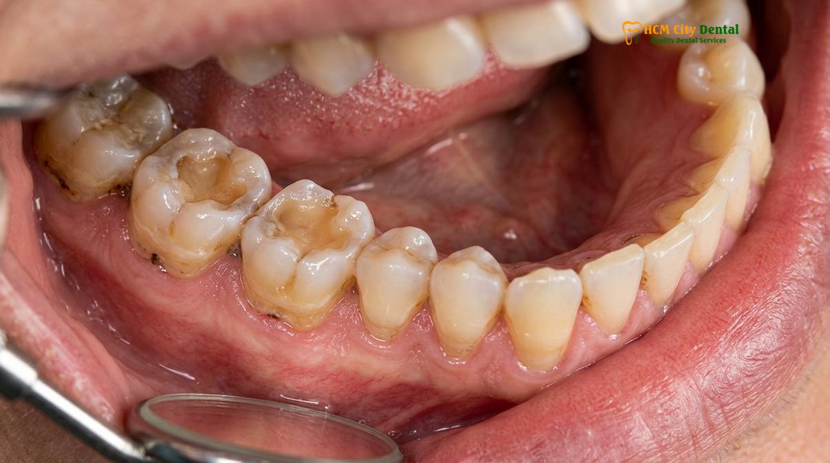
The hallmark of chronic bruxism is abnormal occlusal wear, clinically referred to as attrition. In a healthy mouth, the chewing surfaces of the molars and premolars feature distinct cusps (peaks) and fissures (valleys) that aid in the efficient mastication of food. In a patient with severe bruxism, these anatomical features are systematically ground down, leaving the teeth completely flat. This flattening not only diminishes chewing efficiency but also alters the vertical dimension of occlusion—the resting distance between the upper and lower jaws. As the teeth become shorter, the lower face can take on a collapsed or prematurely aged appearance.
As the protective enamel layer is worn away, the underlying dentin is exposed. Dentin is significantly softer than enamel and wears down at an accelerated rate once exposed [2]. It is also distinctly yellow in color, which is why patients with advanced bruxism often complain that their teeth look discolored or stained, despite rigorous oral hygiene. More importantly, dentin contains thousands of microscopic tubules that communicate directly with the dental pulp (the nerve center of the tooth). When these tubules are exposed to the oral environment, patients experience acute dentin hypersensitivity. A sudden, sharp pain in response to hot coffee, cold water, or sweet foods is one of the most common teeth grinding symptoms that drives patients to seek emergency dental care.
Beyond the chewing surfaces, bruxism exerts immense lateral stress on the teeth, causing them to flex microscopically at the cervical margin (the area where the tooth meets the gumline). This continuous flexing causes the enamel rods at the neck of the tooth to fracture and chip away, creating deep, V-shaped notches known as abfraction lesions.
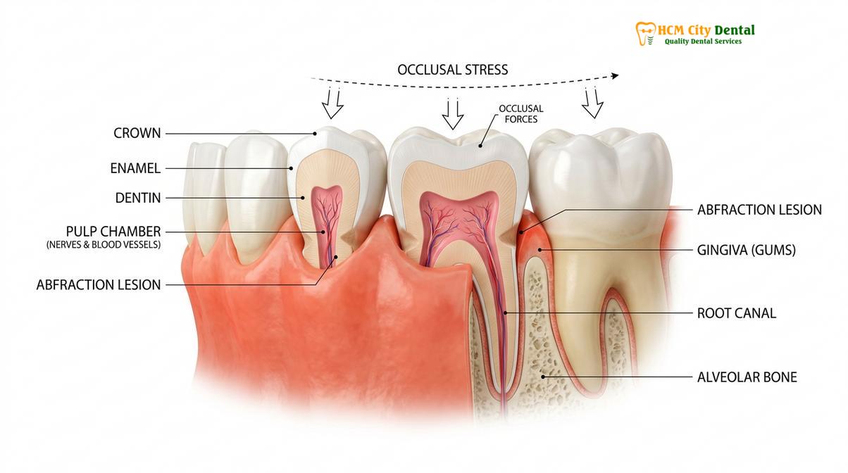
These lesions are often mistakenly attributed to aggressive toothbrushing, but they are a classic biomechanical consequence of occlusal overload. Furthermore, the chronic trauma to the periodontium (the supporting structures of the teeth) can lead to localized gum recession, widened periodontal ligaments visible on X-rays, and increased tooth mobility.
| Clinical Feature | Normal Physiological Wear (Age-Related) | Pathological Bruxism Wear |
|---|---|---|
| Rate of Progression | Slow, gradual over decades. | Rapid, aggressive enamel loss. |
| Wear Pattern | Matches normal chewing pathways. | Flat, matching non-functional grinding patterns. |
| Tooth Sensitivity | Rare; secondary dentin forms slowly to protect the nerve. | Common and severe due to rapid dentin exposure. |
| Cervical Lesions | Typically absent unless caused by abrasive brushing. | Frequent V-shaped abfraction notches at the gumline. |
| Muscle Symptoms | None. | Frequent masseter hypertrophy and TMJ pain. |
Jaw and Facial Pain Indicators
Persistent clenching overworks the masseter and temporalis muscles, resulting in localized facial pain, masseter hypertrophy (enlarged jaw muscles), and temporomandibular joint dysfunction.
The neuromuscular consequences of bruxism are often more debilitating than the dental damage itself. The human jaw is powered by a complex network of muscles, primarily the masseter (located at the angle of the jaw) and the temporalis (fanning out across the side of the head). During an episode of sleep bruxism, these muscles can contract with a force exceeding 250 pounds of pressure. When this level of exertion is sustained night after night, the muscles undergo physiological changes that manifest as prominent jaw clenching signs.
One of the most notable physical adaptations to chronic bruxism is masseter hypertrophy. Just as skeletal muscles increase in size in response to weightlifting, the masseter muscles can become visibly enlarged due to the constant workout of nocturnal grinding [3]. Clinically, this presents as a widening of the lower face, giving the patient a pronounced, square-jawed appearance. While some patients may not notice the gradual change in their facial aesthetics, they almost certainly notice the accompanying myalgia (muscle pain). The overworked muscles become tender to the touch, prone to spasms, and often harbor hyperirritable localized nodules known as trigger points.
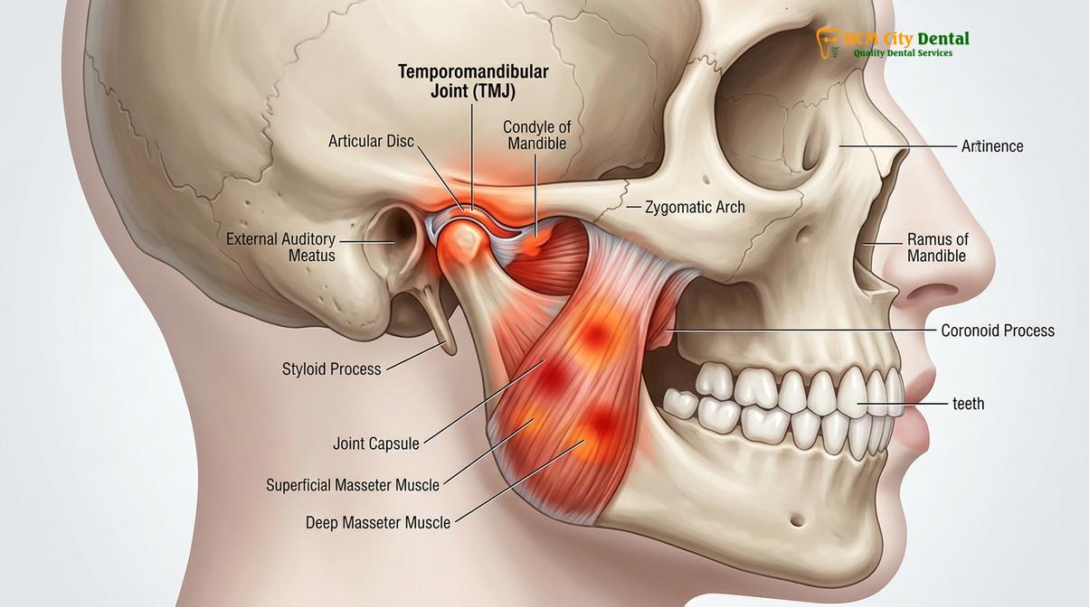
The extreme forces generated by the masticatory muscles are ultimately absorbed by the temporomandibular joint (TMJ), the complex hinge that connects the lower jaw to the skull. The TMJ is a highly specialized joint featuring a cartilaginous disc that acts as a shock absorber between the condyle of the mandible and the articular eminence of the temporal bone. Chronic bruxism compresses this joint space, squeezing the articular disc and surrounding synovial tissues.
Clinical Case Review: Managing TMJ Pain
A 34-year-old patient presented to HCMC Dental Clinic in Ho Chi Minh City complaining of severe, radiating jaw pain and an inability to open her mouth fully in the mornings. Upon clinical examination, Dr. Nguyen Van Cuong noted significant masseter hypertrophy, bilateral TMJ clicking, and severe attrition on the mandibular incisors. The patient was unaware of her nocturnal grinding. Dr. Cuong diagnosed severe sleep bruxism with secondary temporomandibular joint disorder (TMD). A comprehensive treatment plan was initiated, featuring a custom-fabricated hard acrylic occlusal splint to decompress the joint space and protect the remaining dentition. Within three weeks of consistent appliance wear, the patient reported a 90% reduction in morning facial pain and restored normal jaw function.
Over time, the excessive pressure can cause the articular disc to become displaced. When the jaw opens and closes, the condyle slips on and off the displaced disc, producing an audible clicking, popping, or grating sound (crepitus). If left unmanaged, the continuous trauma can lead to degenerative joint disease, osteoarthritis of the TMJ, and severe limitations in jaw mobility, making simple tasks like chewing, speaking, or yawning intensely painful.
Headaches and Ear Pain Connection
Referred pain from overworked jaw muscles frequently causes tension-type headaches in the temples and secondary ear symptoms like aching, fullness, or tinnitus.
Patients suffering from bruxism frequently present to medical doctors with complaints of chronic headaches or ear infections, unaware that the root cause lies within their oral cavity. The phenomenon of referred pain—where pain originates in one anatomical location but is felt in another—is highly prevalent in the maxillofacial region due to the complex web of interconnected cranial nerves.
Tension-type headaches are among the most frequently reported bruxism symptoms warning signs. These headaches typically present as a dull, continuous ache wrapping around the head like a tight band, often concentrated in the temporal regions. This specific location correlates directly with the temporalis muscle, which spans the side of the skull and is heavily involved in jaw clenching. During nocturnal bruxism, the temporalis muscle remains in a state of sustained contraction, leading to muscle fatigue, reduced localized blood flow (ischemia), and the release of inflammatory neuropeptides. The resulting pain is often misdiagnosed as a primary migraine or stress headache, leading to ineffective pharmacological treatments that fail to address the underlying biomechanical cause [4].
“The intersection of dentistry and otolaryngology is most evident in the management of bruxism. Countless patients undergo unnecessary treatments for presumed ear infections or primary migraines, when a simple occlusal evaluation reveals that hyperactive jaw muscles are the true source of their referred pain.”
Similarly, the anatomical proximity of the TMJ to the ear canal explains why bruxism frequently mimics otologic (ear) conditions. The posterior aspect of the TMJ is separated from the auditory canal by only a thin plate of bone. When the joint is inflamed or the condyle is pushed backward due to a collapsed bite from severe tooth wear, it can compress the surrounding tissues and the auriculotemporal nerve. This compression translates into symptoms such as a deep ache within the ear, a sensation of fullness or pressure (as if the ear needs to “pop”), and even tinnitus (a persistent ringing or buzzing sound).
Because these symptoms so closely mimic an ear infection (otitis media), patients often seek care from an ENT specialist. However, when an otoscopic examination reveals a perfectly healthy eardrum and clear ear canal, the diagnostic focus must shift to the masticatory system. Recognizing the connection between jaw clenching signs and secondary ear or head pain is crucial for accurate diagnosis and effective, targeted intervention.
How a Dentist Confirms Bruxism
Clinical diagnosis involves a comprehensive intraoral examination of wear facets, muscle palpation, radiographic analysis of the periodontal ligament, and patient symptom history.
Because sleep bruxism occurs subconsciously, self-reporting is notoriously unreliable. Therefore, confirming the presence of this condition requires a meticulous clinical evaluation by a qualified dental professional. At HCMC Dental Clinic, the diagnostic workflow is designed to identify both the primary structural damage and the secondary neuromuscular adaptations associated with chronic grinding.
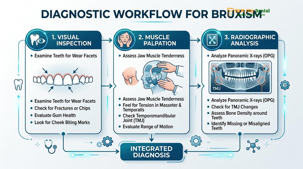
The diagnostic process begins with a thorough medical and dental history. The clinician will inquire about morning symptoms, sleep quality, stress levels, and any history of jaw locking or clicking. Following the interview, a comprehensive intraoral examination is conducted. The dentist looks for specific, undeniable physical markers of bruxism. The most obvious sign is the presence of wear facets—flat, shiny areas on the chewing surfaces of the teeth where the enamel has been ground away. The clinician will often ask the patient to slide their jaw side to side to see if the upper and lower wear facets interlock perfectly, which definitively confirms the grinding pathway.
In addition to examining the hard tissues, the dentist evaluates the soft tissues of the mouth. Chronic clenching often causes the patient to press their cheeks and tongue forcefully against their teeth. This results in a raised, white, hyperkeratotic line along the inside of the cheek (linea alba) and a scalloped appearance along the lateral borders of the tongue (crenated tongue). These soft tissue adaptations are classic indicators of sustained intraoral pressure.
Muscle palpation is another critical component of the diagnostic exam. Dr. Cuong and the clinical team will systematically press on the masseter, temporalis, and medial pterygoid muscles to check for hypertrophy, tenderness, and trigger points. The TMJ is also evaluated for range of motion, deviations upon opening, and the presence of joint sounds like clicking or crepitus.
Finally, radiographic analysis is utilized to assess the health of the underlying bone and supporting structures. On a panoramic or periapical X-ray, chronic bruxism often presents as a widened periodontal ligament space—a sign that the tooth is being rocked back and forth in its socket. The bone surrounding the roots may also appear denser (osteosclerosis) as it attempts to reinforce itself against the excessive occlusal load [5].
Once a definitive diagnosis is established, the primary line of defense is the fabrication of protective oral appliances like custom nightguards. These precision-milled devices provide a smooth, durable barrier that absorbs grinding forces, protects the enamel from further attrition, and helps deprogram the hyperactive jaw muscles, providing significant symptomatic relief.
When Symptoms Require Urgent Care
Immediate dental evaluation is necessary if you experience locked jaw, severe radiating facial pain, fractured teeth, or sudden changes in your dental bite.
While mild bruxism can often be managed with routine monitoring and conservative lifestyle adjustments, certain symptoms indicate that the condition has progressed to a critical stage requiring immediate clinical intervention. Ignoring these red flags can lead to irreversible damage to the dentition, permanent joint dysfunction, and chronic pain syndromes that are exceedingly difficult to treat.
Urgent Clinical Red Flags
Seek immediate dental evaluation if you experience any of the following symptoms:
- Trismus (Lockjaw): An inability to open or close the mouth fully, indicating severe muscle spasm or acute TMJ disc displacement.
- Acute Tooth Fracture: A sudden break or vertical crack in a tooth, often accompanied by sharp pain when biting down.
- Severe, Unrelenting Pain: Radiating facial pain that disrupts sleep and does not respond to over-the-counter analgesics.
- Sudden Bite Changes: A feeling that your upper and lower teeth no longer fit together correctly, suggesting joint inflammation or rapid tooth wear.
A fractured tooth is a common emergency associated with severe bruxism. The extreme forces can cause micro-cracks in the enamel to propagate deep into the dentin and eventually reach the pulp chamber. This condition, known as cracked tooth syndrome, causes excruciating pain upon release of biting pressure and extreme sensitivity to temperature. If the crack extends below the gumline, the tooth may become non-restorable and require extraction. Early intervention with a protective nightguard is the most effective way to prevent this catastrophic outcome.
Similarly, acute TMJ locking requires prompt attention. If the articular disc becomes permanently displaced anteriorly, it acts as a mechanical wedge, preventing the condyle from translating forward and severely restricting jaw opening. This is a highly distressing condition that requires specialized manipulation and appliance therapy to recapture the disc and restore normal function.
If you are experiencing any of these severe bruxism symptoms warning signs, it is imperative to seek professional care without delay. At HCMC Dental Clinic in Ho Chi Minh City, our experienced clinical team, led by Dr. Cuong, utilizes advanced diagnostic imaging and evidence-based therapeutic protocols to manage acute bruxism flare-ups, alleviate pain, and design long-term strategies to protect your oral health.
Frequently Asked Questions
What does bruxism damage look like?
Bruxism damage typically presents as flattened or chipped chewing surfaces, yellowing teeth due to exposed dentin, and receding gums with V-shaped notches at the gumline. Over time, the continuous grinding forces wear away the protective enamel, leaving the teeth looking shorter, structurally compromised, and highly susceptible to decay and fractures. Dentists also look for secondary signs like a scalloped tongue or a white line on the inner cheek caused by chronic clenching pressure.
Can bruxism cause tooth sensitivity?
Yes, bruxism is a leading cause of severe tooth sensitivity because the grinding action strips away the protective enamel layer, exposing the underlying dentin. Dentin contains microscopic tubules that lead directly to the tooth’s nerve center; when exposed to hot, cold, or sweet stimuli, fluid shifts within these tubules trigger sharp, sudden pain. This sensitivity often worsens in the morning immediately following a night of intense grinding.
Why does my jaw hurt every morning?
Morning jaw pain occurs because sleep bruxism forces your masticatory muscles to contract intensely for prolonged periods during the night, leading to lactic acid buildup and muscle fatigue. This sustained hyperactive muscle tension overworks the temporomandibular joint (TMJ) and surrounding tissues, resulting in localized soreness, stiffness, and restricted jaw movement upon waking. The pain typically radiates from the angle of the jaw up toward the temples.
Is ear pain related to teeth grinding?
Ear pain is frequently related to teeth grinding due to the anatomical proximity of the temporomandibular joint (TMJ) to the ear canal and shared neural pathways. Inflammation and muscle spasms in the jaw can refer pain to the ear, causing symptoms like aching, a feeling of fullness, or even tinnitus (ringing in the ears) without any actual ear infection. This referred pain is a common reason bruxism is initially misdiagnosed as an otologic issue.
Should I see a dentist or doctor for bruxism?
You should primarily see a dentist for bruxism, as they are specialists in diagnosing occlusal wear, muscle tension, and TMJ disorders associated with teeth grinding. A dentist can provide custom oral appliances to protect your teeth and joints, though they may collaborate with a sleep physician if an underlying sleep disorder like sleep apnea is suspected. Medical doctors can prescribe muscle relaxants, but they cannot address the biomechanical dental damage.
References
- Journal of Oral Rehabilitation. Sleep architecture and bruxism micro-arousals. (2021).
- American Journal of Dentistry. Enamel attrition rates in chronic bruxism patients. (2020).
- Journal of Clinical Medicine. Masseter hypertrophy and temporomandibular disorders. (2022).
- International Journal of Prosthodontics. Referred pain mechanisms in masticatory muscle myalgia. (2019).
- Journal of the American Dental Association. Diagnostic criteria for sleep and awake bruxism. (2023).