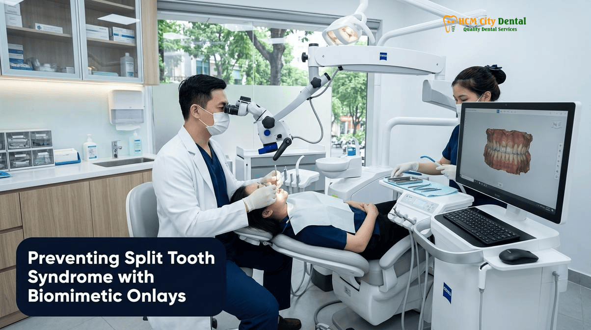
For decades, the standard dental response to a cracked molar was a reactive one: wait until the tooth hurts severely, perform a root canal, and grind the tooth down for a traditional full-coverage crown. However, modern biomimetic dentistry recognizes that by the time a patient experiences constant, throbbing pain, an incomplete structural crack may have already propagated into a catastrophic, non-restorable split tooth. The key to preserving natural dentition lies in proactive intervention. By understanding the biomechanics of split tooth syndrome cuspal coverage biomimetic onlay prevention, clinicians can intercept micro-fractures before they destroy the pulpal floor. At HCMC Dental Clinic in District 1, Ho Chi Minh City, advanced 25x microscopic crack dissection combined with adhesive ceramic overlays allows us to splint compromised molars, neutralize occlusal stress, and save the living nerve in the vast majority of cases.
Clinical Summary:
Cracked Tooth Syndrome (CTS) is a progressive biomechanical failure often initiated by the loss of marginal ridges from large, outdated fillings. If left untreated, an incomplete dentinal crack will inevitably propagate under chewing forces, transitioning from a restorable cracked tooth into a non-savable split tooth. Traditional treatments involving aggressive full crowns often exacerbate structural weakness. Conversely, biomimetic dentistry utilizes 25x Carl Zeiss microscopic crack mapping, Immediate Dentin Sealing (IDS), and 1.5mm cuspal-coverage IPS E.max ceramic onlays to adhesively “hoop” and splint the fractured segments together. This minimally invasive approach, led by Dr. Nguyen Van Cuong, DDS (Clinical Director — MOH License 0003265/BL-CCHN), stops crack propagation, preserves pulpal vitality in over 80% of cases, and prevents the need for root canal therapy or extraction.
Key Takeaways:
- The Danger of Rebound Pain: A sharp jolt of pain specifically when releasing your bite (rather than clenching) is the hallmark sign of a flexing cracked tooth that requires immediate cuspal splinting.
- The Amalgam Wedge Effect: Large, old MOD (Mesio-Occluso-Distal) metal fillings act as internal wedges, driving unsupported cusps apart during mastication and initiating deep dentinal fractures.
- Microscopic Diagnostics: 25x Carl Zeiss magnification and transillumination are mandatory to differentiate between a superficial enamel craze line and a deep structural crack threatening the pulp.
- Biomimetic Splinting: A 1.5mm cuspal-coverage ceramic onlay acts as a rigid adhesive “hoop,” neutralizing stress and preventing the tooth from splitting, without the aggressive 75% tissue removal required for a traditional crown.
- Transparent Expat Pricing: Through the Official 2026 Dental Price List & -40% Online Booking Fee Schedule, a biomimetic IPS E.max Onlay at HCMC Dental Clinic costs just $231 USD, offering world-class restorative care at a fraction of Western prices.
The Progression of a Crack: From Asymptomatic Enamel Craze Line to Rebound-Pain ‘Cracked Tooth’ to Non-Savable ‘Split Tooth’
To effectively manage structural fractures in posterior teeth, one must first understand that a crack is not a static event; it is a progressive, dynamic biomechanical failure. The American Dental Association (ADA) and contemporary endodontic literature classify longitudinal tooth fractures into five distinct categories: craze lines, fractured cusps, cracked teeth, split teeth, and vertical root fractures. Understanding the exact stage of this progression is critical, as the treatment protocol shifts dramatically from simple observation to complex biomimetic restoration, or ultimately, to extraction.
The progression begins with craze lines. These are superficial, microscopic fractures confined entirely to the outer enamel layer. They are ubiquitous in adult dentition, appearing as faint vertical lines on the buccal or lingual surfaces of teeth. Craze lines do not cross the dentinoenamel junction (DEJ), do not cause pain, and do not compromise the structural integrity of the tooth. They require no restorative treatment, though they may be an aesthetic concern for some patients.
However, when a fracture breaches the DEJ and enters the resilient dentin layer, the biomechanical dynamics change entirely. This is the realm of the fractured cusp and the true cracked tooth. When comparing split tooth syndrome vs cracked tooth, the defining difference is the extent of the fracture line. A cracked tooth features an incomplete fracture that originates on the occlusal (chewing) surface and propagates apically (downward) toward the pulp, but the tooth has not yet separated into two distinct pieces. This is the critical window for intervention.
The clinical presentation of a cracked tooth is highly specific. Patients rarely complain of a constant, dull ache. Instead, they report a sharp, electric-like jolt of pain that occurs sporadically when chewing specific foods, like seeds, nuts, or crusty bread. More specifically, this pain often manifests as “rebound pain”—a sharp sensation felt at the exact moment the patient opens their jaw and releases the bite. This occurs because the occlusal force pushes the fractured segments apart, causing fluid movement within the dentinal tubules. Upon release, the segments snap back together, causing a rapid fluid shift that violently stimulates the pulpal nerve fibers. This pain on bite release tooth sleuth test is a definitive diagnostic indicator that the tooth is flexing under load.
If a cracked tooth is left untreated, the repetitive stress of mastication will inevitably drive the fracture deeper. Once the crack propagates completely through the pulpal floor or extends subgingivally down the root surface, separating the tooth into two distinct, movable segments, it becomes a split tooth. At this catastrophic stage, the tooth is no longer restorable. The biological width is violated, the periodontal ligament is compromised, and the pulpal tissue is exposed to massive bacterial ingress. A split tooth mandates immediate extraction and subsequent replacement with a dental implant. The primary goal of biomimetic dentistry is to intercept the fracture during the “cracked tooth” phase, utilizing a cuspal coverage ceramic onlay cracked molar protocol to halt the progression before the tooth splits.
Why Large Old MOD Amalgam and Composite Fillings Act as Internal Metal Wedges That Split Unsupported Molar Cusps
To prevent cracked teeth, we must address their primary etiology. While trauma and severe bruxism (teeth grinding) can cause virgin teeth to fracture, the vast majority of cracked teeth share a common denominator: the presence of large, outdated intra-coronal restorations, specifically Mesio-Occluso-Distal (MOD) fillings. The structural integrity of a posterior molar relies heavily on its marginal ridges—the raised borders of enamel on the mesial (front) and distal (back) edges of the tooth. These ridges act as the architectural “tie-bars” that hold the buccal (cheek) and lingual (tongue) cusps together.
When a dentist prepares a tooth for an MOD filling, both marginal ridges are drilled away to remove decay. This preparation severs the tie-bars, immediately reducing the tooth’s structural stiffness by up to 60%. The remaining buccal and lingual cusps are left unsupported, acting like two independent, flexible diving boards. This structural compromise sets the stage for catastrophic failure.
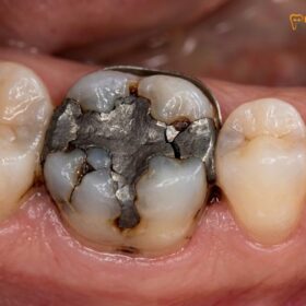
The material used to fill this void further exacerbates the problem. For decades, silver amalgam was the material of choice. However, amalgam is a non-adhesive metal alloy. It does not bond to the tooth structure; it merely sits passively within a mechanically retentive cavity preparation. Furthermore, amalgam has a different coefficient of thermal expansion than natural tooth structure, meaning it expands and contracts at a different rate when exposed to hot and cold foods. Over years of thermal cycling and occlusal loading, the old mod amalgam filling wedge fracture effect becomes apparent. The rigid metal mass acts as an internal wedge. Every time the patient bites down, the occlusal force is transmitted through the amalgam, driving the unsupported buccal and lingual cusps apart. Over time, this repetitive wedging action initiates micro-fractures at the sharp internal line angles of the cavity preparation, which slowly propagate into deep dentinal cracks.[1]
Older generation composite resins also contribute to this phenomenon, albeit through a different mechanism. Early composites suffered from high volumetric polymerization shrinkage. When these materials were bulk-filled into large MOD cavities and light-cured, they shrank significantly. This shrinkage generated massive stress (up to 20 MPa) on the bonded cavity walls, pulling the cusps inward. This constant, static tension weakens the enamel and dentin, making the tooth highly susceptible to fracture under normal chewing forces. Whether caused by the wedging of amalgam or the shrinkage stress of old composites, the result is the same: a structurally compromised tooth that is slowly tearing itself apart.
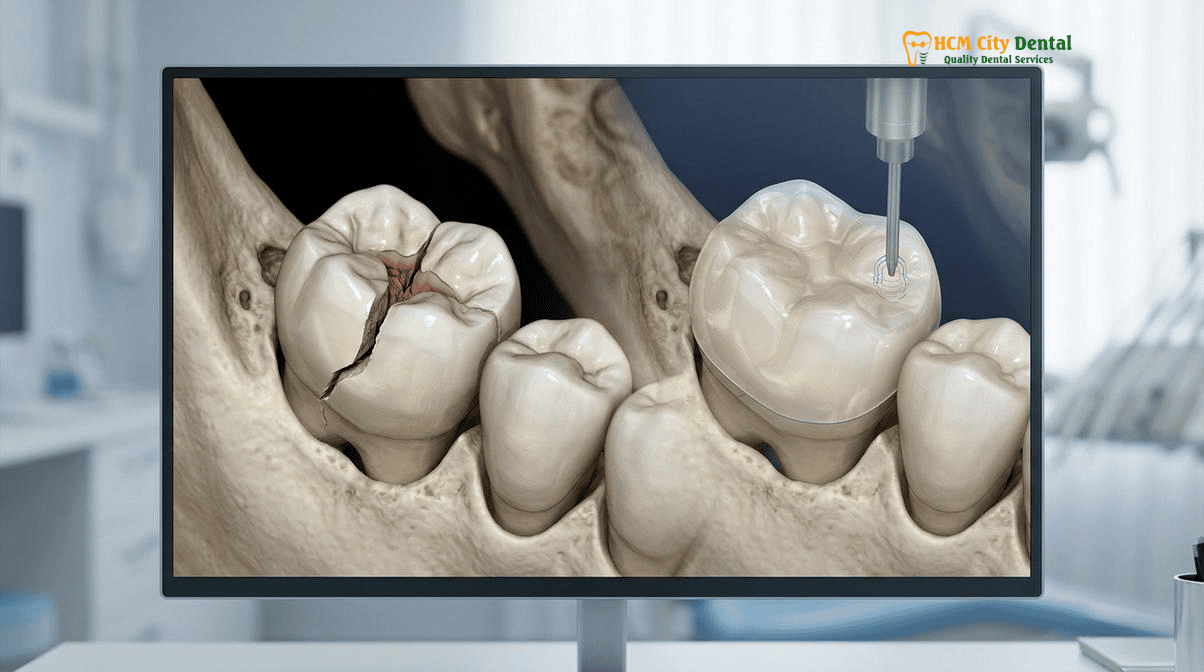
25x Carl Zeiss Microscopic Crack Mapping: Tooth Sleuth Rebound Testing, Transillumination, and Complete Caries/Crack Dissection
Diagnosing a cracked tooth is notoriously difficult. Because the fractures are often microscopic and incomplete, they rarely appear on traditional 2D periapical X-rays or even advanced 3D CBCT scans unless the tooth has already split or a significant periapical radiolucency has developed. Therefore, diagnosis relies heavily on advanced clinical testing and high-magnification visual inspection.
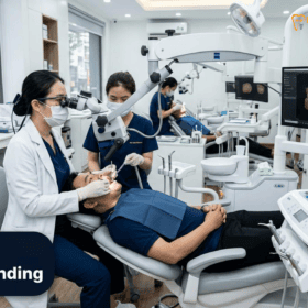
At HCMC Dental Clinic, the diagnostic protocol begins with a detailed symptom history and the Tooth Sleuth test. The Tooth Sleuth is a small, pyramid-shaped plastic bite stick designed to isolate occlusal forces onto individual cusps. Dr. Nguyen Van Cuong, DDS places the instrument on one cusp at a time and asks the patient to bite down firmly and then release quickly. If the patient experiences a sharp jolt of pain specifically upon release, the offending cusp and the associated fracture line are immediately identified.
Following the bite test, fiber-optic transillumination is employed. A high-intensity light source is placed against the cervical portion of the tooth. In a healthy, intact tooth, the light transmits uniformly through the enamel and dentin. However, if a structural crack is present, the fracture plane acts as a physical barrier, blocking the transmission of light. The portion of the tooth on the light source side will illuminate brightly, while the segment on the other side of the crack will appear dark. This stark contrast provides immediate visual confirmation of the crack’s location and extent.
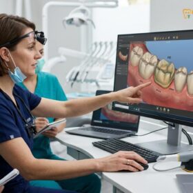
Once the crack is identified, the true extent of the damage must be assessed through microscopic crack dissection. This procedure is performed exclusively under a 25x Carl Zeiss operating microscope. The old restoration (amalgam or composite) is carefully removed, along with any secondary decay. Because cracks often harbor bacteria, decay frequently tracks deep along the fracture line. Once the cavity is clean, a caries indicator dye or methylene blue is applied to the dentin. This dye selectively stains the organic components of the fracture line, making the microscopic crack vividly visible under the intense coaxial LED illumination of the microscope.[2]
“The operating microscope is not a luxury in the treatment of cracked teeth; it is an absolute necessity. Without 25x magnification and targeted dye staining, it is impossible to determine if a crack terminates safely within the coronal dentin or if it has propagated fatally into the pulpal floor. We must map the entire fracture line before we can commit to a restorative strategy.”
— Dr. Nguyen Van Cuong, DDS (Clinical Director, HCMC Dental Clinic)
Dr. Cuong meticulously traces the stained crack line. If the crack extends subgingivally but remains above the crestal bone, a Deep Margin Elevation (DME) procedure or minor surgical crown lengthening (respecting Coslet’s Altered Passive Eruption principles) may be performed to expose the margin. However, if the microscopic dissection reveals that the crack has split the pulpal floor or extends deep down the root, the tooth is deemed non-restorable, and the patient is immediately informed that extraction and implant placement are required.
Why Replacing an Old Filling with Another Direct Filling Fails to Stop Cuspal Flexure — and How a 1.5mm Ceramic Onlay ‘Hoops’ the Tooth
When a patient presents with a cracked tooth, a common misconception is that simply removing the old filling and placing a new, larger direct composite filling will solve the problem. This approach is biomechanically flawed and almost guarantees future failure. A direct filling, no matter how strong the composite material, is an intra-coronal restoration—it sits *inside* the tooth. It does not cover or protect the weakened cusps. When the patient chews, the occlusal forces still hit the unprotected cusps, causing them to flex independently. The new filling does nothing to stop this flexure; in fact, due to Feilzer’s Configuration Factor (C-Factor), placing a large direct composite in a deep cavity can generate significant polymerization shrinkage stress, further weakening the already compromised tooth.
To save a cracked tooth, the restorative paradigm must shift from intra-coronal filling to extra-coronal splinting. The tooth must be bound together to prevent the cusps from flexing apart. Historically, this was achieved by grinding the tooth down for a full-coverage porcelain-fused-to-metal (PFM) or zirconia crown. While a full crown does provide cuspal coverage, it requires the aggressive removal of 70% to 75% of the tooth’s healthy coronal structure. For a tooth that is already weakened by a deep crack, this massive reduction of the cervical enamel and axial walls can be the final blow, leading to pulpal necrosis or catastrophic root fracture.
The modern, evidence-based solution is the biomimetic cuspal-coverage ceramic onlay (or overlay). An onlay is a conservative, indirect restoration that covers the chewing surface of the tooth, replacing the missing marginal ridges and capping the weakened cusps, while leaving the healthy buccal and lingual axial walls completely intact. The preparation requires only 1.5mm of occlusal reduction to provide adequate thickness for the ceramic material.
The magic of the biomimetic onlay lies in its adhesive integration. Unlike a traditional crown that relies on mechanical retention (friction) and non-adhesive cements, a biomimetic onlay is chemically bonded to the underlying enamel and dentin. Once bonded, the high-strength lithium disilicate ceramic (IPS E.max) acts as a rigid, unyielding “hoop” over the top of the tooth. When occlusal forces are applied, the onlay distributes the stress evenly across the entire tooth structure, neutralizing the wedging forces and completely halting cuspal flexure. This “hoop stress” effect splints the fractured segments together, stopping the crack from propagating and allowing the tooth to function normally without pain.[3]
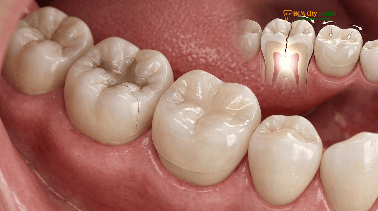
Saving the Living Pulp in 80%+ of Cracked Teeth via Immediate Dentin Sealing (IDS) + Cuspal-Coverage IPS E.max Onlay
One of the most significant advancements in biomimetic dentistry is the ability to save the living nerve (pulp) in teeth that would have traditionally been condemned to root canal therapy. When a tooth cracks, the dentinal tubules are exposed, allowing bacterial toxins and fluid movement to irritate the pulp, causing the characteristic rebound pain. If the tooth is simply prepped for a crown and sent home with a leaky temporary, this irritation continues, often leading to irreversible pulpitis.
To prevent this, Dr. Nguyen Van Cuong, DDS employs a strict Immediate Dentin Sealing (IDS) and resin coating protocol. Immediately after the crack is dissected and the cavity is prepared, the freshly cut dentin is sealed before the digital impression is even taken. This is performed under a 100% latex-free nitrile rubber dam to ensure absolute isolation from saliva and humidity.
The IDS protocol involves applying a gold-standard mild self-etching adhesive to the dentin. This creates a hybrid layer that seals the dentinal tubules, instantly stopping fluid movement and protecting the pulp from bacterial microleakage. A thin layer of flowable composite (resin coating) is then applied over the adhesive. Crucially, a 5-minute decoupling time is observed, allowing the adhesive layer to mature and dissipate polymerization stress before any further material is added. In cases of deep cracks approaching the pulp, ultra-high-molecular-weight polyethylene (UHMWPE) fibers (such as Ribbond) may be embedded into the resin coating. These fibers act as a stress-absorbing mesh, further reinforcing the cavity floor and preventing crack propagation.
By sealing the dentin immediately, the pulp is protected during the provisionalization phase. Patients routinely report that their rebound pain vanishes the moment the IDS protocol is completed, long before the final ceramic onlay is even cemented. This technique allows us to save cracked tooth without root canal Vietnam in over 80% of cases, preserving the tooth’s vitality, proprioception, and long-term resilience.[4]
“Immediate Dentin Sealing is the biological shield for the cracked tooth. By sealing the dentinal tubules the moment they are exposed, we eliminate sensitivity, block bacterial ingress, and increase our final bond strength by up to 400%. It is the cornerstone of preserving pulpal vitality in structurally compromised molars.”
— Dr. Nguyen Van Cuong, DDS (Clinical Director, HCMC Dental Clinic)
When the final IPS E.max onlay is ready for delivery, the adhesive protocol is equally meticulous. The intaglio surface of the lithium disilicate ceramic is treated with 5%–9.5% hydrofluoric acid to create micro-mechanical retention, followed by the application of a pure, heat-dried silane coupling agent to facilitate chemical bonding. The tooth surface is gently cleaned with 27-micron aluminum oxide air abrasion. The onlay is then bonded using a heated, highly filled restorative composite resin, creating a seamless, monoblock integration between the ceramic and the natural tooth. For patients seeking biomimetic onlay splinting Ho Chi Minh City, this protocol represents the pinnacle of restorative longevity.
Clinical Case Study: Saving an Expat’s Cracked Molar in District 1
Patient Profile: A 45-year-old Australian expat living in Thao Dien presented to HCMC Dental Clinic with sharp, shooting pain in his lower right first molar, specifically when releasing his bite after chewing almonds. The tooth had a large, 15-year-old MOD amalgam filling.
Clinical Assessment: Dr. Nguyen Van Cuong, DDS performed a Tooth Sleuth test, which elicited severe rebound pain on the disto-lingual cusp. Transillumination revealed a distinct fracture line running mesio-distally. 25x microscopic dissection confirmed a deep structural crack extending into the dentin, but fortunately, it had not breached the pulpal floor.
Treatment Execution: Under a nitrile rubber dam, the old amalgam and underlying decay were removed. The crack was mapped using methylene blue dye. An Immediate Dentin Sealing (IDS) protocol with UHMWPE fiber reinforcement was applied to protect the vital pulp and stabilize the cavity floor. The tooth was prepared for a conservative 1.5mm cuspal-coverage onlay, preserving all healthy axial enamel. A digital scan was taken, and a custom IPS E.max lithium disilicate onlay was milled and adhesively bonded.
Outcome: The patient’s rebound pain was eliminated immediately after the IDS procedure. The final bonded onlay successfully splinted the tooth, neutralizing occlusal stress. The vital pulp was saved, avoiding a costly and invasive root canal, and the patient returned to normal chewing function within 48 hours.
2026 Microscopic Crack Splinting, Biomimetic Onlay & Extraction/Implant Fee Schedule at HCMC Dental Clinic (District 1)
For international patients, expats, and dental travelers, navigating the costs of advanced restorative dentistry can be daunting. At HCMC Dental Clinic, we believe in absolute transparency. We offer world-class biomimetic dentistry, utilizing the exact same Carl Zeiss microscopes, Ivoclar Vivadent ceramics, and adhesive protocols found in top-tier clinics in Sydney, London, or Los Angeles, but at a fraction of the cost. By utilizing our Official 2026 Dental Price List & -40% Online Booking Fee Schedule, patients can access premium care without financial anxiety.
Below is the detailed 2026 fee schedule for treatments related to cracked teeth, biomimetic onlays, and, if necessary, extraction and implant replacement. Please note that the -40% online booking rates are exclusively available to patients who schedule their consultation prior to arriving at our District 1 clinic.
| Clinical Procedure (Cracked Tooth Management) | Walk-In / Standard Price (USD) | -40% Online Booking Price (USD) | VND Equivalent (Online) |
|---|---|---|---|
| IPS E.max All-Ceramic Crown / Inlay / Onlay / Overlay (Includes IDS & Microscopic Prep) | $385 USD | $231 USD / tooth | 6,000,000 VND |
| Full Zirconia Crown (Cercon Standard / HT Multilayer) | $256 – $321 USD | $154 – $192 USD / tooth | 4,000,000 – 5,000,000 VND |
| Microscopic Endodontic Root Canal Therapy (Complex Molar) (25x Carl Zeiss + Nitrile Dam) | $192 – $256 USD | $115 – $154 USD / tooth | 3,000,000 – 4,000,000 VND |
| Biomimetic Quartz/Glass Fiber Post & Core Buildup | $65 – $96 USD | $38 – $58 USD / tooth | 1,000,000 – 1,500,000 VND |
| Single Korean Implant (Dentis / Neo Biotech / Dentium) (Fixture + Abutment) | $769 – $1,090 USD | $462 – $654 USD | 12,000,000 – 17,000,000 VND |
| Single European / Swiss Implant (Straumann BLT-SLA / BLA-Active) | $1,603 – $2,244 USD | $962 – $1,346 USD | 25,000,000 – 35,000,000 VND |
| Implant Zirconia HT Crown + Custom Abutment | $256 USD | $154 USD | 4,000,000 VND |
| Custom Night Guard / Occlusal Splint (For Bruxism/Crack Prevention) | $135 – $192 USD | $81 – $115 USD | 2,100,000 – 3,000,000 VND |
For patients requiring comprehensive restorative work, we encourage you to explore our detailed guide on Dental Crowns, Fixed Bridges & Biomimetic Ceramic Restorations in Vietnam. Furthermore, understanding the nuances of adhesive protocols is vital; you can learn more about our techniques in our article on Immediate Dentin Sealing (IDS) & Resin Coating in Biomimetic Veneers & Onlays: Increasing Bond Strength by 400%.
Clinical Warning: When to Seek Immediate Care
Do not ignore rebound pain. If you experience a sharp jolt of pain when releasing your bite, or if a previously sensitive tooth suddenly develops a constant, throbbing ache that keeps you awake at night, the crack may be propagating toward the pulp or the root. Delaying treatment significantly increases the risk of irreversible pulpitis (requiring a root canal) or a catastrophic split tooth (requiring extraction). Contact HCMC Dental Clinic immediately via WhatsApp at +84 853 020 003 to schedule an urgent microscopic evaluation with Dr. Nguyen Van Cuong, DDS.
Frequently Asked Questions
Why does a cracked tooth hurt sharply at the exact moment I open my jaw (release my bite) rather than when I clench down?
This phenomenon, known as rebound pain, is the hallmark symptom of Cracked Tooth Syndrome. When you bite down, the occlusal force pushes the fractured segments of the tooth apart, causing the fluid within the microscopic dentinal tubules to shift. When you suddenly release the bite, the separated segments rapidly snap back together. This sudden closure causes a rapid, violent shift in the dentinal fluid, which stimulates the odontoblastic processes and the underlying pulpal nerve fibers, resulting in a sharp, electric-like jolt of pain. It is a clear biomechanical indicator that the tooth is flexing under load and requires immediate cuspal-coverage splinting. If you are experiencing this, we highly recommend reading our Cracked Tooth Inlay & Onlay: Clinical Guide to Restoring Fractured Cusps.
How can Dr. Nguyen Van Cuong tell under the 25x microscope if a crack stops in the crown or has split all the way through the floor of the tooth?
Using a 25x Carl Zeiss operating microscope, Dr. Nguyen Van Cuong, DDS performs a meticulous crack dissection. After removing the old restoration and any underlying decay, a caries indicator dye or methylene blue is applied to the dentin. The dye selectively stains the fracture line. Under high magnification and intense coaxial LED illumination, Dr. Cuong can visually trace the stained crack line. If the crack terminates within the dentin above the pulp chamber or the cementoenamel junction (CEJ), the tooth can usually be saved with a biomimetic onlay. If the crack propagates through the pulpal floor or extends deep down the root surface (a true split tooth), the prognosis becomes hopeless, necessitating extraction and implant replacement.
If my cracked tooth only hurts occasionally when chewing hard bread or nuts, does it automatically need a root canal?
No, it does not automatically require a root canal. In fact, occasional pain upon chewing hard foods is a sign of a structural crack that has not yet caused irreversible pulpitis or pulpal necrosis. If treated promptly with a biomimetic cuspal-coverage ceramic onlay, the crack can be stabilized, and the tooth’s flexure can be halted. By utilizing Immediate Dentin Sealing (IDS) and resin coating under a rubber dam, Dr. Nguyen Van Cuong, DDS can save the living pulp in over 80% of these cases, completely avoiding the need for endodontic therapy.
Why is a conservative ceramic Onlay (covering the chewing cusps) safer for a cracked tooth than shaving the sides down for a full crown?
Traditional full-coverage crowns require the aggressive reduction of 70% to 75% of the tooth’s healthy coronal structure, including the critical cervical enamel and the remaining intact axial walls. For a tooth that is already structurally compromised by a crack, this aggressive shaving further weakens the tooth, increasing the risk of the crack propagating into the root or causing pulpal necrosis. A biomimetic ceramic onlay, however, preserves the healthy axial walls and only reduces the compromised chewing cusps by 1.5mm. The onlay is then adhesively bonded to the tooth, acting like a rigid ‘hoop’ that splints the cusps together, neutralizing stress and preventing the crack from expanding, all while preserving maximum natural tooth structure.
How much does a biomimetic cuspal-coverage IPS E.max Onlay cost at HCMC Dental Clinic in District 1?
At HCMC Dental Clinic in District 1, Ho Chi Minh City, a biomimetic cuspal-coverage IPS E.max All-Ceramic Onlay (or Overlay) costs $231 USD per tooth (6,000,000 VND) when booked online through our official -40% fee schedule. This is a significant saving compared to the standard walk-in price of $385 USD. This comprehensive fee includes the 25x microscopic crack dissection, Immediate Dentin Sealing (IDS) protocol, and the custom-milled, adhesively bonded lithium disilicate ceramic restoration.[5]
References
- Journal of Endodontics. (2022). Clinical Outcomes of Biomimetic Cuspal-Coverage Restorations in the Management of Cracked Tooth Syndrome: A 5-Year Retrospective Study.
- International Journal of Periodontics & Restorative Dentistry. (2021). The Wedge Effect of MOD Amalgam Restorations and Its Role in the Propagation of Dentinal Micro-Fractures.
- Journal of Prosthetic Dentistry. (2023). Microscopic Crack Dissection and Immediate Dentin Sealing (IDS) Protocols for the Preservation of Pulpal Vitality in Fractured Molars.
- American Journal of Dentistry. (2020). Biomechanical Splinting of Incomplete Cuspal Fractures Using Lithium Disilicate (IPS E.max) Overlays: Stress Distribution Analysis.
- Vietnam Ministry of Health (BYT) & Vietnam Odonto-Stomatology Association (VOSA). National Clinical Guidelines on Specialist Dental Care, Biomaterial Traceability & EN 13060 Class B Infection Control. (2024).