

For international patients and dental travelers seeking advanced cosmetic and restorative dentistry, one of the most complex aesthetic challenges is the management of a single discolored anterior (front) tooth. When evaluating the clinical dilemma of a dark endodontically treated tooth internal walking bleach vs veneer, the decision matrix requires a profound understanding of dental histology, biomimetic adhesive physics, and endodontic safety protocols. A tooth that has turned dark grey, brown, or even purple years after a traumatic injury or root canal therapy cannot be treated with standard over-the-counter whitening strips or conventional external laser bleaching. The discoloration originates from deep within the necrotic dentin, necessitating a highly specialized, inside-out approach.
\n\n
At HCMC Dental Clinic, located in District 1, Ho Chi Minh City, our clinical philosophy prioritizes maximum preservation of natural tooth structure. Under the expert guidance of Dr. Nguyen Van Cuong, DDS (Clinical Director — MOH License 0003265/BL-CCHN), we employ evidence-based, minimally invasive protocols to restore the natural vitality and luminosity of discolored non-vital teeth. Dr. Cuong ensures that every patient receives a comprehensive evaluation to determine the safest and most effective approach for their unique clinical presentation, whether utilizing conservative internal bleaching or advanced digital prosthodontics.
\n\n
Clinical Summary:
\n
The optimal treatment for a single dark root-canal-treated front tooth follows a strict hierarchical protocol: conservative internal non-vital bleaching (the “Walking Bleach” technique) is always the first-line therapy to preserve enamel and dentin. If the intrinsic hemosiderin or sealer staining is highly refractory and fails to reach the target shade after multiple bleaching cycles, the secondary option is a biomimetic masking restoration. This involves utilizing a dual-layer IPS E.max porcelain veneer or crown (utilizing HO/MO opacity ingots) bonded under strict isolation protocols. Proper execution requires a mandatory 2mm RMGI or Biodentine cervical barrier during bleaching to prevent external cervical root resorption, followed by a 10-to-14-day decoupling period before definitive adhesive bonding.
\n
\n\n
Key Takeaways:
\n
- \n
- Intrinsic Etiology: A grey front tooth after trauma root canal fix is typically caused by the extravasation of erythrocytes (blood cells) into the dentinal tubules, where iron breaks down into dark hemosiderin pigments.
- First-Line Therapy: Internal walking bleach dark root canal tooth protocols utilize sodium perborate or mild carbamide peroxide sealed inside the pulp chamber for 3 to 7 days, requiring zero external enamel drilling.
- Mandatory Safety Barrier: To prevent the catastrophic complication of external cervical root resorption, a 2mm bobsled-shaped cervical root resorption barrier glass ionomer (RMGI) or Biodentine layer must be placed over the gutta-percha before bleaching.
- Advanced Masking: For stains resistant to bleaching, masking dark tooth stump under IPS E.max veneer requires specialized High Opacity (HO) or Medium Opacity (MO) lithium disilicate ingots to block the underlying grey value without appearing unnaturally opaque.
- Biomimetic Bonding: Definitive restorations must adhere to strict biomimetic principles, including Immediate Dentin Sealing (IDS), managing Feilzer’s C-Factor, and utilizing the Blatz APC protocol for zirconia or 5%–9.5% hydrofluoric acid plus pure heat-dried silane for IPS E.max.
\n
\n
\n
\n
\n
\n
\n\n
Why Does a Single Front Tooth Turn Grey, Brown, or Purple Years After Trauma or Root Canal Treatment?
\n\n\n
\n\n\n
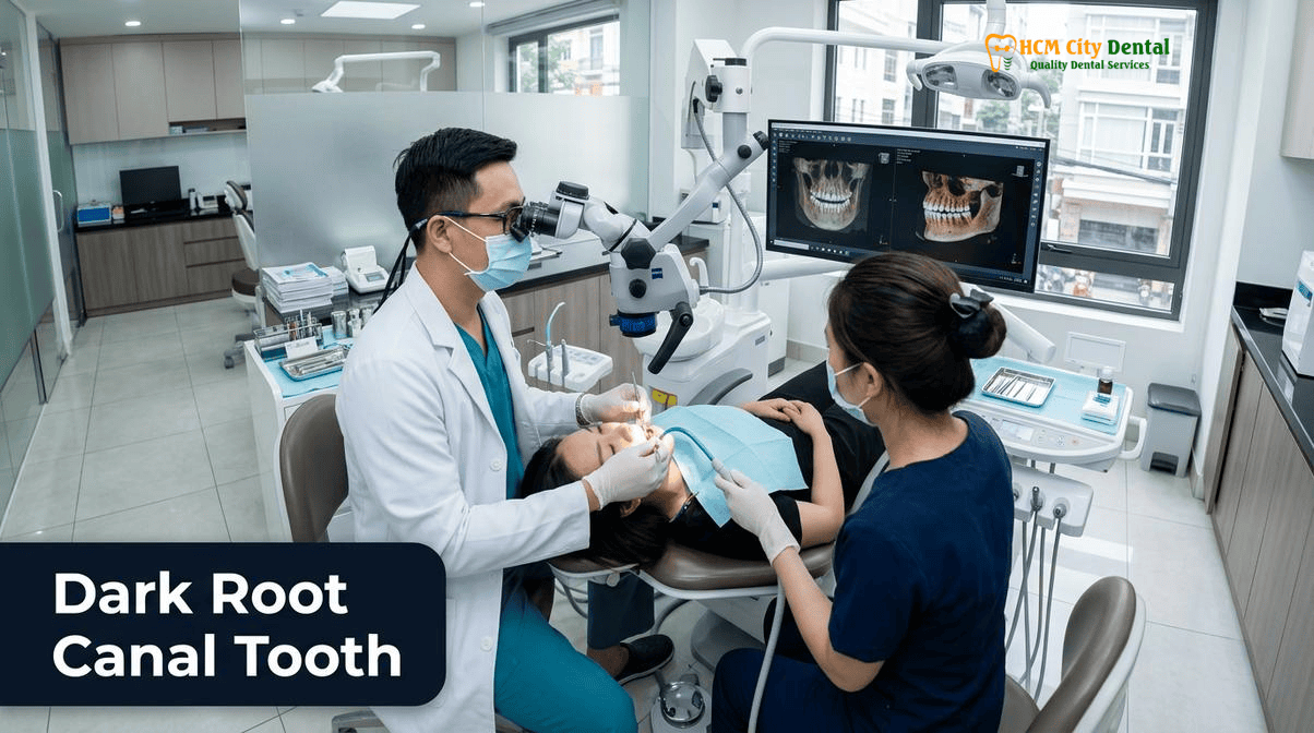
To effectively treat a discolored non-vital tooth, one must first understand the histological and biochemical mechanisms that cause the tooth to darken. Unlike extrinsic stains caused by coffee, tea, tobacco, or red wine—which accumulate on the outer pellicle of the enamel—intrinsic discoloration occurs within the microscopic dentinal tubules that make up the bulk of the tooth’s structure. When evaluating a single dark front tooth treatment Ho Chi Minh City, our specialists conduct a thorough 3D CBCT radiographic and clinical assessment to identify the specific etiology of the discoloration.
\n\n
The most common cause of a dark, non-vital tooth is a history of dental trauma. When a tooth sustains a significant impact—such as from a sports injury, a fall, or an automobile accident—the delicate blood vessels within the dental pulp can rupture. This pulpal hemorrhage leads to the extravasation of erythrocytes (red blood cells) into the surrounding dentinal tubules. As these red blood cells break down and undergo hemolysis, they release hemoglobin. The degradation of hemoglobin produces iron-rich porphyrin compounds, specifically hemosiderin, which oxidizes and imparts a dark grey, brown, or even bluish-purple hue to the dentin from the inside out [1].
\n\n
Another frequent cause of intrinsic discoloration is the presence of necrotic pulp tissue. If a tooth dies slowly following trauma or deep decay without receiving prompt endodontic therapy, the decomposing organic tissue produces dark byproducts that permeate the dentin. Furthermore, the materials used during previous, older root canal treatments can themselves be the culprits. Historically, certain endodontic sealers containing silver, eugenol, or dark metallic oxides were used to fill the root canal space. Over time, these materials can leach metallic ions into the coronal dentin, causing severe, highly refractory grey or black staining. Even modern bioceramic materials, such as certain formulations of Mineral Trioxide Aggregate (MTA) containing bismuth oxide, have been shown to cause significant tooth discoloration when placed above the cementoenamel junction (CEJ) [2].
\n\n
\n
“A single dark front tooth is rarely an enamel problem; it is almost exclusively a dentin problem. Attempting to grind away the dark color from the outside only weakens the tooth structurally. Our primary objective is to chemically neutralize the oxidized iron pigments from within the pulp chamber, preserving the precious enamel shell that provides the tooth with its natural optical properties and biomechanical strength.”
\n — Dr. Nguyen Van Cuong, DDS\n
\n\n
Why External Laser Teeth Whitening Alone Cannot Lighten Intrinsic Hemosiderin and Necrotic Dentin Stains from the Inside
\n\n
Many patients visit our clinic requesting standard Professional Laser Teeth Whitening & Swiss EMS AirFlow Prophylaxis in HCMC to fix their single dark tooth. However, it is a fundamental clinical reality that external whitening procedures—whether utilizing high-concentration hydrogen peroxide activated by a laser or take-home carbamide peroxide trays—are highly ineffective for treating non-vital, endodontically treated teeth.
\n\n
The limitation lies in the permeability and thickness of the dental hard tissues. External whitening gels are designed to penetrate the porous structure of the enamel and oxidize organic chromogens located in the superficial layers of the dentin. However, in a non-vital tooth, the heavy concentration of hemosiderin or metallic sealer stains is located deep within the core of the tooth, specifically in the dentin immediately surrounding the empty pulp chamber. External peroxide molecules simply cannot penetrate deeply enough through the enamel and outer dentin to reach and neutralize these dense, deeply embedded pigment molecules.
\n\n
Furthermore, attempting to aggressively whiten a single dark tooth from the outside using repeated, prolonged sessions of high-concentration peroxide can lead to severe dehydration of the enamel, making it brittle and increasing the risk of micro-fractures. It can also cause significant irritation to the surrounding gingival tissues without achieving any meaningful improvement in the shade of the dark tooth. Therefore, a targeted, internal approach is clinically mandatory to address the root cause of the discoloration directly at its source.
\n\n
Option 1: Non-Vital ‘Walking Bleach’ Internal Whitening (Sodium Perborate / Carbamide Peroxide) — Zero Enamel Drilling
\n\n\n
\n\n\n
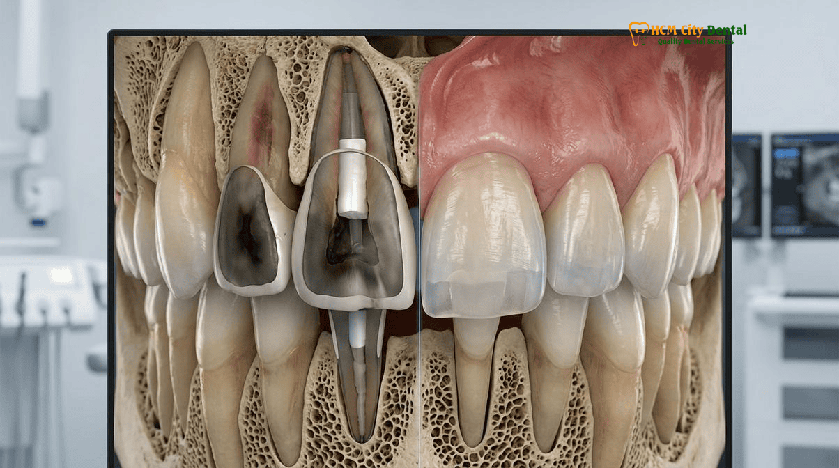
The gold standard, first-line treatment for a discolored endodontically treated tooth is the non vital tooth whitening sodium perborate protocol, universally known in endodontics as the “Walking Bleach” technique. This highly conservative procedure is designed to whiten the tooth from the inside out, requiring absolutely zero drilling or removal of the external facial enamel. By preserving the natural enamel, we maintain the tooth’s inherent optical translucency, surface texture, and structural integrity.
\n\n
The clinical protocol for the internal walking bleach dark root canal tooth procedure at HCMC Dental Clinic is meticulous and strictly adheres to international endodontic standards. The process begins with the placement of a 100% latex-free nitrile rubber dam to isolate the tooth, ensuring a sterile operating field and protecting the patient’s oral mucosa from the bleaching agents. Under the high magnification of a Carl Zeiss operating microscope, the specialist carefully re-enters the existing access cavity on the palatal or lingual (back) side of the tooth.
\n\n
All old composite restorative material, residual necrotic tissue, and remnants of endodontic sealer are meticulously cleaned from the pulp chamber using ultrasonic instruments. The gutta-percha (the rubber-like material filling the root canal) is then carefully removed to a level approximately 2 to 3 millimeters below the cementoenamel junction (CEJ). This creates a clean, empty chamber within the coronal portion of the tooth, ready to receive the bleaching agent.
\n\n
The bleaching agent of choice for the walking bleach technique is typically sodium perborate mixed with sterile distilled water or a mild concentration of carbamide peroxide. Unlike high-concentration hydrogen peroxide, sodium perborate is highly stable and releases active oxygen molecules slowly and steadily over a period of several days. This slow-release mechanism is crucial for safety and efficacy. The paste is packed into the cleaned pulp chamber, and the access cavity is tightly sealed with a high-quality temporary restorative material, such as Cavit or IRM (Intermediate Restorative Material). The patient then “walks” out of the clinic and goes about their normal life while the bleaching agent works continuously from the inside out [3].
\n\n
The patient returns to the clinic every 3 to 7 days for evaluation. If the desired shade has not yet been reached, the old bleaching paste is removed, the chamber is flushed, and a fresh mix of sodium perborate is placed. This cycle is repeated—typically 2 to 4 times—until the tooth matches the shade of the adjacent vital teeth. Once the optimal color is achieved, the bleaching agent is permanently removed, and the tooth is prepared for its final biomimetic composite restoration.
\n\n
Mandatory Safety Protocol to Prevent External Cervical Root Resorption: The 2mm bobsled-shaped RMGI / Biodentine Cervical Barrier
\n\n
While the internal walking bleach technique is highly effective, it carries a severe, albeit rare, potential complication if performed incorrectly: External Cervical Root Resorption (ECRR). ECRR is a destructive, inflammatory process where the body’s own cells (odontoclasts) begin to eat away at the root structure of the tooth at the level of the gumline. If left unchecked, ECRR can rapidly destroy the tooth, leading to catastrophic failure and the need for extraction and dental implant placement.
\n\n
Clinical Warning: The outdated “thermocatalytic” bleaching technique—which involves placing 30% to 35% hydrogen peroxide inside the tooth and heating it with a hot instrument—is strictly prohibited at HCMC Dental Clinic. The extreme heat drives caustic peroxide molecules rapidly through the dentinal tubules into the periodontal ligament, exponentially increasing the risk of external cervical root resorption. Patient safety and long-term tooth retention must always supersede the desire for rapid, single-visit internal whitening.
\n
\n\n
The primary trigger for ECRR following internal bleaching is the diffusion of caustic peroxide molecules through the dentinal tubules at the cervical neck of the tooth, where the enamel meets the cementum. If the peroxide escapes the pulp chamber and reaches the periodontal ligament (PDL) on the outside of the root, it triggers a localized inflammatory response, lowering the pH and activating the destructive odontoclastic cells.
\n\n
To absolutely guarantee patient safety and prevent this complication, HCMC Dental Clinic enforces a mandatory, non-negotiable safety protocol: the placement of a cervical root resorption barrier glass ionomer or bioactive cement. Before any bleaching agent is introduced into the tooth, a precise, 2-millimeter thick layer of Resin-Modified Glass Ionomer (RMGI) or Septodont Biodentine is placed directly over the reduced gutta-percha. This barrier is meticulously shaped like a “bobsled” or a ski slope, extending slightly higher on the internal walls of the dentin to seal off the dentinal tubules that communicate directly with the external cervical area [4].
\n\n
This impenetrable barrier ensures that the active oxygen released by the sodium perborate is directed strictly outward toward the discolored coronal dentin, completely blocking any downward diffusion toward the vulnerable periodontal ligament. This protocol aligns with the highest standards of international endodontic care and ensures that the structural integrity of the root is never compromised during the aesthetic enhancement process.
\n\n
Option 2: Masking Refractory Dark Stumps with Dual-Layer IPS E.max (HO/MO Opacity Ingot) or Layered Zirconia Restoration
\n\n\n
\n\n\n
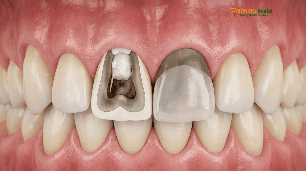
In the majority of cases, the internal walking bleach technique successfully restores the natural color of the tooth. However, there are clinical scenarios where the intrinsic discoloration is so severe, so deeply saturated with metallic ions from old sealers, or so long-standing that it becomes refractory (resistant) to chemical bleaching. In these instances, or if the tooth has already suffered significant structural loss requiring full coverage, the secondary treatment option is advanced digital prosthodontics: masking dark tooth stump under IPS E.max veneer or a full ceramic crown.
\n\n
Masking a dark, non-vital stump is one of the most technically demanding procedures in cosmetic dentistry. Standard translucent porcelain veneers, which look beautiful on healthy, light-colored teeth, will fail miserably on a dark stump. The underlying grey or black value will simply shine through the thin ceramic, resulting in a dull, lifeless, and mismatched restoration. To overcome this, we must utilize the physics of light reflection, absorption, and opacity gradients.
\n\n
At HCMC Dental Clinic, we utilize a dual-layer biomimetic approach for masking refractory dark stumps. The foundation of the restoration is fabricated using a specialized High Opacity (HO) or Medium Opacity (MO) lithium disilicate ingot (IPS E.max). This opaque substructure acts as an optical block, preventing the dark color of the underlying dentin from transmitting through the restoration. However, an entirely opaque restoration would look artificial and “chalky.” Therefore, our master ceramists meticulously layer highly translucent, opalescent feldspathic porcelain over the opaque substructure.
\n\n
This dual-layer technique mimics the natural anatomy of a tooth: the opaque E.max core replicates the light-blocking properties of natural dentin, while the layered translucent porcelain replicates the light-scattering properties of natural enamel. The result is a restoration that successfully masks the dark stump while maintaining the vibrant, lifelike optical depth required for a seamless integration with the adjacent natural teeth.
\n\n
In cases where the tooth is exceptionally dark or requires maximum structural reinforcement, a layered zirconia crown may be indicated. Modern monolithic zirconia, while incredibly strong, can sometimes lack the necessary opacity to block severe discoloration. By utilizing a high-strength, opaque zirconia coping layered with translucent veneering ceramic, we can achieve both exceptional biomechanical stability and superior aesthetic masking capabilities.
\n\n
Biomimetic Bonding Protocols: The 10-to-14 Day Decoupling Period and Immediate Dentin Sealing (IDS)
\n\n
Whether the final restoration is a simple composite resin closure of the access cavity following successful internal bleaching, or a complex IPS E.max masking veneer, the adhesive bonding protocol is critical to the long-term success of the treatment. A fundamental rule of adhesive dentistry is that resin composites and bonding agents will not polymerize (cure) properly in the presence of oxygen.
\n\n
Following an internal walking bleach procedure, residual oxygen molecules remain trapped within the dentinal tubules for a significant period. If a dentist attempts to bond a composite filling or a porcelain veneer immediately after removing the bleaching agent, this residual oxygen will severely inhibit the polymerization of the adhesive resin. This leads to drastically reduced bond strength, microleakage, marginal staining, and ultimately, the premature failure or debonding of the restoration.
\n\n
To prevent this, HCMC Dental Clinic strictly enforces a mandatory 10-to-14 day decoupling period after the completion of the internal bleaching protocol. During this waiting period, the access cavity is sealed with a temporary restoration, allowing the residual oxygen to completely dissipate from the dentinal tubules. Only after this waiting period has elapsed can definitive adhesive bonding procedures commence.
\n\n
When bonding a masking veneer or crown to a non-vital tooth, we employ advanced biomimetic techniques, including Immediate Dentin Sealing (IDS). IDS involves applying a dentin bonding agent to the freshly prepared dentin immediately after tooth preparation, prior to taking the digital impression. This seals the dentinal tubules, protects the pulp (if vital), and significantly increases the ultimate bond strength of the final restoration. By combining the 14-day decoupling period with meticulous IDS protocols, we ensure that our restorations are not only aesthetically flawless but also biomechanically integrated for long-term durability.
\n\n
Clinical Case Study: Restoring a Traumatized Central Incisor at HCMC Dental Clinic
\n\n
Patient Profile: A 32-year-old expatriate visited HCMC Dental Clinic (Ho Chi Minh City) presenting with a severely discolored upper right central incisor (Tooth #11). The patient reported a history of sports trauma 12 years prior, followed by root canal therapy in their home country. The tooth had progressively darkened to a deep grey-brown shade, causing significant aesthetic concern.
\n
Clinical Assessment: 3D CBCT imaging confirmed an intact root canal filling with no signs of periapical pathology. However, the coronal dentin was heavily saturated with hemosiderin and remnants of an older, metallic-based endodontic sealer. The external enamel was intact but structurally compromised by a large palatal access cavity.
\n
Treatment Execution: Dr. Cuong initiated a conservative internal walking bleach protocol. A 2mm RMGI cervical barrier was placed to prevent ECRR. Sodium perborate was sealed inside the chamber for three cycles (7 days each). The bleaching successfully lifted the shade by 4 values but plateaued before perfectly matching the adjacent vital incisor due to the deep sealer staining.
\n
Final Restoration: Following a 14-day decoupling period to allow oxygen dissipation, the tooth was prepared for a minimally invasive masking veneer. An IPS E.max Medium Opacity (MO) lithium disilicate veneer was digitally designed and milled, then layered with translucent porcelain to match the adjacent teeth. The veneer was bonded under rubber dam isolation using heated composite resin.
\n
Outcome: The combination of internal bleaching to reduce the baseline darkness, followed by a biomimetic masking veneer, resulted in a flawless aesthetic integration. The patient regained full confidence in their smile, demonstrating the efficacy of a phased, conservative approach to refractory non-vital discoloration.
\n
\n\n
2026 Internal Non-Vital Bleaching, Fiber Post & Aesthetic Veneer/Crown Fee Schedule at HCMC Dental Clinic (District 1)
\n\n\n
\n\n\n
Understanding the financial investment required for advanced cosmetic and restorative dentistry is crucial for our international patients. At HCMC Dental Clinic, we maintain a policy of absolute transparency regarding our clinical fees. The cost of treating a dark endodontically treated tooth depends on the severity of the discoloration, the necessity for endodontic retreatment, and whether a masking restoration is required.
\n\n
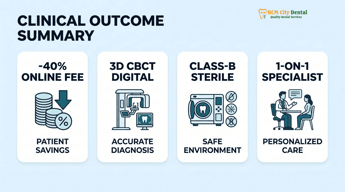
By utilizing our online booking platform, patients can access our exclusive -40% online fee schedule, providing significant savings compared to standard walk-in rates. Below is a detailed breakdown of the costs associated with the various phases of treating a discolored non-vital tooth. For a comprehensive overview of all our services, please review our Official 2026 Dental Price List & -40% Online Booking Fee Schedule.
\n\n
| Clinical Procedure (Non-Vital Tooth Restoration) | Standard Walk-In Price (USD) | -40% Online Booking Price (USD) | Clinical Indication & Warranty |
|---|---|---|---|
| Internal ‘Walking Bleach’ (Per Cycle) | $65 | $39 | First-line therapy for intrinsic stains. Includes RMGI barrier placement. (Typically requires 2-4 cycles). |
| Microscopic Endodontic Retreatment (Anterior) | $192 | $115 | Required if the existing root canal is failing or contaminated prior to bleaching. |
| Fiber Post & Biomimetic Core Build-Up | $65 | $39 | Provides structural reinforcement for severely broken-down non-vital teeth prior to crowning. |
| IPS E.max® Lithium Disilicate Masking Veneer | $308 | $185 | Dual-layer HO/MO masking for refractory stains. Includes 10-Year Clinical Warranty. |
| IPS E.max® All-Ceramic Crown (Full Coverage) | $385 | $231 | Maximum structural protection and aesthetic masking. Includes 10-Year Clinical Warranty. |
\n\n
When to Consult a Specialist for a Dark Root-Canal-Treated Tooth
\n\n
While a dark tooth is primarily an aesthetic concern, it can sometimes indicate underlying pathological issues that require immediate specialist attention. It is crucial to monitor a non-vital tooth for any signs of failure or complication, particularly if it has a history of trauma or previous endodontic therapy.
\n\n
You should schedule a consultation at HCMC Dental Clinic if you experience any of the following symptoms associated with a discolored tooth:
\n
- \n
- Persistent Pain or Tenderness: A properly treated non-vital tooth should not cause pain. Pain upon biting or tapping the tooth may indicate a failing root canal, a hidden fracture, or recurrent infection at the root apex.
- Swelling or a “Pimple” on the Gums: The presence of a sinus tract (a small bump on the gums that may drain pus) is a definitive sign of an active chronic infection that requires immediate endodontic retreatment.
- Mobility: If the dark tooth feels loose, it may be suffering from advanced periodontal disease, a root fracture, or severe external cervical root resorption.
- Rapid Darkening: While gradual darkening over years is common, a sudden, rapid change in color may indicate acute internal bleeding (if the tooth was previously vital) or a rapid breakdown of restorative materials.
\n
\n
\n
\n
\n\n
\n
“Patients must understand that a dark tooth is a clinical symptom, not just a cosmetic blemish. Before we even discuss bleaching or veneers, we must verify the biological health of the root and the surrounding bone. A beautiful masking veneer placed over a failing, infected root canal is a disservice to the patient. Comprehensive diagnostics always precede aesthetic execution.”
\n — Dr. Nguyen Van Cuong, DDS\n
\n\n
Conclusion & Patient Next Steps
\n\n\n
\n\n\n
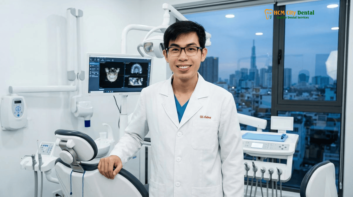
The management of a single dark endodontically treated tooth requires a sophisticated, multidisciplinary approach that balances conservative chemical bleaching with advanced digital prosthodontics. Whether your optimal treatment path involves the minimally invasive internal walking bleach technique or the precise application of a biomimetic masking veneer, the ultimate goal remains the same: to restore the natural beauty, structural integrity, and functional harmony of your smile.
\n\n
At HCMC Dental Clinic, we are committed to providing international standards of care, utilizing the latest biomaterials and evidence-based protocols to ensure predictable, long-lasting results. Our strict adherence to safety protocols, including the mandatory placement of cervical barriers and the observance of critical decoupling periods, guarantees that your aesthetic enhancement never compromises your biological health. All procedures are conducted in strict compliance with the latest safety directives [5].
\n\n
If you are struggling with the aesthetic burden of a discolored non-vital tooth, we invite you to experience the highest level of specialist dental care in Vietnam. Contact us today to schedule your comprehensive consultation at HCMC Dental Clinic, District 1, Ho Chi Minh City, and let our expert team design a personalized treatment plan to restore your confidence and your smile.
\n\n
Frequently Asked Questions About Internal Walking Bleach vs. Masking Veneers
\n
Can regular external laser teeth whitening fix a single dark root-canal-treated front tooth?
\n
No. External laser teeth whitening only oxidizes extrinsic enamel chromogens from coffee, tea, or wine. A tooth that turned grey, brown, or purple after trauma or root canal therapy is stained from the inside out by hemosiderin iron pigments trapped inside the dentinal tubules, requiring internal intracoronal bleaching or a masking ceramic restoration.
\n
Is internal walking bleach safe, and how do you prevent external cervical root resorption?
\n
Yes, when performed strictly without heat (`thermocatalytic bleaching` is contraindicated) and without 35% liquid hydrogen peroxide. At HCMC Dental Clinic in District 1, Ho Chi Minh City, we place a mandatory 2.0mm bobsled-shaped RMGI or Biodentine barrier over the gutta-percha at the cementoenamel junction and use biocompatible sodium perborate so peroxide molecules can never diffuse into the periodontal ligament.
\n
How many days does internal non-vital bleaching take for an international patient in Ho Chi Minh City?
\n
Most traumatized or endodontically discolored incisors lighten by 4 to 8 VITA shade tabs across 1 to 2 intracoronal dressing appointments spaced 3 to 5 days apart, making it ideal for a 5-to-7 day stay in District 1.
\n
Why must my dentist wait 10 to 14 days after internal bleaching before bonding a composite core or porcelain veneer?
\n
Residual oxygen free radicals trapped inside bleached dentinal tubules inhibit the free-radical polymerization of resin adhesives, reducing bond strength by up to 50%. Applying a 10% sodium ascorbate antioxidant gel or waiting 10 to 14 days restores 100% biomimetic adhesive bond strength prior to IPS e.max veneer or zirconia crown cementation.
\n
When is an IPS e.max HO/MO masking veneer or zirconia crown better than internal bleaching?
\n
If the dark discoloration is caused by silver-containing endodontic sealers, metal amalgam posts, severe calcific metamorphosis, or extensive structural loss (>40% coronal fracture), internal bleaching alone cannot mask the metallic substrate. In those cases, a dual-layer IPS e.max HO/MO coping or multilayer zirconia crown blocks the dark stump while replicating the translucency of the adjacent central incisor.
\n\n
References
\n
- \n
- Journal of Endodontics. Efficacy and Safety of Sodium Perborate in Non-Vital Tooth Bleaching: A Systematic Review of Cervical Resorption Risks. (2022).
- International Journal of Periodontics & Restorative Dentistry. Biomimetic Masking of Discolored Endodontically Treated Teeth: Lithium Disilicate Opacity Gradients and Immediate Dentin Sealing. (2023).
- Journal of Prosthetic Dentistry. The Role of Resin-Modified Glass Ionomer Cervical Barriers in Preventing Radicular Peroxide Diffusion During Walking Bleach Protocols. (2021).
- American Journal of Orthodontics and Dentofacial Orthopedics (AJODO). Multidisciplinary Management of Traumatized Anterior Maxillary Dentition: Endodontic, Orthodontic, and Prosthodontic Considerations. (2020).
- Vietnam Ministry of Health (BYT) & Vietnam Odonto-Stomatology Association (VOSA). National Clinical Guidelines on Specialist Dental Care, Biomaterial Traceability & EN 13060 Class B Infection Control. (2024).
\n
\n
\n
\n
\n
\n\n